The Classic Tetrad Plus Nephropathy
- Classic Tetrad: Nails, Patellae, Iliac horns, Elbows.
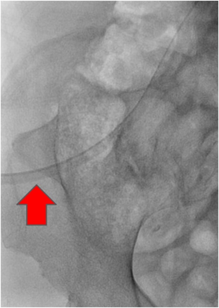
- Iliac Horns: Pathognomonic - bony spurs off the posterior ilium.
- Patellar Instability: From absent/hypoplastic patellae plus dysplastic trochlea.
- Nephropathy: Determines long-term prognosis - screen urine for proteinuria.
- Glaucoma: Also associated - needs ophthalmology screening.
- “Iliac horns are pathognomonic
- “LMX1B autosomal dominant
- “Patella absent or hypoplastic and laterally displaced
- “Nephropathy determines prognosis
Overview and Epidemiology
Nail-patella syndrome (NPS), also called hereditary onycho-osteodysplasia or Fong disease, is a rare connective-tissue and developmental disorder. It is caused by mutations in LMX1B on chromosome 9q34, which encodes a LIM-homeodomain transcription factor, and its prevalence is approximately 1 in 50,000.
Inheritance. NPS is autosomal dominant, with high penetrance but very variable expression, even within one family.
The defining concept. A single transcription factor defect produces both the dorsoventral limb patterning errors, in the nails, patella and elbow, and the glomerular basement membrane defect that causes the nephropathy.
Pathophysiology and Mechanisms
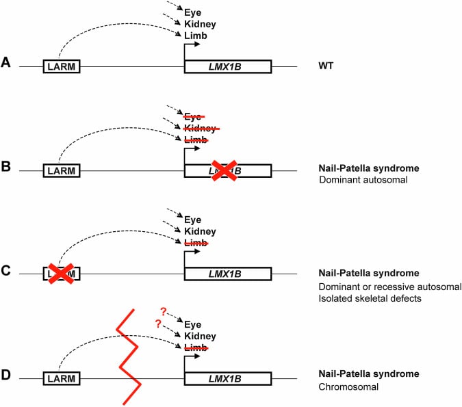
One gene, two pathways. LMX1B is a transcription factor required for normal dorsoventral patterning of the limb during development, and for normal podocyte and glomerular basement membrane formation. Loss of one functional copy (haploinsufficiency) disrupts both pathways, which is why one gene causes such varied features.
The limb. Without normal dorsalisation the dorsal limb structures are underdeveloped: the nails, the dorsal-most ectodermal structure, and the patella, a dorsal sesamoid. The trochlear groove also forms abnormally, shallow and dysplastic, so even a patella that is present is unstable.
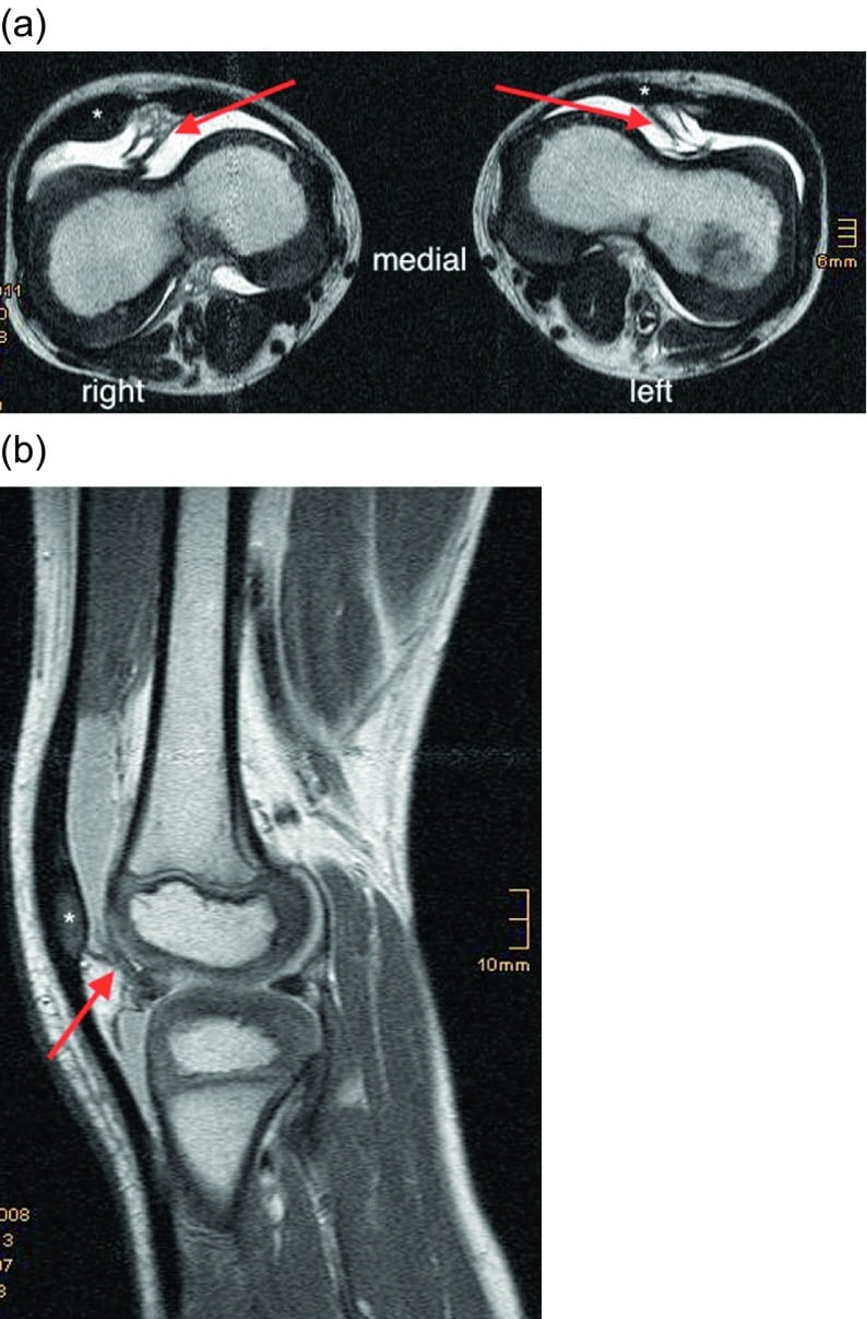
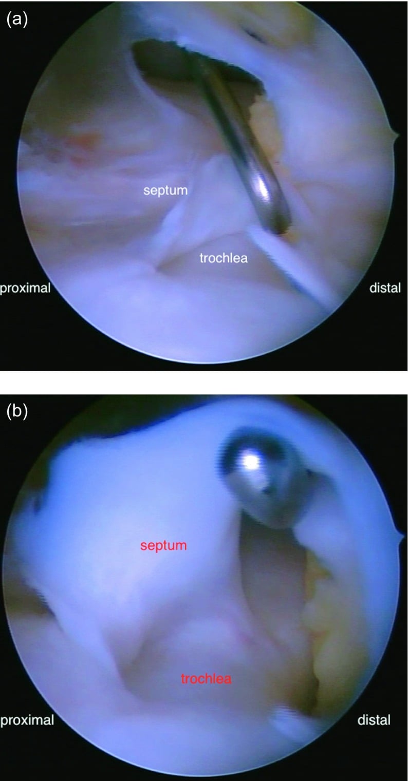
The knee. An absent or small patella, a shallow dysplastic trochlea and abnormal extensor mechanism alignment combine to drive lateral subluxation or dislocation. A distinctive feature reported in NPS is a synovial or fibrous band that can physically prevent the patella from engaging the groove, a pathology unlike ordinary patellofemoral instability.
The kidney. LMX1B controls expression of podocyte genes and type IV collagen chains. Its loss produces a defective glomerular basement membrane that manifests as proteinuria; the ultrastructural lesion is described with the renal biopsy below.
Classification Systems
NPS is described by the classic tetrad, by where the knee sits on the patellofemoral spectrum, and by the systems involved beyond the skeleton.
The tetrad. The frequency and management of each feature are in the table below.
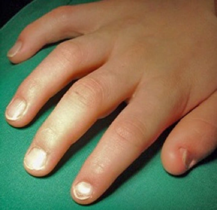
- Nails - the most constant feature: hypoplastic and dystrophic, with longitudinal ridging and triangular lunulae
- Patellae - absent or hypoplastic, and laterally displaced
- Iliac horns - posterior bony spurs off the ilium, and pathognomonic
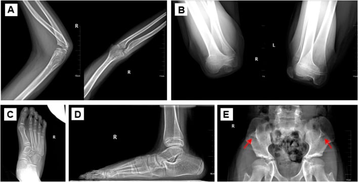
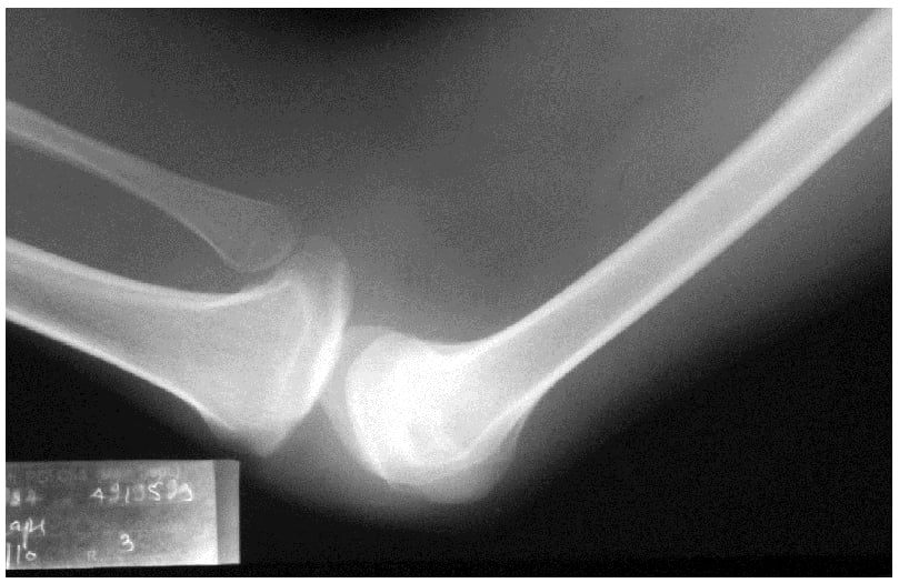
- Elbows - radial head dislocation, limited extension and supination, antecubital pterygia
- Frequency
- ~98%
- Management
- Cosmetic only
- Frequency
- ~90%
- Management
- Realignment if recurrent instability
- Frequency
- ~70% (pathognomonic)
- Management
- No treatment
- Frequency
- ~70%
- Management
- Usually non-operative
- Frequency
- ~30-50%
- Management
- Nephrology referral, surveillance
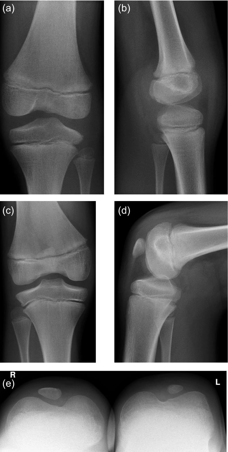
The knee spectrum. The patella may be completely absent as an ossified structure (aplasia), or small and often riding laterally (hypoplasia). Recurrent dislocation is the most common symptomatic presentation, and secondary patellofemoral osteoarthritis follows late in long-standing maltracking.
Beyond the skeleton. The associated systems:
- Renal (LMX1B nephropathy) - proteinuria and haematuria, which may progress to ESRD
- Ophthalmic - open-angle glaucoma and ocular hypertension
- Neurological and other - variable, with sensorimotor and gastrointestinal features reported
What the Iliac Horns Actually Are
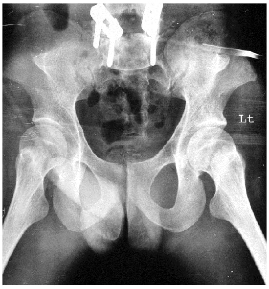
What they are. Bilateral, roughly symmetric, conical bony processes arising from the external (posterior) surface of the iliac wings and projecting posteriorly, and slightly laterally. A horn may carry its own secondary ossification centre, a separate epiphysis.
Finding them. They are seen on an AP pelvis radiograph, are present in around 70 percent of patients, and are asymptomatic.
Why they matter. Iliac horns are pathognomonic: their presence is essentially diagnostic of NPS. They were among the first radiological signs described, by Fong in 1946, hence the eponym "Fong disease".
Clinical Assessment
The skeletal features are cosmetic or functional; nephropathy is what drives morbidity, and it can progress to end-stage renal failure. Renal involvement may even precede overt orthopaedic signs. A young patient referred for patellar instability with odd nails deserves renal screening and a family history, not just a knee work-up.
History. Inheritance is autosomal dominant, so take a family history and examine the relatives' nails. Ask about recurrent giving way, dislocation and anterior knee pain, about any known proteinuria, hypertension or renal disease, and about visual symptoms or a history of glaucoma.
Examination. Work through the tetrad, then check the systemic signs:
- Nails - inspect every fingernail for hypoplasia, ridging and triangular lunulae, worst at the thumb
- Knees - palpate for the patella, which may be impalpable; assess tracking, apprehension, the J-sign, effusion and range of motion
- Elbows - loss of extension and supination, a palpable dislocated radial head, cubitus valgus, pterygia
- Pelvis - the iliac horns are not usually palpable and are found on imaging
- General - blood pressure and, where possible, a urine dipstick for proteinuria and haematuria
Investigations
Radiographs. Image each skeletal site of the tetrad:
- AP pelvis - iliac horns
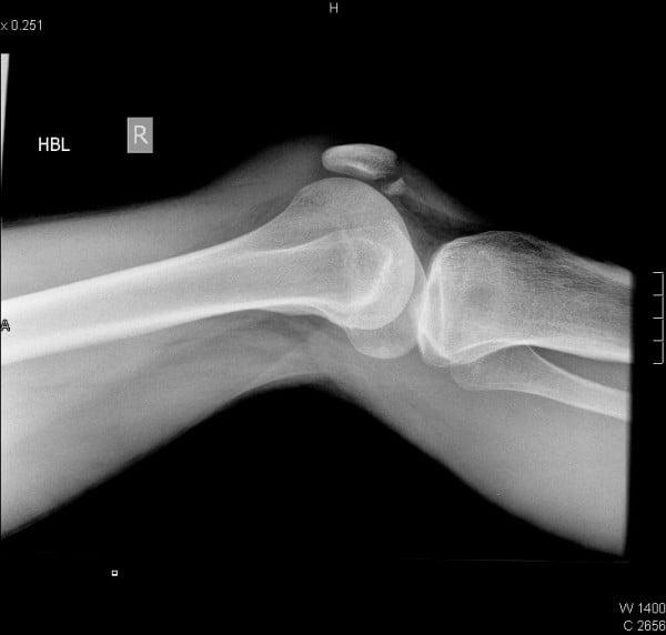
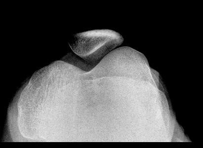
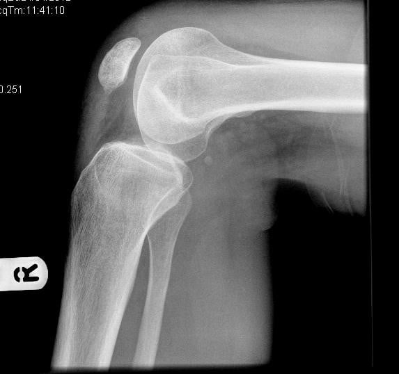
- Knee, AP, lateral and skyline - absent or hypoplastic patella, lateral subluxation, trochlear dysplasia
- Elbow - radial head dislocation, hypoplastic capitellum
MRI of the knee. Before realignment surgery, MRI shows the cartilaginous patellar anlage, the trochlear morphology and the extensor mechanism, and finds any synovial band.
Renal and systemic. Urinalysis looks for proteinuria and haematuria, and a protein:creatinine ratio quantifies the protein. Measure blood pressure and renal function, and reserve renal biopsy for significant nephropathy that needs characterisation. Arrange an ophthalmology review for glaucoma.
Genetics. LMX1B sequencing confirms the diagnosis and aids family counselling.
Differential Diagnosis
- Isolated trochlear dysplasia / patellofemoral instability: No nails, iliac horns, or renal features.
- Small patella syndrome (ischiopubic-patellar): Patella hypoplasia with pelvic anomalies but no iliac horns or nail changes.
- Patella aplasia-hypoplasia (PTLAH): Isolated patellar maldevelopment without the full tetrad.
- Iliac horns on pelvis radiograph (pathognomonic).
- Triangular lunulae and dystrophic nails.
- Family history (autosomal dominant) and proteinuria.
- Proteinuria or hypertension → nephrology referral (prognosis driver).
- Visual symptoms → glaucoma assessment.
- Anterior knee instability with intact cruciates → consider absent patella.
- Open-angle glaucoma.
- LMX1B-associated isolated nephropathy in relatives.
Management Algorithm
The knee. Asymptomatic or mild instability is treated with activity modification, quadriceps (VMO) strengthening and physiotherapy. Recurrent dislocation is the indication for realignment surgery, with counselling beforehand that results are variable and instability may persist.
What makes NPS different. The operation should address the underlying pathology, resecting any obstructing synovial band and recentring the extensor mechanism, not just perform a generic soft-tissue procedure.
Late arthritis. It is managed as patellofemoral osteoarthritis; the salvage options are under surgical techniques.
The elbow. Most cases are managed non-operatively, because function is usually acceptable despite the limited extension and supination. A dislocated radial head is generally left alone in children, since relocation is unreliable.
The kidney, the eye and the family.
- Nephropathy - nephrology referral; monitor proteinuria and blood pressure; ACE inhibitors or ARBs for proteinuria; dialysis or transplantation if it progresses
- Glaucoma - ophthalmology surveillance and control of intraocular pressure
- Genetic counselling - a 50 percent transmission risk; offer LMX1B testing and discuss testing for children
Surgical Techniques
Indications for realignment. Recurrent dislocation with a patella present, even a hypoplastic one, and a functional extensor mechanism.
Principles. Maltracking is corrected distally and proximally, and in NPS the crucial step is releasing the band:
- Distal realignment - tubercle transfer in the skeletally mature
- Proximal soft-tissue balancing - lateral release, medial reefing or MPFL reconstruction
- Release of any synovial band blocking trochlear engagement
Counselling. Persistent instability is reported in roughly 40 percent after realignment, and in the largest series that is the same rate as in patients who were never operated on. The figure is therefore the background rate of instability in a knee whose trochlea, patella and extensor mechanism are all malformed, and it does not measure how often the operation fails. Surgery is offered for pain and function, which do improve, and not on a promise of a stable patella.
Salvage for late arthritis. In historically advanced or end-stage cases patellectomy has been described, accepting that complete patellar loss can itself cause anterior knee instability. Total patellar absence reduces the extensor lever arm and has been linked to anterior knee instability mimicking ACL insufficiency.
Complications
- Risk
- Common (NPS pathomorphology)
- Prevention
- Address synovial band + realignment
- Management
- Revision realignment
- Risk
- ~40%, the same as unoperated NPS knees
- Prevention
- Treat underlying pathology, not generic release
- Management
- Counsel before surgery; revise only if disabling
- Risk
- Long-standing maltracking
- Prevention
- Early stabilisation, strengthening
- Management
- OA management, patellectomy late
- Risk
- If patella fully absent
- Prevention
- Avoid unnecessary patellectomy
- Management
- Bracing, rehab
- Risk
- Rare; small patella complicates fixation
- Prevention
- Confirm extensor disruption and displacement on orthogonal views
- Management
- Fix a displaced fracture with extensor disruption; protect the repair with staged mobilisation
Beyond the knee. Progressive nephropathy, managed as above, can reach end-stage renal disease. Untreated glaucoma costs vision. Elbow contracture limits function but is usually accepted and rarely operated on.
Before any elective operation. Check renal function and proteinuria, and adjust nephrotoxic drugs and NSAIDs accordingly. Screen the family too: other affected relatives may have undiagnosed nephropathy.
Postoperative Care
After patellar realignment. Brace the knee in extension initially, then progress flexion as the construct allows. Rehabilitation starts with early quadriceps activation and VMO-focused strengthening, with a gradual return to activity over 3-6 months. Watch for redislocation and effusion, and reassess tracking.
Long-term surveillance. Care is coordinated between orthopaedics, nephrology and ophthalmology:
- Interval
- Periodic urinalysis + BP
- Purpose
- Detect/track nephropathy
- Interval
- Regular IOP checks
- Purpose
- Detect glaucoma
- Interval
- As symptomatic
- Purpose
- Instability, early OA
Outcomes and Prognosis
The knee. Mild instability treated with physiotherapy is often manageable non-operatively. Realignment with band release for recurrent dislocation gives good pain relief and satisfaction, with the persistent instability rate discussed under counselling. The outcome of patellofemoral OA is variable, and patellectomy is a late salvage.
The skeleton overall. The skeletal features are stable and non-progressive, and function is usually preserved. The iliac horns and nails are diagnostic but clinically inconsequential.
The kidney. Nephropathy is highly variable: many patients have only intermittent proteinuria, but a subset progress to end-stage renal failure, and this is the principal determinant of long-term prognosis. Life expectancy is normal in most, dominated by renal status. Renal surveillance is the single most important long-term action.
The Renal Biopsy in NPS Nephropathy
Light microscopy is often near-normal early, with focal segmental glomerulosclerosis (FSGS)-like or mesangial changes later, and is non-specific.
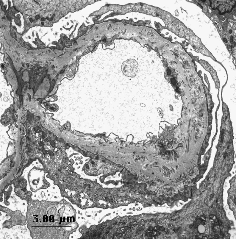
Electron microscopy is diagnostic. It shows irregular thickening of the glomerular basement membrane with electron-lucent ("moth-eaten") areas containing bundles of fibrillar (type III) collagen, together with podocyte foot-process effacement.
Immunostaining shows reduced alpha-3, alpha-4 and alpha-5 chains of type IV collagen in the basement membrane. LMX1B regulates these collagen genes, as well as podocin (NPHS2) and CD2AP, which is why the membrane is structurally defective and leaks protein. The resulting proteinuria can be nephrotic-range.
Guidelines, Registries & Global Practice
Global epidemiology
- NPS is rare, around 1 in 50,000, with worldwide distribution and autosomal dominant inheritance; roughly half of cases may be new (de novo) mutations.
- The skeletal tetrad is highly penetrant (nails ~98 percent, patellar changes ~90 percent, iliac horns ~70 percent, elbow dysplasia ~70 percent); nephropathy affects a substantial minority and drives prognosis.
Side-by-side guidance
- Practice emphasis
- Clinical tetrad + iliac horns on pelvis film; confirm with LMX1B sequencing
- Rationale
- Iliac horns are pathognomonic; genetics aids family counselling
- Practice emphasis
- Periodic urinalysis/BP; ACEi/ARB for proteinuria; nephrology referral
- Rationale
- Nephropathy is the main prognostic determinant
- Practice emphasis
- Physiotherapy first; surgery targets the malformative pathology, counsel on ~40 percent persistent instability
- Rationale
- Generic realignment underperforms
- Practice emphasis
- Glaucoma screening
- Rationale
- Open-angle glaucoma association
Registry and outcome notes
- No NPS-specific registry exists given the rarity; the strongest orthopaedic evidence is a single large questionnaire-based series and small surgical case series, so recommendations are necessarily low-level.
High- vs limited-resource practice
- Where available, MRI and LMX1B genetic testing refine the knee plan and confirm diagnosis, and multidisciplinary clinics coordinate renal/ophthalmic care.
- In limited-resource settings, the diagnosis can still be made on clinical examination plus a plain AP pelvis (iliac horns), and the priority is urine dipstick surveillance and blood pressure to catch treatable nephropathy early.
Controversies & Areas of Uncertainty
1. Surgery versus conservative management of the knee
- The largest series found surgically treated patients had lower KOOS/Kujala scores and the same rate of persistent instability as non-operated patients - yet reported good satisfaction. Whether and when to operate, and which procedure to choose, remains debated.
2. What "nail-patella syndrome" even means
- Discovery that some LMX1B homeodomain mutations cause isolated nephropathy without any nail, patellar or skeletal signs ("LMX1B-associated nephropathy") challenges the classic eponymous definition and blurs the boundary of the diagnosis.
3. Genotype-phenotype correlation
- There is no reliable correlation between LMX1B mutation and severity of renal or skeletal disease, so risk cannot be predicted from the genotype alone - mandating lifelong surveillance.
4. Role of patellectomy
- Historically used for end-stage patellofemoral arthritis, but complete patellar loss can itself cause anterior knee instability, so its role is increasingly limited.
MCQ Practice Points
Q: What radiological finding is pathognomonic of nail-patella syndrome? A: Iliac horns - posterior bony spurs off the iliac wings.
Q: What gene and inheritance pattern underlie NPS? A: LMX1B on chromosome 9q34, autosomal dominant.
Q: What determines long-term prognosis in NPS? A: Nephropathy (proteinuria, may progress to end-stage renal failure).
Q: What proportion have persistent patellar instability after realignment in NPS? A: Around 40 percent - and the point of the figure is that it is the same rate as in patients managed non-operatively, so it is the background rate of a malformed patellofemoral joint rather than a surgical failure rate. Pain relief and satisfaction are generally good, which is why the operation is offered.
Q: What NPS-specific structure can block patellar reduction? A: An obstructing synovial band preventing trochlear engagement.
Q: What ophthalmic condition is associated with NPS? A: Open-angle glaucoma / ocular hypertension.
Self-Assessment Quiz
Additional Quiz Questions
Viva Scenarios
Practise clinical reasoning and management decisions out loud
“A 14-year-old presents with recurrent patellar dislocations. Their AP pelvis shows bilateral bony spurs projecting posteriorly from the iliac wings. What is the unifying diagnosis and what else do you assess?”
“A 16-year-old with confirmed NPS has recurrent patellar dislocation despite physiotherapy. How do you plan surgery and what do you tell them about outcome?”
“A young adult complains of the knee giving way with a positive anterior drawer sensation, but MRI shows intact cruciates. On examination you cannot palpate a patella. What is going on?”
TETRAD
- Nails ~98%
- Patellae absent/hypoplastic
- Iliac horns pathognomonic
- Elbow/radial head dislocation
GENETICS
- LMX1B gene 9q34
- Autosomal dominant
- Variable expression
- ~1 in 50,000
KNEE
- Lateral patella
- Shallow trochlea
- Synovial band can block reduction
- ~40% unstable after surgery = same as unoperated
PROGNOSIS
- Nephropathy drives prognosis
- Proteinuria to ESRD
- Glaucoma association
- Screen urine + BP
MANAGEMENT
- Physio first for knee
- Address malformative pathology
- ACEi/ARB for proteinuria
- MDT: ortho/nephro/eye
EXAM PEARLS
- Iliac horns = diagnosis
- Absent patella mimics ACL instability
- Avoid needless patellectomy
- LMX1B can cause isolated nephropathy
Evidence Base
Bongers EMHF, Gubler MC, Knoers NVAM - Nail-patella syndrome: overview on clinical and molecular findings
- Defines NPS as autosomal dominant pleiotropic disorder: nail dysplasia, patellar aplasia/hypoplasia, iliac horns, elbow dysplasia, plus glaucoma and progressive nephropathy
- LMX1B is a LIM-homeodomain transcription factor controlling dorsoventral limb patterning and early glomerular basement membrane morphogenesis
- No reliable genotype-phenotype correlation for severity of renal or extrarenal anomalies
Harita Y, Kitanaka S, Isojima T, Ashida A, Hattori M - Spectrum of LMX1B mutations: from nail-patella syndrome to isolated nephropathy
- Renal involvement is the major determinant of prognosis in NPS
- Some LMX1B homeodomain mutations cause isolated nephropathy with no nail, patellar or skeletal signs (LMX1B-associated nephropathy)
- Patients present with varying proteinuria/haematuria and may progress to chronic renal failure