Ligamentous Laxity and Orthopaedic Challenges
- Atlantoaxial Instability: ADI greater than 5mm on lateral flexion-extension.
- Ligamentous Laxity: Universal - affects all joints.
- Hip Instability: Late onset common. May develop in childhood.
- Cervical Surveillance: No routine radiographic clearance when asymptomatic; use clinical assessment and neutral-position precautions.
- Os Odontoideum: Associated with AAI.
- “ADI greater than 5mm is abnormal
- “Neutral cervical positioning during intubation
- “Hip instability is late onset
- “Ligamentous laxity universal
Overview and Epidemiology
Down syndrome (trisomy 21) is the most common chromosomal abnormality: an extra chromosome 21, in 1 in 700-1000 live births. Associated conditions include cardiac defects, hypothyroidism, leukaemia and cognitive impairment.
One cause, several joints. The key feature is generalised ligamentous laxity affecting all joints, and the orthopaedic problems follow from it:
- Atlantoaxial instability - radiographic in 10-20%
- Hip instability - 10-20%
- Patellar instability - common
- Pes planovalgus - 90%, nearly universal
Pathophysiology and Mechanisms
Ligamentous laxity. Collagen abnormalities lead to the laxity, and all joints are hypermobile. In the upper cervical spine the lax structure is the transverse ligament of C1, and the result is atlantoaxial instability.
Why the hip is different. DDH is present at birth; hip instability in Down syndrome may develop later. The lax capsule and ligaments allow progressive subluxation, and the child may present at 2-10 years with a painful limp.
Classification Systems
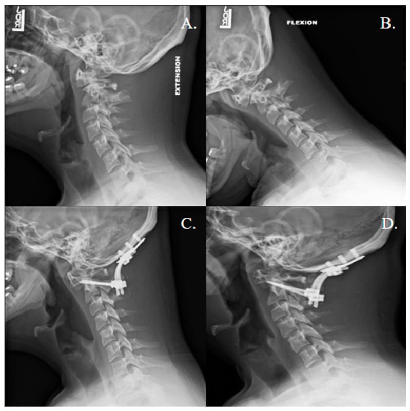
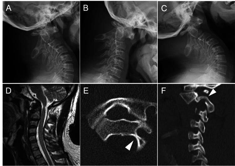
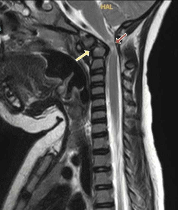
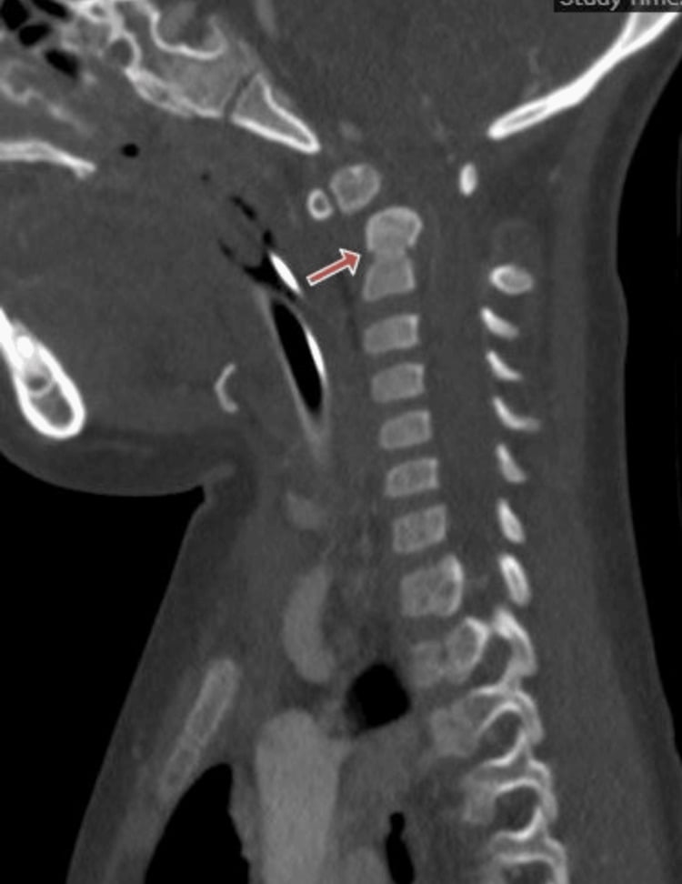
Cervical spine. Atlantoaxial instability is defined radiographically as an atlantodental interval (ADI) greater than 5mm on lateral flexion-extension radiographs, and os odontoideum is an associated finding. Most children with AAI are asymptomatic, and they are still at risk. The symptomatic child presents with myelopathy, gait changes or torticollis.
Hip. The unstable hip in Down syndrome takes several forms:
- Habitual dislocation - voluntary
- Subluxation or dislocation - progressive
- Dysplasia - secondary acetabular changes
- AVN - can occur after reduction, and the risk is high
Keep slipped capital femoral epiphysis on the list. Down syndrome carries a high rate of hypothyroidism, and endocrinopathy is an established SCFE risk factor. A limp or knee pain in an older child with Down syndrome therefore deserves hip radiographs and a thyroid check rather than attribution to laxity.
Predicting the other side. The standard tools for predicting a contralateral slip, such as the modified Oxford bone age score, were derived in cohorts that explicitly excluded children with Down syndrome and endocrine disorders. They do not transfer to this population, and the threshold for prophylactic fixation of the other side has to be judged, not calculated.
Knee and foot. Patellar instability presents as recurrent dislocation and has its own section below. Pes planovalgus is usually asymptomatic. Metatarsus primus varus is seen, with hallux valgus.
Clinical Assessment
History. Ask about developmental milestones and walking age, then specifically about neck pain, gait changes, and hip or knee symptoms.
Examination. Look first for hypotonia and short stature; the Beighton score for generalised laxity is often high. Then examine each region:
- Cervical spine - range of movement, where limitation may indicate instability, and a neurological examination
- Hips - stability and range of movement
- Knees - patellar tracking and apprehension
- Feet - arch and alignment
Investigations
Cervical spine. Symptoms and examination decide who is imaged, and the sequence runs:
- Asymptomatic child - no routine screening radiographs; a focused history and neurological examination instead
- Symptoms or an abnormal examination - a neutral cervical radiograph first, and specialist review; flexion-extension views may be considered with the specialist if the neutral film is not significantly abnormal
- CT - defines os odontoideum or other bony anomalies and plans fixation
- MRI - assesses cord compression, the cerebrospinal-fluid space and cord signal when symptoms, proven instability or specialist concern exists
Hips. An AP pelvis and a lateral radiograph, with MRI if AVN is suspected.
Differential Diagnosis
- Ehlers-Danlos Syndrome: Skin hyperelasticity, joint hypermobility, different genetics.
- Marfan Syndrome: Tall stature, arachnodactyly, aortic root dilatation.
- Larsen Syndrome: Multiple joint dislocations at birth.
- Characteristic facial features.
- Cognitive impairment.
- Chromosome analysis confirms Trisomy 21.
- Cardiac defects (40-50%).
- Any neurological symptoms → urgent cervical spine assessment.
- New limp → hip instability developing.
- Patellar dislocation → realignment may be needed.
- Cardiac defects (40-50%) - AV canal defects.
- Hypothyroidism - screen regularly.
- Leukemia - increased risk.
- Hearing impairment - assess.
Management Algorithm
Cervical surveillance. Surveillance of the neck is clinical: ask about myelopathic symptoms, examine neurologically, use neutral positioning during procedures and image selectively. Management then follows the clinical picture:
- Asymptomatic with no known pathology - clinical surveillance; no routine radiographic clearance
- Proven radiographic instability - paediatric spine review, assessment of PADI, MRI findings and progression, and individualised activity advice
- Neurological symptoms or cord compression - urgent specialist assessment and stabilisation
- Before anaesthesia - focused clinical assessment and neutral-position precautions; image only when symptoms, known instability or another clinical concern exists
Symptomatic instability. Neck pain, torticollis or myelopathy start the work-up above: a neutral radiograph and urgent specialist review. Symptomatic AAI is then treated by C1-C2 fusion to prevent cord injury.
Hip instability. An asymptomatic, stable hip is observed. A hip that is reducible, with intact cartilage, is reconstructed by open reduction with pelvic and/or femoral osteotomy. Reconstruction has a high failure rate, and the failed hip may need salvage by a Girdlestone or McHale procedure. The AVN risk is high after any procedure.
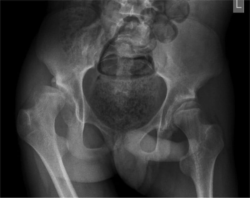
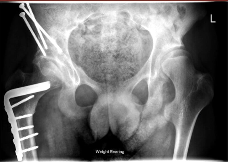
Knee and foot. Patellar instability is managed as set out in its own section below. Planovalgus is treated with orthotics, and surgery is rarely needed.
Scoliosis in Down Syndrome
A syndromic curve. Scoliosis is more common in Down syndrome than in the general population. It is driven by the same generalised ligamentous laxity and hypotonia as the other manifestations, so it behaves like a syndromic or neuromuscular curve rather than an idiopathic one.
Two clinical settings. The curve appears in one of two ways:
- An idiopathic-like curve arising in the ambulatory child or adolescent
- A curve following cardiac (open-heart) surgery, a recognised post-thoracotomy association in this population, given how many children with Down syndrome have congenital heart disease
Watching and bracing. Hypotonia and laxity can let curves progress, so monitor clinically and radiographically through growth. Bracing is often poorly tolerated and less effective than in idiopathic scoliosis, because of body habitus, hypotonia, and compliance and cognitive factors, and it has a limited role.
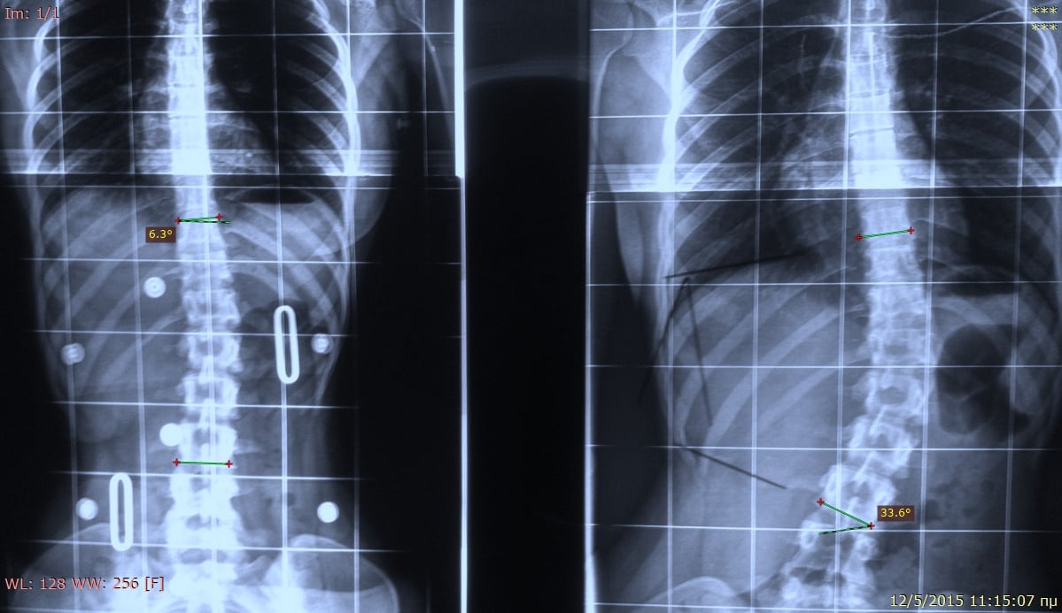
Surgery. Posterior instrumented fusion is reserved for progressive curves, and the complication burden is markedly higher than in idiopathic scoliosis:
- Infection and wound problems - immune dysfunction
- Implant or fixation failure and pseudarthrosis - hypotonia and low bone mass
- A higher neurological risk
Before a prone operation. Two pre-conditions are non-negotiable: a focused cervical history and neurological examination, with targeted imaging when clinically indicated, and a neutral-position airway and positioning strategy planned before prone spinal surgery and intubation. General correction technique is developed in the idiopathic and neuromuscular scoliosis topics; here the point is the syndromic behaviour and the perioperative caveats.
Patellar Instability in Down Syndrome
Why it dislocates. Recurrent, and sometimes habitual or fixed, patellar dislocation is common in Down syndrome because three factors stack up. It is frequently bilateral.
- Ligamentous laxity
- Hypotonia - a weak extensor mechanism and vastus medialis
- Valgus malalignment - genu valgum, planovalgus and external tibial torsion
The spectrum. It is worse the more fixed the patella and the earlier the problem appears. Lateral subluxation progresses to recurrent dislocation, then habitual dislocation, where the patella dislocates with every flexion, and finally fixed or obligate dislocation. Fixed dislocation in a young child drives a crouch gait, frequent falls and functional loss.
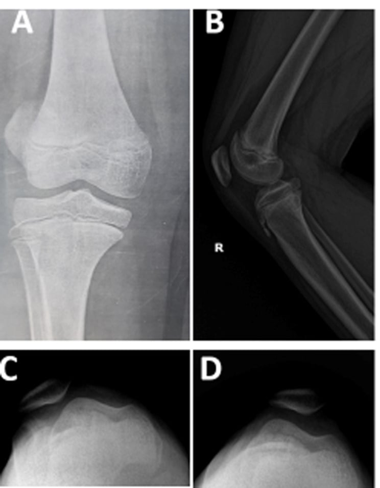
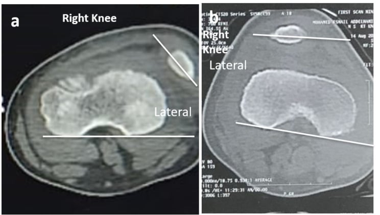
Who needs surgery. Many, particularly the milder cases, are surprisingly functional and are managed non-operatively with activity modification, bracing and physiotherapy to strengthen the extensor mechanism. Operate for instability that impairs walking or causes falls, or for a fixed or obligate dislocation.
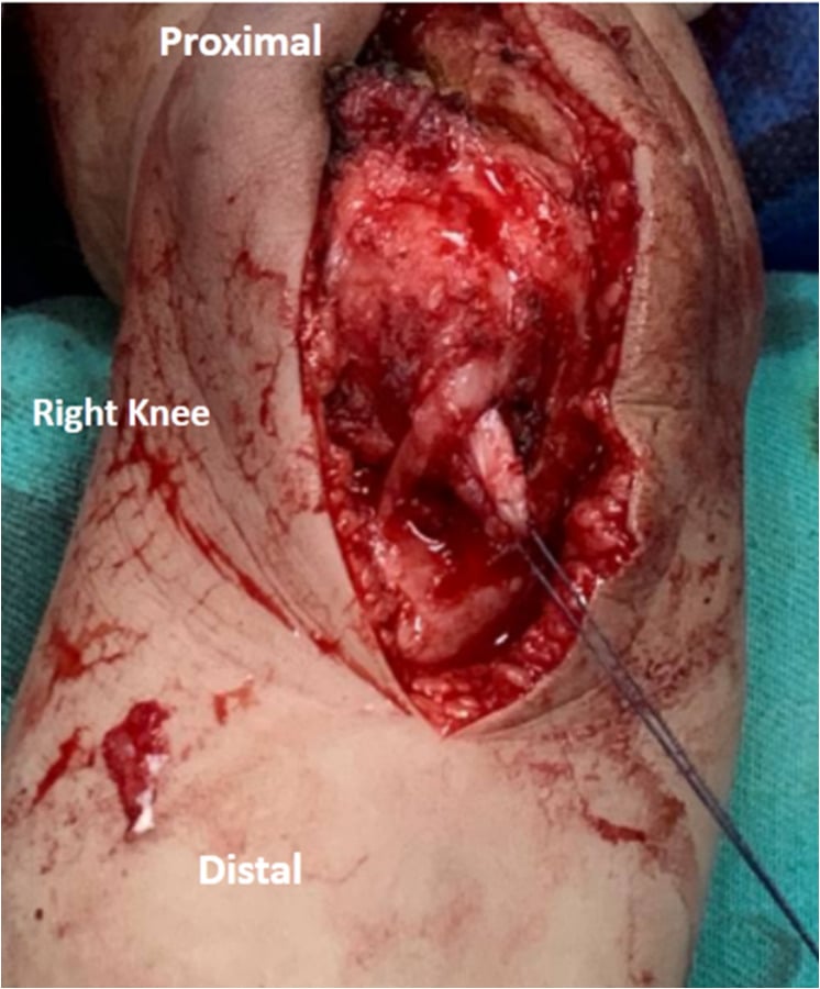
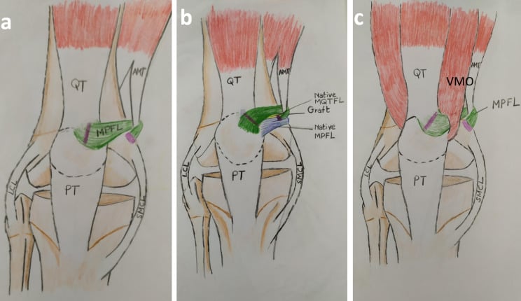
Why surgery is hard. Recurrence is high because the underlying laxity persists, and soft-tissue realignment alone (lateral release with medial reefing or MPFL reconstruction) commonly fails. A comprehensive combined soft-tissue and bony realignment is usually needed, and families should be counselled about the high recurrence rate.
The growing knee. Realignment options include MPFL reconstruction and tibial tubercle transfer, but in the skeletally immature child the tibial tubercle must be protected. Use a distal soft-tissue realignment such as a Roux-Goldthwait-type procedure, or guided-growth or distal-femoral options, rather than a tubercle transfer, with a distal femoral osteotomy for marked valgus. General MPFL and realignment technique is developed in the paediatric patellar-instability topic.
Surgical Techniques
Indications. Symptomatic AAI and progressive instability.
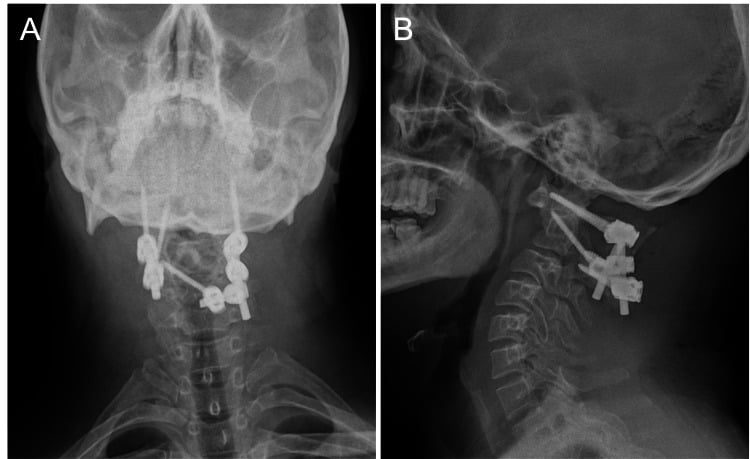
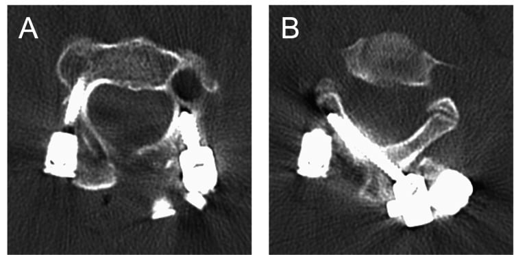
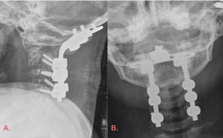
Technique. A posterior approach, with Gallie or Brooks wiring or a Harms construct (C1 lateral mass screws, C2 pedicle screws), and bone graft. A halo or rigid collar follows.
Extending to the occiput. Fusion should extend to the occiput only when anatomy or instability prevents a reliable isolated C1-C2 reconstruction.
Complications
Cervical Spine Complications
- Risk
- With AAI + trauma/intubation
- Prevention
- Activity restriction, careful intubation
- Management
- Fusion, rehabilitation
- Risk
- Progressive myelopathy
- Prevention
- Early fusion for symptomatic AAI
- Management
- Emergency stabilisation
- Risk
- Post-trauma
- Prevention
- Avoid high-risk activities
- Management
- Traction, fusion if recurrent
- Risk
- After fusion
- Prevention
- Proper technique, bone graft
- Management
- Revision fusion
- Risk
- Osteoporosis, hypotonia
- Prevention
- Adequate fixation
- Management
- Revision with reinforcement
Hip Surgery Complications
The incidence ranges below are conventional teaching ranges drawn from the Down-syndrome hip-surgery literature (Peterlein 2013 and related retrospective series); they are not single-study measured rates.
- Incidence
- 20-40%
- Risk Factors
- Open reduction, age over 2
- Management
- Monitor, salvage procedures
- Incidence
- 15-30%
- Risk Factors
- Inadequate soft tissue, hypotonia
- Management
- Revision, muscle transfers
- Incidence
- Variable
- Risk Factors
- Prolonged immobilisation
- Management
- Physiotherapy, releases
- Incidence
- Common
- Risk Factors
- Inadequate correction
- Management
- Pelvic osteotomy
- Incidence
- Higher than typical
- Risk Factors
- Immune dysfunction
- Management
- Aggressive treatment
Anaesthetic and perioperative risks. These considerations are critical for surgery in Down syndrome:
- Difficult airway - macroglossia, a small trachea and atlantoaxial instability
- Cardiac disease - 40-50% have congenital heart defects, and a preoperative echo is essential
- Respiratory problems - subglottic stenosis and sleep apnoea, so postoperative monitoring is crucial
- Immune dysfunction - a higher infection risk, demanding meticulous sterile technique
- Osteoporosis - hardware complications are more common, so fixation must be adequate
Long-term orthopaedic issues. Several problems persist or progress:
- Progressive hypotonia, which may affect rehabilitation outcomes
- Joint laxity, leading to instability and recurrent dislocations
- Accelerated osteoarthritis of the hip and knee in young adults
- Cervical degeneration, with early spondylosis due to laxity
- Progression of pes planus, which may require lifelong orthotic support
Postoperative Care
Cervical Fusion Rehabilitation
Halo or rigid collar. Neutral neck positioning at all times.
Continue halo or rigid collar; confirm radiographic union before weaning.
No contact sports or other high-risk activities indefinitely.
Clinical and radiographic follow-up until fusion confirmed, then annual surveillance for adjacent segment disease.
Hip Surgery Recovery
Spica cast or abduction brace.
Walking aids and physiotherapy.
Progressive muscle strengthening.
Monitor for AVN and residual dysplasia.
Beyond the protocol. Adapt instructions to the child's cognitive level, and enlist the family, whose support is essential for compliance with bracing. Coordinate with school and therapy services, and monitor the heart where there is cardiac disease.
Follow-up Protocol
Wound check and neurological status.
Radiographic healing assessment.
Skeletal maturity and late complications.
Plan transition to adult services.
Outcomes/Prognosis
How firm the numbers are. The outcome percentages in this section are conventional teaching estimates. The Down-syndrome-specific evidence base is small retrospective series plus one systematic review (Sha 2019), so treat the numbers as approximate.
Cervical spine. Fusion gives good outcomes when it is done for symptomatic patients before a permanent deficit.
- Treatment
- Activity restriction
- Expected Outcome
- Usually stable, rare progression
- Treatment
- Fusion
- Expected Outcome
- 70-80% neurological improvement
- Treatment
- Fusion
- Expected Outcome
- Stabilisation, limited recovery
- Treatment
- Supportive
- Expected Outcome
- Poor prognosis
Hip. Open reduction under 2 years of age is satisfactory in the long term in 60-70%; over 2 years the AVN and redislocation rates are higher. Pelvic osteotomy improves coverage when the hip is reducible, and salvage procedures are palliative for failed reconstruction. Reconstruction has a high failure rate (30-40%) and AVN is common, but function is often maintained.
Function. Early intervention gives better outcomes for both spine and hip, and most orthopaedic issues are manageable, with a good quality of life achievable.
- Expectation
- 90% community ambulators
- Factors
- Hip stability, hypotonia degree
- Expectation
- Variable
- Factors
- Cognitive level, orthopaedic status
- Expectation
- Generally good
- Factors
- Family support, access to services
Guidelines, Registries & Global Practice
Global epidemiology
- Down syndrome occurs in roughly 1 in 700-1000 live births worldwide; live-birth prevalence varies with maternal age distribution and the availability and uptake of prenatal screening.
- Symptomatic atlantoaxial instability is rare (around 1-2%) even though radiographic instability is reported in 10-20%; pes planovalgus is near-universal.
Side-by-side guidance
- Cervical screening stance
- No routine asymptomatic radiographs; symptom-based surveillance
- Practical emphasis
- Neurological history/exam at every health visit
- Cervical screening stance
- Documented neurological clearance required before high-risk events
- Practical emphasis
- Activity-related certification, not population screening
- Cervical screening stance
- No routine pre-anaesthetic radiographs when asymptomatic; image for symptoms, known pathology or clinical concern
- Practical emphasis
- Airway and neutral neck-positioning care perioperatively
- Cervical screening stance
- Stabilize neurologic or cord-threatening instability; consider asymptomatic proven AAI individually
- Practical emphasis
- Rigid screw-rod fixation preferred when surgery is indicated
Registry and outcome notes
- No DS-specific arthroplasty registry exists, but national joint registries (NJR UK, AOANJRR Australia, AJRR US, SHAR Sweden) capture small numbers of DS arthroplasties; pooled data show higher revision and complication rates than matched controls.
High- vs limited-resource practice
- In well-resourced settings, MRI, CT-based screw planning and rigid instrumentation are standard, and multidisciplinary clinics coordinate cardiac, airway and orthopaedic care.
- In limited-resource settings, plain radiography and clinical surveillance predominate; the priority is recognising myelopathic symptoms early and avoiding avoidable cervical trauma (careful intubation, neutral positioning), since access to complex paediatric spine surgery may be limited.
Controversies & Areas of Uncertainty
1. Routine cervical screening radiographs
- The single largest controversy. The AAP (2022) and most paediatric bodies do not recommend routine asymptomatic screening films because the atlantodental interval is a poor predictor of who develops myelopathy and is stable over time in most children.
- The Special Olympics, however, still mandates a documented neurological clearance (historically a screening lateral radiograph) before athletes participate in high-risk events. This creates a real-world tension: clinicians may be asked to obtain films that their own national guideline no longer endorses.
- Pragmatic position: prioritise a careful neurological history and examination at every visit; use neutral-position precautions for procedures; image when symptoms, known instability or another clinical concern exists; and separately check the sporting body's current participation policy.
2. ADI thresholds vs neural canal width
- Numbers vary between sources (ADI greater than 4.5mm, greater than 5mm, or greater than 10mm). Increasingly the space available for the cord (SAC, posterior interval) and dynamic MRI cord signal are regarded as more meaningful than ADI alone.
3. Surgical timing in asymptomatic AAI
- Whether to fuse a markedly increased ADI in an asymptomatic child is unresolved. Prophylactic fusion carries a high complication and nonunion rate, so most surgeons reserve operation for symptomatic instability, progressive radiographic change, or cord signal change.
4. Hip reconstruction vs early arthroplasty
- Reconstruction has high redislocation and revision rates; some advocate accepting a stable subluxation and planning for later arthroplasty rather than repeated failed childhood reconstructions.
MCQ Practice Points
Q: What ADI is abnormal in Down Syndrome? A: Greater than 5mm on lateral flexion-extension X-ray.
Q: How does hip instability in DS differ from DDH? A: It is late-onset (develops in childhood), unlike DDH which is present at birth.
Q: Should an asymptomatic child with Down syndrome have routine cervical radiographs before anaesthesia or sport? A: No. Use focused clinical surveillance and neutral-position precautions; image when symptoms, known instability or another clinical concern is present.
Q: What is the common foot deformity in DS? A: Pes planovalgus (flatfoot). Usually managed with orthotics.
Q: What is the prognosis for hip surgery in Down Syndrome? A: High failure rate with AVN common. Salvage procedures often needed.
Q: What cervical anomaly is associated with AAI in DS? A: Os odontoideum - separate ossicle at tip of odontoid.
Self-Assessment Quiz
Additional Quiz Questions
Viva Scenarios
Practise clinical reasoning and management decisions out loud
“5-year-old with Down Syndrome requires general anesthesia for dental work. How do you assess cervical spine?”
“8-year-old with DS presents with a limp. X-ray shows subluxated left hip. Walking milestone was normal. No trauma.”
“Same child develops neck pain and gait changes. ADI is 8mm. What is your management?”
CERVICAL
- AAI 10-20% radiographic
- Symptoms are far less common
- No routine clearance films when asymptomatic
- Stabilize cord-threatening instability
HIP
- Late onset
- High failure rate
- AVN common
- Salvage may be needed
KNEE/FOOT
- Patella instability
- Pes planovalgus 90%
- Orthotics usually enough
- Surgery rarely needed
KEY CONCEPT
- Ligamentous laxity
- All joints affected
- Generalized hypermobility
- Beighton score high
SCREENING
- Focused history and neurological examination
- Neutral-position procedural precautions
- Targeted imaging for clinical concern
- Check the sport programme's current policy
EXAM PEARLS
- Os odontoideum association
- Hip differs from DDH
- MRI for cord compression
- Multidisciplinary care
Evidence Base
Bull, Trotter, Santoro et al — Health Supervision for Children and Adolescents With Down Syndrome
- AAP does not recommend routine cervical screening radiographs in asymptomatic children
- Plain radiographs are poor predictors of neurological risk; a normal film does not guarantee future safety
- Symptoms prompt neutral radiography and specialist assessment; procedural care should avoid excessive cervical flexion or extension
Pueschel SM, Scola FH, Pezzullo JC — Longitudinal study of atlanto-dens relationships
- 141 individuals with Down syndrome with serial radiographs followed over time
- 130 (92%) showed only minor ADI change (1-1.5mm); 11 (8%) changed 2-4mm but none became symptomatic
- Atlantodental interval is largely stable over time and a single measurement has limited predictive value
Yang BW, Hedequist DJ, Proctor MR et al — Screw-rod fixation for upper cervical instability in paediatric DS
- 12 paediatric DS patients undergoing cervical fusion; mean age 9.3 years; os odontoideum in 7
- Overall complication rate 41.7%, with 4 reoperations for nonunion, but union eventually achieved in 11/11 with adequate follow-up
- Only 3/12 were identified through asymptomatic screening and none of these had cord signal change
Peterlein CD, Schofer MD, Fernandez FF et al — Hip surgery in Down syndrome
- 166 DS patients reviewed; hip problems in 63; 31 underwent surgery (49 hip operations)
- Isolated femoral varus derotation osteotomy failed in half, requiring later pelvic osteotomy
- Best clinical and radiological results came from complete redirectional acetabular osteotomy combined with capsular plication
Sha S, Board T, Alshryda S et al — THA in Trisomy 21: systematic review
- 9 studies, 321 DS patients undergoing total hip arthroplasty pooled
- Harris and WOMAC scores improved substantially; 5-year cumulative revision 7.5% (twice age-matched controls)
- Medical and surgical complications roughly 3 times higher than matched controls