Hydroxyapatite and Osteoconduction
- Definition: Synthetic bone void fillers that mimic the mineral phase of bone (Hydroxyapatite)
- Definition: They are Osteoconductive (scaffold) but not Osteoinductive (no growth factors)
- Mechanism: Sets via an isothermic (non-exothermic) reaction
- Management: Must usually be protected with hardware (plate) as it has no shear/tensile strength
- “High Compressive Strength (20-50 MPa - greater than cancellous bone)
- “Low Tensile Strength (Brittle)
- “Excellent biocompatibility
- “Replaced by bone over 6-18 months
Calcium Phosphate Cements
CaP: Isothermic (Cool setting) - No necrosis risk. PMMA: Exothermic (Hot setting) - Risk of thermal necrosis.
CaP: Osteoconductive & Resorbable (replaced by bone). PMMA: Inert (fibrous encapsulation) & Permanent.
Compression: CaP greater than cancellous bone (prevents subsidence). Shear: CaP is BRITTLE (fails catastrophically). Needs plate protection.
Overview/Introduction
Calcium phosphate cements (CPCs) are synthetic, injectable bone-void fillers whose set product is the same mineral phase as bone — carbonated apatite or brushite. First described by Brown and Chow in 1983 and brought to clinical use as the Skeletal Repair System (Norian SRS) in the 1990s, they fill the gap between inert acrylic cement (PMMA) and biological graft. They are osteoconductive but not osteoinductive or osteogenic, set at body temperature by a dissolution–precipitation (isothermic) reaction with no toxic monomer, develop high compressive but very low tensile/shear strength, and are gradually resorbed and replaced by host bone (osteotransduction). Their niche is the contained metaphyseal void — buttressing an elevated articular fragment against subsidence while internal fixation neutralises bending and shear.
Concepts & Mechanism: Core Principles
The four properties that define any bone-graft substitute
- Osteoconduction — passive scaffold for ingrowth. CPC has this.
- Osteoinduction — signalling (BMPs) that recruits/differentiates osteoprogenitors. CPC lacks this.
- Osteogenesis — living transplanted cells. CPC lacks this.
- Structural support — immediate mechanical load-sharing. CPC provides this in compression only.
Why CPC works as a strut, not a plate CPC behaves like a ceramic: strong when squeezed (20-50 MPa, exceeding cancellous bone) but brittle and weak in tension/shear (2-5 MPa). It therefore supports a subarticular fragment against axial collapse but will fracture under bending — so it is always combined with neutralising/buttress hardware, never used alone in a load-bearing site.
Material Science Overview
Composition
Powder Components
- Tetracalcium phosphate (TTCP): Ca₄(PO₄)₂O
- Dicalcium phosphate anhydrous (DCPA): CaHPO₄
- α-Tricalcium phosphate (α-TCP): α-Ca₃(PO₄)₂
- Calcium carbonate, calcium oxide (modifiers)
Liquid Phase
- Water or sodium phosphate solution
- pH modifiers
- Accelerators (e.g., citric acid)
Setting Reaction
Mechanism
- Powder dissolves in liquid (acidic microenvironment)
- Supersaturation of calcium and phosphate ions
- Precipitation of new crystalline phase
- Interlocking crystal network provides mechanical strength
Key Characteristics
- Isothermic: No heat generated (unlike PMMA)
- Time: 10-30 minutes working time, 24 hours for full strength
- Environment: Sets in aqueous (wet) environment
Microstructure
Porosity
- Macropores (100-500 μm): Created by incorporation techniques
- Micropores (1-10 μm): Inherent to setting reaction
- High surface area enhances osteoconduction
Crystal Structure
- Hydroxyapatite: Hexagonal crystals
- Brushite: Monoclinic crystals
- Similar to biological bone mineral
Osteotransduction and the Resorption-Formation Balance
The topic invokes "osteotransduction" repeatedly and the controversies note that "rapid resorption may outpace bone formation", but the mechanism that makes a calcium phosphate cement different from an inert filler is never spelled out. It is the single concept that separates CaP from PMMA.
Osteotransduction = creeping substitution. Unlike PMMA (which is simply encapsulated and stays forever), a CaP cement is biologically turned over and replaced by host bone. The process couples two activities at the cement surface:
- Cell-mediated resorption - osteoclasts (and giant cells) resorb the cement, much as they resorb bone, supplemented by simple chemical dissolution;
- Appositional bone formation - osteoblasts lay down new bone onto the osteoconductive cement surface as it is resorbed.
The result is gradual, front-by-front replacement of cement by living bone - the same "creeping substitution" seen with structural allograft.
Why the rate must match. The clinical performance of a cement depends on whether resorption and bone formation stay in step:
- Cement type
- Ideal (rarely achieved)
- Consequence
- Smooth hand-off of load from cement to new bone
- Cement type
- Apatite (HA)
- Consequence
- Cement persists for years/decades; mostly asymptomatic but can block remodelling and obstruct future drilling/revision
- Cement type
- Brushite
- Consequence
- Cement may disappear before bone replaces it, leaving a transient void and risking loss of support
Resorption rate is tunable through chemistry and structure - higher porosity and surface area, smaller crystals and the brushite (vs apatite) phase all resorb faster. Brushite is designed for speed but can paradoxically convert to poorly-resorbing apatite in vivo, which is why the "ideal" match remains unsolved.
CaP cement is replaced by host bone through osteotransduction (creeping substitution) - coupled osteoclastic resorption and osteoblastic bone formation at the surface - whereas PMMA is inert and permanent. The art is matching resorption to bone formation: apatite resorbs too slowly (persists, can hinder later revision) and brushite can resorb too fast (transient defect, loss of support). That balance, not raw strength, is what makes a "good" cement.
Clinical Indications
Primary Indications
Metaphyseal Fractures
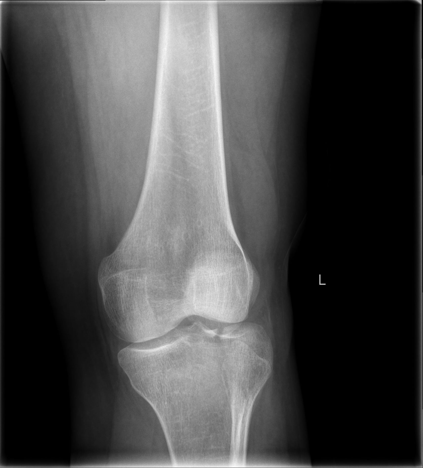
- Tibial plateau: Schatzker II, III, VI with articular depression
- Distal radius: Metaphyseal void after reduction in osteoporotic bone
- Calcaneal fractures: Structural support of posterior facet
- Proximal humerus: Metaphyseal void filling
Tumour Surgery
- Curettage of benign bone tumours (GCT, ABC, unicameral bone cyst)
- Filling defect after tumour removal
- May be combined with autograft/allograft
Vertebral Augmentation
- Alternative to PMMA for kyphoplasty/vertebroplasty
- Lower exothermic risk but higher cost
Contraindications
Absolute
- Active infection
- Uncontained defects (cement will leak)
- Load-bearing diaphyseal sites
Relative
- Large defects requiring structural support
- Poor soft tissue coverage
- Immunocompromised patients
Cement Augmentation of Screw and Implant Fixation
The Larsson review, the controversies section and an explicit viva follow-up ("how does cement augmentation improve screw pull-out in osteoporotic bone?") all invoke cement augmentation of metalwork, yet the concept is never developed. It is the second major use of CaP cement beyond void filling, and a recurring exam question in osteoporotic fixation.
The problem. In osteoporotic bone the trabeculae are sparse, so a screw thread engages little bone and pull-out and cut-out are the dominant failure modes - the metalwork is fine, the bone holds it poorly.
How cement augmentation works. Injecting a small volume of cement around the screw tip (often through a cannulated/fenestrated screw) before final tightening:
- Increases the effective bone-implant interface - the cured cement interdigitates with surrounding trabeculae and converts a few thread-bone contacts into a large, load-distributing cement-bone block;
- Spreads load over a wider volume, lowering the peak stress at any single trabecula and resisting the toggling that causes cut-out;
- Raises pull-out force, stiffness and fatigue life of the construct - cadaveric work (Larsson & Bauer) consistently shows cement-augmented metal fixation is stiffer and stronger than metal alone in the distal radius, tibial plateau, proximal femur and calcaneus.
CaP versus PMMA for augmentation. Both are used; the trade-off mirrors the void-filling story:
- CaP cement augmentation
- Isothermic, no monomer
- PMMA augmentation
- Exothermic; monomer; small embolic/BCIS risk
- CaP cement augmentation
- Osteoconductive, remodels
- PMMA augmentation
- Inert, permanent
- CaP cement augmentation
- Resorbs/incorporates - easier later revision
- PMMA augmentation
- Permanent block - complicates revision/removal
- CaP cement augmentation
- Good in compression, brittle
- PMMA augmentation
- Tougher
Caveats. Augmentation is an adjunct that buys early fixation in poor bone; it does not correct malreduction, the evidence is largely biomechanical (cadaveric) rather than high-level clinical, and leakage into a joint or fracture gap must be avoided. A permanent PMMA mass in particular can complicate any future revision.
In osteoporotic bone a screw engages too few trabeculae, so it pulls out or cuts out. Cement (CaP or PMMA), often via a fenestrated screw, interdigitates with the trabeculae and enlarges the bone-implant interface into a load-distributing block, raising pull-out strength, stiffness and fatigue life. Prefer CaP when you want later remodelling and easier revision; PMMA is tougher but permanent and complicates revision.
Mechanical Properties
Compressive Strength
- Compressive Strength (MPa)
- 30-50
- Compressive Strength (MPa)
- 15-25
- Compressive Strength (MPa)
- 2-12
- Compressive Strength (MPa)
- 100-200
- Compressive Strength (MPa)
- 70-100
Tensile/Shear Strength
- CaP Cement: 2-5 MPa (very low)
- PMMA: 25-40 MPa
- Cortical Bone: 50-150 MPa
Clinical Implication: CaP cements are brittle; require hardware protection (plate, screws) in fracture treatment
Modulus of Elasticity
- CaP cements: 5-15 GPa
- Cancellous bone: 0.1-1 GPa
- Cortical bone: 15-20 GPa
Fatigue Properties
- Limited fatigue resistance
- Catastrophic failure under cyclic loading
- Not suitable for high-stress cyclical loading
Classification
By End Product
- End Product
- Hydroxyapatite (HA)
- Ca/P Ratio
- 1.67
- Resorption
- Slow (years)
- Strength
- Higher (50+ MPa)
- End Product
- Dicalcium phosphate dihydrate
- Ca/P Ratio
- 1.0
- Resorption
- Fast (months)
- Strength
- Lower (20 MPa)
By Form
Injectable
- Paste form, delivered via syringe
- Sets in situ after injection
- Ideal for minimally invasive application
- Examples: Norian SRS, HydroSet
Pre-formed
- Blocks, granules, or putty
- Shaped before or during surgery
- Higher initial strength
By Application
- Product Type
- Injectable HA
- Key Property
- Structural support
- Product Type
- Low viscosity paste
- Key Property
- Injectability
- Product Type
- Granules/blocks
- Key Property
- Volume filling
- Product Type
- Fast-setting brushite
- Key Property
- Rapid integration
Commercial Products
- Norian SRS/CRS: Apatite cement, high strength
- ChronOS: β-TCP based, resorbable
- α-BSM: Injectable, fast-setting
- HydroSet: Brushite based, faster resorption
Choosing a Graft / Void Filler (Differential)
When a metaphyseal or cavitary defect must be filled, the realistic options are compared below. The "diagnosis" the examiner wants is matching the right filler to the defect (containment, load, biology needed).
Bone-graft options for a contained metaphyseal void
Con-Ind-GenThe 3 O's of Bone Graft
Hook:Conducive scaffold, Induction signals, Genesis cells
Postoperative Management

Immediate
Weight-Bearing
- Protected weight-bearing initially
- Progressive loading as bone heals
- Hardware provides protection during healing
Monitoring
- Standard wound care
- Watch for signs of extravasation
- Imaging at 2 and 6 weeks
Medium-Term
Rehabilitation
- Range of motion as tolerated
- Strengthening when fracture stable
- Progress based on clinical and radiographic healing
Imaging Follow-up
- X-rays at 6, 12 weeks
- Assess fracture healing and cement integration
- CT if concern about resorption
Long-Term
Cement Remodeling
- Brushite: 6-12 months
- Apatite: Years to decades
- Gradual replacement by host bone
Hardware Removal
- Consider once fracture healed
- Cement usually incorporated or resorbed
- Not routinely required
Complications
Material-Related
Cement Extravasation
- Leakage into soft tissues or joint
- More common with uncontained defects
- Usually resorbs without issue (unlike PMMA)
Incomplete Fill
- Air pockets reduce strength
- May require reoperation
- Prevented by proper technique
Brittleness/Fracture
- Catastrophic failure under shear
- Requires hardware protection
- More common in brushite cements
Clinical Complications
Infection
- Not inherent to material
- Biofilm formation possible
- Requires debridement if occurs
Delayed Resorption
- Apatite cements may persist for years
- Usually asymptomatic
- May interfere with future surgery
Subsidence
- Despite cement support
- Usually due to poor technique or osteoporosis
- Hardware failure common cause
Comparison to Alternatives
- CaP Cement
- No
- PMMA
- Yes
- Autograft
- No
- CaP Cement
- No
- PMMA
- No
- Autograft
- Yes
- CaP Cement
- No (resorbs)
- PMMA
- Yes
- Autograft
- No (remodels)
- CaP Cement
- Low
- PMMA
- Low
- Autograft
- Low
Outcomes
Clinical Outcomes
Tibial Plateau Fractures
- Reduced articular subsidence vs autograft
- Equivalent functional outcomes
- No donor site morbidity
Distal Radius Fractures
- Maintains reduction in osteoporotic bone
- Faster return to function
- Equivalent long-term outcomes
Radiographic Outcomes
Cement Incorporation
- Evidence of bone ingrowth at 6-12 months
- Progressive replacement by host bone
- Apatite cements may remain visible longer
Subsidence Prevention
- Superior to autograft for structural support
- 2-3 mm less subsidence in tibial plateau studies
- Maintains articular congruity
Functional Outcomes
Patient-Reported Outcomes
- No difference in pain scores
- Equivalent range of motion
- No donor site morbidity (vs autograft)
Return to Activity
- Similar timeframes to other grafts
- Hardware removal rates similar
- Long-term function maintained
Clinical Applications
- Elevate depressed articular surface.
- Fill the metaphyseal void with CaP cement.
- Advantage: Unlike cancellous chips, it provides immediate structural support (high compression strength) to prevent re-collapse/subsidence before the plate takes full load.
- Void filler in elderly osteoporotic bone.
CaP cement vs iliac autograft in tibial plateau fractures (landmark RCT)
- Multicentre, prospective RCT: 120 acute tibial plateau fractures in 119 adults, 12 North American sites, randomised 2:1 to calcium phosphate cement (82) vs autogenous iliac graft (38)
- Significantly higher rate of articular subsidence at 3-12 months in the autograft group (p = 0.009)
- Union rates and time to union were equivalent between groups
- No donor-site morbidity with the synthetic cement
Clinical Use Guidelines
Pre-operative Planning
Patient Selection
- Contained metaphyseal defect
- Adequate soft tissue coverage
- No active infection
Defect Assessment
- Size and containment
- Load-bearing requirements
- Need for structural vs void-filling
Intraoperative Considerations
Preparation
- Read manufacturer instructions carefully
- Ensure powder/liquid ratio correct
- Prepare before need (limited working time)
Working Time
- Typically 10-15 minutes
- Temperature affects setting (faster if warm)
- Must be injected before setting begins
Adjuncts
Hardware Protection
- Buttress plating for metaphyseal fractures
- Prevents shear/tensile failure
- Essential for weight-bearing bones
Combination with Biologics
- May add autograft for osteoinduction
- Platelet-rich plasma (theoretical benefit)
- BMP addition (research stage)
Application Technique
Tibial Plateau Example
Step 1: Fracture Reduction
- Elevate depressed articular segment
- Use bone tamp or elevator through cortical window
- Confirm reduction under fluoroscopy
Step 2: Cement Preparation
- Mix powder and liquid per manufacturer
- Achieve paste consistency
- Work within time window
Step 3: Cement Application
- Inject through cortical window or cannula
- Fill void completely (no air pockets)
- Overfill slightly (will compress)
Step 4: Hardware Application
- Apply buttress plate before cement sets
- Screws through or around cement
- Provides protection against shear forces
Step 5: Confirmation
- Fluoroscopy to confirm fill and reduction
- Check cement containment
- Assess hardware position
Key Technical Points
Containment
- Create cortical window if needed
- Block significant egress points
- May use small bone graft to contain
Bleeding Control
- Lavage defect before injection
- Blood dilutes cement, weakens setting
- Tourniquet useful if applicable
Setting Confirmation
- Wait for initial set before wound closure
- Typically 15-30 minutes
- Test with probe
Guidelines, Registries & Global Practice
Global Epidemiology & Use
- Bone-graft substitutes are used in a large minority of fracture and reconstructive procedures worldwide; CaP cements are the dominant injectable, resorbable, structural option for contained metaphyseal voids.
- Highest-volume indications globally: tibial plateau, distal radius (osteoporotic), calcaneus and proximal humerus metaphyseal voids, and curettage cavities after benign bone lesions.
- Uptake tracks with availability of fluoroscopy, theatre cost tolerance and surgeon familiarity rather than any single national guideline.
Society Guidance, Side by Side
- Position on CaP cement
- Recognised bone-graft substitute; no procedure-specific mandate — choice individualised to defect containment and load environment
- Position on CaP cement
- Void fillers permitted within fracture-management standards; emphasis on contained defects and adequate skeletal fixation
- Position on CaP cement
- Teaches CaP cement for subarticular metaphyseal void support combined with neutralising/buttress fixation; not for diaphyseal or uncontained defects
- Position on CaP cement
- Apatite vs brushite selection driven by required resorption rate and strength; injectability valued for minimally invasive augmentation
No major society endorses CaP cement as a stand-alone load-bearing construct; all frame it as an adjunct to internal fixation.
Regulatory & Registry Notes
- Regulated as implantable medical devices (e.g. FDA in the US, CE-mark/MDR in Europe). Norian SRS holds long-standing approval for selected distal radius and other metaphyseal fractures.
- There is no dedicated international registry for bone-graft substitutes comparable to arthroplasty registries (NJR, AJRR, AOANJRR); evidence rests on RCTs and meta-analysis (Bajammal 2008; Russell & Leighton 2008; Cassidy 2003) rather than registry survival data.
High- vs Limited-Resource Practice
- Well-resourced settings: ready access to injectable apatite/brushite cements and intra-operative imaging; cement chosen to avoid iliac-crest harvest morbidity.
- Limited-resource settings: autograft (iliac crest, RIA) and allograft remain first-line because synthetic cement cost and supply are limiting; the structural advantage of cement is weighed against expense.
Controversies & Areas of Uncertainty
- Apatite vs brushite resorption. Brushite is designed to resorb faster, but in vivo it can convert to poorly-resorbing apatite, and rapid resorption may outpace bone formation — leaving a transient defect. The "ideal" resorption-to-formation match is not solved.
- Extraosseous extravasation. In the distal radius RCT (Cassidy 2003), cement was extraosseous in 70% of wrists and that subgroup had the highest loss of reduction — the clinical significance of leakage versus a marker of poor containment is debated.
- Does it accelerate union or just resist subsidence? Trials consistently show better maintenance of reduction, but a true acceleration of fracture healing is not established; benefit may be purely mechanical.
- Stand-alone augmentation in osteoporotic fixation. Screw-tip cement augmentation improves pull-out in cadaver studies, but high-quality clinical evidence for routine use (and concern over complicating revision) remains limited.
- Vertebroplasty/kyphoplasty. CaP avoids PMMA's exotherm and monomer but has lower fatigue strength and higher cost; whether it improves outcomes over PMMA in the spine is unresolved.
- Function vs radiographs. Several RCTs show radiographic superiority (less subsidence) without a durable difference in patient-reported function at one year — the patient-relevant value is questioned.
MCQ Practice Points
Q: What are the two main types of calcium phosphate cement and their key differences?
A: (1) Apatite cement (Hydroxyapatite, HA): Sets to crystalline hydroxyapatite Ca₁₀(PO₄)₆(OH)₂, very slow resorption (years), excellent biocompatibility, used for bone void filling. (2) Brushite cement (DCPD): Sets to CaHPO₄·2H₂O, faster resorption (months), lower compressive strength. Both set via dissolution-precipitation reactions at body temperature.
Q: What is the mechanism of setting for calcium phosphate cements?
A: Acid-base or dissolution-precipitation reaction at room/body temperature (no exothermic heat unlike PMMA). Powder phase dissolves, supersaturates, and precipitates as new calcium phosphate crystite. Setting time: 10-30 minutes. No toxic monomer released. Final product resembles bone mineral (hydroxyapatite or brushite phase).
Q: What are the clinical advantages of calcium phosphate cement over PMMA bone cement?
A: (1) Osteoconductive - bone grows directly onto/into it. (2) Bioactive - integrates with host bone. (3) Resorbable (brushite) or slowly remodeled (HA). (4) No exothermic setting - no thermal necrosis. (5) No toxic monomer. Disadvantages: Weak in tension and shear, only suitable for compression loading (metaphyseal fractures), cannot be used for arthroplasty fixation.
Q: What is the compressive strength of calcium phosphate cements and how does this influence clinical applications?
A: Compressive strength: 20-50 MPa (similar to cancellous bone). Tensile/shear strength: Very low (2-5 MPa). Applications: Metaphyseal fractures (tibial plateau, distal radius, vertebral augmentation where compression dominates). Not suitable for: Diaphyseal fractures, arthroplasty fixation, or any load-bearing without metallic supplementation.
Q: How does calcium phosphate cement resorb and remodel?
A: Osteoclasts resorb the cement (cell-mediated resorption) similar to bone remodeling. Brushite cements: 6-12 months, faster resorption, replaced by woven bone. Apatite cements: Years to decades, very slow remodeling. Rate depends on porosity, surface area, and Ca/P ratio. Ideal for augmenting metaphyseal fractures where gradual load transfer to healing bone is desired.
At a Glance
Calcium phosphate cements are synthetic bone void fillers that mimic the mineral phase of bone (hydroxyapatite) and are osteoconductive but not osteoinductive. They set via an isothermic (non-exothermic) precipitation reaction, unlike PMMA which generates thermal necrosis risk. Key properties include high compressive strength (20-50 MPa, greater than cancellous bone) but low tensile/shear strength, making hardware protection essential. Primary applications include metaphyseal void filling in tibial plateau fractures and distal radius fractures, where they prevent articular subsidence. Over 6-18 months, osteoclasts remodel the cement and replace it with host bone through osteotransduction.
CaP vs PMMA at a Glance
CaP vs PMMA
CAPCaP vs PMMA: the C-A-P contrast
Hook:Cool, Absorbed, Plate-dependent
Clinical Decision Scenarios
Practise clinical reasoning and management decisions out loud
Clinical Decision Scenarios
Practise clinical reasoning and management decisions out loud
Clinical Decision Scenarios
Practise clinical reasoning and management decisions out loud
Science
- Hydroxyapatite or Brushite
- Isothermic (Cool)
- Osteoconductive (Scaffold)
Uses
- Metaphyseal voids (Tibial Plateau)
- Tumour voids (GCT)
- Not for infection (biofilm risk)
Evidence Base
Calcium phosphate cement in fracture treatment — meta-analysis of RCTs
- Meta-analysis of 14 randomised trials (11 published, 3 unpublished) of metaphyseal fractures — distal radius, hip, tibial plateau, calcaneus
- Versus autograft: 68% relative risk reduction in loss of fracture reduction (95% CI 29-86%)
- Versus no graft: 56% relative risk reduction in fracture-site pain (95% CI 14-77%)
- Three trials independently showed improved functional outcomes vs no grafting
Norian SRS cement vs conventional fixation in distal radius fractures (RCT)
- Prospective, randomised, multicentre study of 323 distal radial fractures
- Cement group had better grip strength, wrist/digit motion and earlier function at 6-8 weeks; clinical differences had largely equalised by 1 year
- Extraosseous cement seen in 70% of treated wrists; this subgroup had the highest loss of reduction (37%), and supplemental K-wires were recommended
- No increase in total complications; lower infection rate than the externally-fixed/pinned controls
Injectable calcium phosphate cement for fracture fixation — review
- Cements harden with little heat, develop compressive strength and remodel slowly in vivo
- Primary role is filling metaphyseal voids and augmenting screw/device purchase in osteoporotic bone
- Cadaveric work shows cement-augmented metal fixation is stiffer and stronger than metal alone in distal radius, tibial plateau, proximal femur and calcaneus
- Early clinical series report reduced time to full weight-bearing after augmentation
Physical and chemical basis of calcium phosphate cements
- Traces the field to Brown and Chow's first calcium phosphate cement (1983)
- Sets by dissolution and precipitation to either apatite (slow-resorbing) or brushite (fast-resorbing) end products
- Setting time, porosity, strength and resorption are all tunable through powder/liquid chemistry
- Clarifies the trade-off between injectability, strength and resorption rate
References
- Larsson S, Bauer TW. Use of injectable calcium phosphate cements for fracture fixation: a review. Clin Orthop Relat Res. 2002.
- Bajammal SS, et al. The use of calcium phosphate bone cement in fracture treatment. JBJS Am. 2008.