L4-5 Slip in the Elderly
- Intact Pars: Distinguishes from Isthmic Spondylolisthesis
- L4-5: most common level due to sagittal facet orientation
- Measurement: Greater than 4mm translation on flexion-extension = Unstable
- Treatment: Decompression ALONE risks iatrogenic instability and reoperation
- Standard: Decompression PLUS Fusion (Ghogawala 2016)
- “Look for 'Fluid Sign' in facets on MRI T2 = Instability
- “Degenerative = L4-5 (Women), Isthmic = L5-S1 (Men)
- “Pars is INTACT in degenerative type
- “Surgery requires fusion to prevent progression
The key definition of Degenerative Spondylolisthesis is an INTACT Pars Interarticularis.
If a pars defect (lysis) is present, it is Isthmic, not Degenerative. Degenerative slips rarely exceed Grade II.
Overview and Epidemiology
Degenerative Spondylolisthesis (DS) is a disorder of segmental instability producing forward slippage of a vertebra. Unlike isthmic spondylolisthesis, which involves a defect in the pars interarticularis, DS results from progressive degeneration of the facet joints and intervertebral discs. It typically occurs in the lumbar spine and is sometimes termed "pseudospondylolisthesis" because the neural arch remains intact.
Demographics
- Prevalence: Increases markedly with age. In the community-based Framingham cohort imaged with CT, the prevalence of DS rose progressively from the fifth through the eighth decades of life (Kalichman 2009).
- Gender: Female predominant. Large population data show a male-to-female ratio of approximately 1:3 (Kalichman 2009); surgical/clinical series often quote a higher female predominance (up to ~5:1). This is attributed to ligamentous laxity, smaller facet joints, and post-menopausal hormonal effects on connective tissue.
- Age: Peak incidence 50-70 years; extremely rare under 40.
- Ethnicity: Reported more frequently in those with larger pelvic incidence.
Level of Involvement
- L4-5: ~80% of cases. L4 slips forward on L5; this is the most susceptible level due to the transition between the mobile lumbar spine and the stable lumbosacral junction.
- L3-4: Second most common.
- L5-S1: Rare — protected by the iliolumbar ligaments and deep seating in the pelvis below the intercristal line.
Risk Factors
- Anatomy: Sagittally oriented facet joints (permit forward translation).
- Hormonal: Post-menopausal state (reduced ligamentous restraint).
- Medical: Diabetes (accelerated disc and facet degeneration).
- Spinopelvic: High pelvic incidence correlates with slip severity.
- Pregnancy: Multiple pregnancies may contribute via abdominal muscle stretching and hormonal laxity.
Anatomical Considerations
The Facet Joint (Z-Joint)
The zygapophysial joint is a synovial joint formed by the superior articular process (SAP) of the level below and the inferior articular process (IAP) of the level above.
- Capsule: richly innervated (medial branch of the same level and the level above) — the source of facetogenic pain.
- Weight bearing: normally ~20%; increases toward ~70% with disc degeneration.
- Orientation: lumbar facets are predominantly sagittal (flex/ext); thoracic are coronal (rotation). At L5-S1 the facets are more coronally oriented (resist anterior shear), whereas at L4-5 they are more sagittal (susceptible to anterior shear).
Ligaments
- Iliolumbar ligament: connects the L5 transverse process to the ilium, stabilising L5 on the sacrum — explaining why L5-S1 degenerative slips are rare.
- Ligamentum flavum: the elastic yellow ligament buckles anteriorly into the canal with disc collapse, contributing to stenosis along with facet hypertrophy.
Neurovascular Anatomy
- The L4 nerve root exits below the L4 pedicle; the L5 root traverses the L4-5 disc space and is the root at risk.
- The aorta and vena cava lie anterior to the disc; ALIF at L4-5 risks the iliac bifurcation, which is why PLIF/TLIF is preferred at L4-5.
Pathophysiology
Biomechanics of Slip
The lumbar spine resists anterior shear through three mechanisms:
- Disc: The annulus fibrosus provides tensile restraint against translation.
- Facets: A coronal orientation of the joint surfaces provides a bony block to anterior translation.
- Ligaments: The iliolumbar and longitudinal ligaments provide tethering.
In DS the failure is primarily at the facet joints. As disc height is lost, the axis of rotation shifts posteriorly and loads the facets. The facets remodel, lose their coronal orientation, and become more sagittal. Once sagittal they offer little resistance to anterior shear, allowing the slip.
The Degenerative Cascade
The process follows the Kirkaldy-Willis cascade with a specific vector of anterolisthesis:
- Disc degeneration: Loss of disc height and nucleus turgor reduces resistance to shear and settles the motion segment.
- Facet loading: Loss of anterior column height transfers load to the posterior elements. Facets normally bear ~20% of load; with disc degeneration this can rise toward ~70%.
- Facet remodeling: Chronic loading causes cartilage wear, subluxation, and effusion; facets remodel from coronal (resisting slip) to sagittal.
- Instability: The incompetent disc and sagittal facets allow the superior vertebra to translate anteriorly. The iliolumbar ligaments stabilise L5, making L4 the "victim" level above.
- Stenosis ("napkin-ring" effect): the canal is narrowed anteriorly by disc bulge and osteophytes, and posteriorly by hypertrophied facets and buckling ligamentum flavum — producing central stenosis (claudication) and lateral recess stenosis (radiculopathy).
Pelvic Parameters (Spinopelvic Balance)
- Pelvic Incidence (PI): A high PI is a predisposing factor; high PI demands high lumbar lordosis to balance, increasing shear at L4-5.
- Compensation: As the slip and stenosis develop, patients compensate by retroverting the pelvis (increasing pelvic tilt) and flexing the knees (simian stance) to remain upright.
Fluid Sign: On axial T2-weighted MRI, a large facet joint effusion (greater than 1.5mm) is highly predictive of degenerative spondylolisthesis at L4-5 and indicates dynamic instability — even when the supine MRI shows no measurable slip. Chaput (2007) found that 22% of slips were not visible on supine MRI, so a facet effusion mandates standing flexion-extension films.
Spinopelvic Parameters and Sagittal Balance
The section above refers to pelvic incidence and compensation; examiners increasingly expect the formal parameters, the fundamental equation, and the PI-LL mismatch that drives modern alignment and fusion decisions.
- Definition
- Angle between a line perpendicular to the sacral endplate at its midpoint and a line from that midpoint to the femoral head axis
- Nature and approximate normal
- FIXED (morphological), constant after skeletal maturity; normal about 50 to 55 degrees
- Definition
- Angle between the vertical and the line from the sacral-endplate midpoint to the femoral head axis
- Nature and approximate normal
- POSITIONAL; normal under about 20 degrees; rises as the pelvis retroverts to compensate
- Definition
- Angle between the sacral endplate and the horizontal
- Nature and approximate normal
- POSITIONAL; normal about 40 degrees
The fundamental equation: PI = PT + SS. Because PI is fixed, any loss of sacral slope (for example with pelvic retroversion) must be matched by a rise in pelvic tilt - the geometric basis of the compensatory retroversion and simian stance seen in stenosis.
Pelvic incidence and lumbar lordosis must match. Lumbar lordosis (LL) should be tuned to the PI; a PI-LL mismatch (commonly taken as a difference of more than about 10 degrees) signifies under-correction of lordosis and is associated with sagittal imbalance, worse patient-reported outcomes, and a higher rate of adjacent segment disease after fusion. The SRS-Schwab criteria add pelvic tilt (over about 20 degrees) and sagittal vertical axis (over about 5 cm) as the other alignment targets.
Memorise PI = PT + SS and that PI is fixed while PT and SS are positional. The surgical target after a lumbar fusion is to restore lordosis so that PI minus LL is within about 10 degrees; leaving a large PI-LL mismatch predicts ongoing sagittal imbalance and adjacent-segment breakdown. A high PI also predisposes to the slip in the first place.
Classification
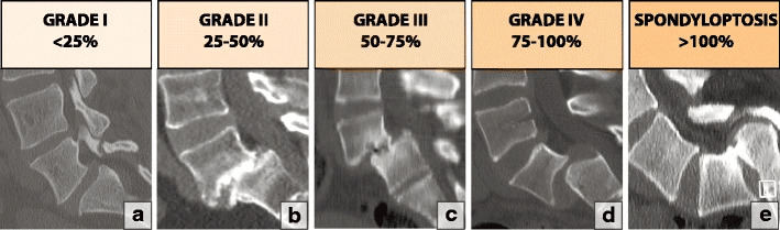
Meyerding Classification — based on the percentage slip of the superior vertebra over the inferior one on lateral X-ray.
- Slip Percentage
- 0 - 25%
- Note
- Most degenerative cases
- Slip Percentage
- 25 - 50%
- Note
- Maximum typical for degenerative
- Slip Percentage
- 50 - 75%
- Note
- Rare in degenerative (think isthmic)
- Slip Percentage
- 75 - 100%
- Note
- Very rare
- Slip Percentage
- Over 100%
- Note
- Spondyloptosis (vertebra falls off)
DITPPIWiltse Types
Hook:Do It To Perfect People's Images
Clinical Presentation
History
- Neurogenic Claudication (~90%): heaviness, fatigue, or pain in buttocks/legs on walking, relieved by sitting or leaning forward (the "shopping-cart sign"). Flexion opens the canal diameter and relieves venous congestion.
- Radiculopathy (~50%): dermatomal leg pain. An L4-5 slip typically causes L5 radiculopathy as the L5 root is compressed in the lateral recess.
- Back Pain: mechanical, lower lumbar, worse with extension (standing/walking) and relieved by flexion.
- Night Pain: may reflect instability as muscles relax during sleep.
- Worse with standing: static standing is often worse than walking due to sustained extension load and venous engorgement.
Physical Examination
- Inspection: flattened lumbar lordosis ("flat back"), flexed posture, simian stance (hips and knees flexed) to open the canal.
- Palpation: a "step-off" may be palpable. With an L4 anterior slip the L4 spinous process moves anteriorly (deep) while L3 remains prominent, so the step is felt between L3 and L4.
- Neurology:
- L5 motor: EHL (great-toe extension) and hip abduction (gluteus medius).
- Sensory: dorsum of foot and first web space.
- Reflexes: ankle (S1) usually normal; no reliable L5 reflex.
- Provocative: extension reproduces back/leg pain (Kemp's test). Femoral stretch may be positive with significant foraminal stenosis.
Investigations
Imaging Algorithm
1. Plain Radiographs
- Standing AP/Lateral: essential — assess slip grade and facet sclerosis.
- Flexion/Extension Views: critical for stability.
- Instability criteria: greater than 4mm translation OR greater than 10 degrees angular motion.
- Demonstrated instability dictates fusion.
- Spinous process view: look for kissing spines (Baastrup disease).
2. MRI (Gold Standard)
- Central canal area (critical stenosis under 75mm2). With facet hypertrophy and ligamentum flavum buckling the canal loses its normal oval shape and becomes trefoil/omega-shaped; CSF signal around the cauda equina is effaced and roots become crowded or redundant (serpiginous) proximal to the block.
- Lateral recess (L5 root compression) — bordered by the posterior vertebral body/disc anteriorly, the pedicle laterally and the superior articular process posteriorly; loss of perineural fat around the traversing root signals impingement.
- Facet effusion: T2 "fluid sign" indicates instability (a facet effusion mandates standing flexion-extension films, as up to 22% of slips reduce when supine).
- Disc quality (Pfirrmann grade).
3. CT Scan
- Confirms pars integrity — if a defect is present the diagnosis becomes isthmic spondylolisthesis.
- Pre-operative planning for pedicle screw trajectory.
- Assesses facet tropism (asymmetry).
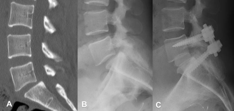
Case Example

Differential Diagnosis
- Degenerative
- Elderly (over 50)
- Isthmic
- Young (20-40)
- Degenerative
- Female
- Isthmic
- Male
- Degenerative
- L4-5
- Isthmic
- L5-S1
- Degenerative
- INTACT
- Isthmic
- DEFECT (Lysis)
- Degenerative
- Central Stenosis
- Isthmic
- Foraminal Stenosis
- Degenerative
- Low (I/II)
- Isthmic
- High (III+) possible
Neurogenic versus Vascular Claudication
The leg symptoms of degenerative spondylolisthesis are usually neurogenic claudication, but distinguishing this from vascular (arterial) claudication is a classic examiner discriminator - and the two often coexist in the elderly.
- Neurogenic (spinal stenosis)
- Standing and walking (lumbar extension)
- Vascular (peripheral arterial disease)
- Any exertion of the muscle (walking)
- Neurogenic (spinal stenosis)
- FLEXION - sitting, leaning forward, squatting (not merely stopping)
- Vascular (peripheral arterial disease)
- Stopping and STANDING STILL (rest), within a few minutes
- Neurogenic (spinal stenosis)
- Variable day to day
- Vascular (peripheral arterial disease)
- Fixed, reproducible claudication distance
- Neurogenic (spinal stenosis)
- Easier UPHILL (trunk flexed)
- Vascular (peripheral arterial disease)
- Worse uphill (higher muscle demand)
- Neurogenic (spinal stenosis)
- Usually pain-free (spine flexed)
- Vascular (peripheral arterial disease)
- Provokes pain (muscle demand)
- Neurogenic (spinal stenosis)
- Normal pulses and skin
- Vascular (peripheral arterial disease)
- Absent/reduced pulses, cool foot, trophic changes, low ABPI
- Neurogenic (spinal stenosis)
- Buttock/thigh/leg heaviness, often bilateral, dermatomal
- Vascular (peripheral arterial disease)
- Cramp in the working muscle (typically calf)
The cleanest discriminators are the relief posture and the pulses/ABPI: neurogenic claudication is relieved by flexion (sitting, the shopping-cart sign) and the patient can usually cycle, whereas vascular claudication is relieved by standing still, is worse uphill, comes on at a fixed distance, and is accompanied by diminished pulses and a low ankle-brachial pressure index. In an elderly patient the two often coexist, so check pulses and ABPI before attributing all leg pain to the spine.
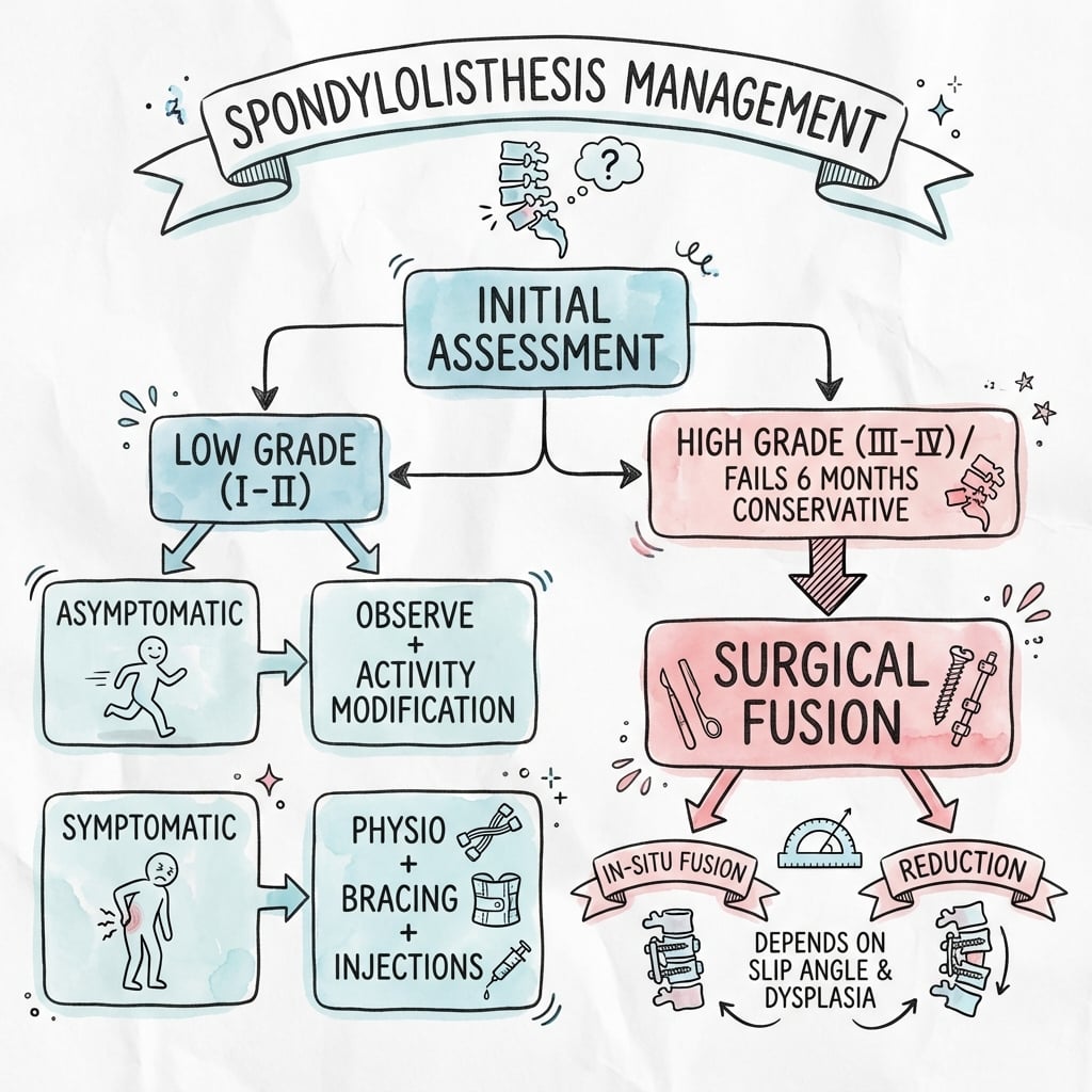
Management
UNI-PSurgical Indications
Hook:UNIque Problem
Complications
Surgical Risks
- Dural tear: higher risk than in simple stenosis due to adhesions and slippage (incidence ~5-10%). Repair primarily; consider flat bed rest ~24 hours.
- Implant failure: screw loosening or pull-out, common in osteoporotic bone — use cement-augmented or fenestrated screws.
- Adjacent segment disease (ASD): symptomatic ASD ~2-3% per year (roughly 25% at 10 years); radiographic ASD is far more common (up to ~50% at 10 years). Fusion increases stress on the level above (L3-4).
- Pseudarthrosis: failure to fuse causes loose hardware and recurrent pain. Risk factors: smoking (doubles risk), NSAIDs, diabetes.
- Infection: ~1-2%; usually Staphylococcus aureus/epidermidis — prompt washout required.
- Neurological injury: L5 root injury during screw placement or reduction; foot drop (L5) is the classic deficit.
Outcomes
Expected Results
- ~80% good-to-excellent results; leg pain relief is more reliable than back pain relief.
- Instrumented fusion rate over 90%.
- Walking tolerance and ODI improve substantially; ~70-80% patient satisfaction.
- Pre-op
- 50-60
- Post-op
- 20-30
- Pre-op
- 7-8
- Post-op
- 2-3
- Pre-op
- Limited
- Post-op
- Unlimited in ~70%
Prognostic Factors
- Favourable: claudication as the primary symptom, single-level disease, no prior surgery, non-smoker.
- Unfavourable: predominantly back pain, multi-level disease, obesity/diabetes, workers' compensation claims.
Guidelines, Registries & Global Practice
Global Epidemiology
- Community-based CT imaging (Framingham, USA) shows DS prevalence rising progressively from the fifth through the eighth decades, with a male-to-female ratio of ~1:3 (Kalichman 2009).
- Surgical case series across regions consistently report L4-5 as the dominant level (~80%) and a female predominance.
Side-by-Side Society Guidance
- Position on DS surgery
- Decompression with fusion suggested for DS with stenosis and instability; decompression alone an option for stable slips.
- Position on DS surgery
- Surgery favoured over nonoperative care for refractory symptomatic DS (SPORT); fusion for demonstrated instability.
- Position on DS surgery
- Decompression for stenotic symptoms; fusion reserved for clear instability or deformity, reflecting SSSS-driven caution about routine fusion.
- Position on DS surgery
- Heterogeneous; growing emphasis on reserving fusion for unstable or deformity cases after SSSS (Försth 2016).
Registry & Trial Signals
- The two 2016 NEJM RCTs (Ghogawala/SLIP and Försth/SSSS) reached opposing conclusions on routine fusion, largely explained by case mix — SLIP enrolled grade I DS, while SSSS included many stable stenosis cases. The practical synthesis: fuse demonstrated dynamic instability; decompress alone for stable slips.
- National spine registries (e.g. Swespine in Sweden, the UK British Spine Registry) track reoperation and patient-reported outcomes and inform the trend away from universal instrumented fusion in stable disease.
High- vs Limited-Resource Variation
- Where MRI and intraoperative navigation/fluoroscopy are limited, reliance on standing radiographs and meticulous clinical examination increases; cost and implant availability shift practice toward decompression or non-instrumented fusion.
- Implant selection (interbody cages, cement augmentation) is influenced by availability and funding, not only biomechanics.
Referral & Red Flags (universal)
- Immediate referral: cauda equina syndrome or progressive severe weakness.
- Surgical candidates: correlating MRI/dynamic-radiograph pathology after a failed 3-6 month active rehabilitation program.
- Plain standing radiographs (including flexion-extension) are the first-line screening tool; CT excludes a pars defect; MRI characterises stenosis and root compression.
MCQ Practice Points
Q: What distinguishes degenerative from isthmic spondylolisthesis? A: Degenerative spondylolisthesis has an INTACT pars interarticularis - the slip occurs through facet and disc degeneration. Isthmic spondylolisthesis has a pars defect (spondylolysis).
Q: What is the most common level for degenerative spondylolisthesis and why? A: L4-5 (~80%). L4-5 has more sagittally oriented facets permitting translation, and L5 is stabilised by the iliolumbar ligaments and deep pelvic seating.
Q: What dynamic measurement indicates surgical fusion is required? A: Greater than 4mm translation (or greater than 10 degrees angulation) on flexion-extension radiographs defines dynamic instability and indicates fusion in addition to decompression.
Q: What is the facet fluid sign and why does it matter? A: A large facet effusion (greater than 1.5mm) on supine T2 MRI is highly predictive of DS at L4-5. Up to 22% of slips reduce when supine, so the fluid sign should prompt standing flexion-extension films (Chaput 2007).
Patient Education
Understanding Your Condition
What is a 'slip'? It is the bone (vertebra) shifting forward, not your spinal cord. This narrows the canal and pinches the nerves, causing the "heavy legs" feeling when walking.
Will it paralyse me? Degenerative spondylolisthesis rarely causes paralysis. Untreated, walking distance may gradually shrink until you are housebound.
Recovery timeline
- Hospital: ~3-5 days.
- Walking: from day 1.
- Driving: ~4-6 weeks.
- Full recovery: 6-12 months for the fusion to knit.
Red flags - go to Emergency if you experience:
- Loss of bowel or bladder control (cauda equina).
- Numbness in the saddle area (groin/buttocks).
- Leg weakness preventing walking.
At a Glance
Degenerative spondylolisthesis (DS) is anterolisthesis caused by facet and disc degeneration with an INTACT pars interarticularis (the key feature distinguishing it from isthmic spondylolisthesis). It represents failure of the "three-joint complex" (the intervertebral disc and the two facet joints) to maintain alignment under load. Classic patient: female over 50 years with a slip at L4-5 (sagittally oriented facets). It rarely exceeds Grade II. The decisive investigation is the standing flexion-extension radiograph to assess dynamic instability (greater than 4mm translation or greater than 10 degrees angulation = unstable). Management: a 3-6 month trial of conservative treatment; surgery for refractory symptoms, progressive deficit, or cauda equina. Surgical standard is decompression plus instrumented fusion — the SPORT and Ghogawala trials support surgery over conservative care and fusion over decompression alone for durable outcomes and lower reoperation.
- Key Information
- Anterolisthesis from facet/disc degeneration (Intact Pars)
- Key Information
- Female, over 50 years, L4-5 level
- Key Information
- Sagittal facet orientation, loss of disc height
- Key Information
- Flexion-Extension X-rays (stability check)
- Key Information
- Greater than 4mm translation or 10 degrees angulation
- Key Information
- Conservative trial (3-6 months), then surgery if refractory
- Key Information
- Decompression + instrumented fusion
- Key Information
- SPORT (surgery superior); Ghogawala (fusion superior)
SAD-CClinical Flags (Red and Yellow)
Hook:DS makes you SAD-C
Clinical Decision Scenarios
Practise clinical reasoning and management decisions out loud
“A 65-year-old female presents with L4-5 Degenerative Spondylolisthesis, Grade I. She has failed physiotherapy. MRI shows severe stenosis. Flexion-Extension X-rays show 6mm of translation.”
“A patient undergoes L4-5 PLF for Spondylolisthesis. 6 months later, she returns with recurrent back pain and new L3 radiculopathy.”
Key Definitions
- **Degenerative**: Intact pars interarticularis
- **Isthmic**: Pars defect (lysis)
- **Unstable**: Translation greater than 4mm
Epidemiology
- **Level**: L4-5 (~80%)
- **Gender**: Female predominant
- **Age**: Over 50 years, rises 5th-8th decade
Surgery Evidence
- **SPORT 2007**: Surgery superior to conservative
- **Ghogawala 2016**: Fusion superior to decompression alone
- **Reoperation**: 34% decompression alone vs 14% fusion
Evidence Base
- Year
- 1991
- Comparison
- Decompression vs Decompression + Fusion
- Outcome
- Fusion superior (better pain relief)
- Year
- 2007
- Comparison
- Surgery vs Conservative
- Outcome
- Surgery superior (maintained to 2-4 years)
- Year
- 2016
- Comparison
- Decompression vs Decompression + Fusion
- Outcome
- No clinical difference (many stable slips)
- Year
- 2016
- Comparison
- Laminectomy vs Laminectomy + Fusion
- Outcome
- Fusion superior (less reoperation)
SPORT Trial (Degenerative Spondylolisthesis)
- Randomized and observational cohorts (n=607 total) of surgery vs nonoperative care
- High crossover in the randomized cohort (~40% each direction)
- As-treated analysis: significant surgical advantage in bodily pain and physical function at 2 years
- Treatment effect: +18 bodily pain, +18 physical function, -17 Oswestry vs nonoperative
- Little evidence of harm from either treatment
Ghogawala (SLIP) Trial
- RCT (n=66) of laminectomy alone vs laminectomy + posterolateral instrumented fusion for grade I DS
- Fusion group: greater SF-36 physical-component improvement at 2 years (15.2 vs 9.5; difference 5.7), sustained to 4 years
- Cumulative reoperation: 14% fusion vs 34% decompression alone
- Fusion involved more blood loss and longer hospital stay
Swedish Spinal Stenosis Study (SSSS)
- RCT (n=247) of decompression alone vs decompression + fusion for lumbar stenosis (135 with DS)
- No difference in Oswestry at 2 years (24 decompression vs 27 fusion) or in 6-minute walk test
- Results similar with or without spondylolisthesis, and at 5 years
- Fusion group: longer stay (7.4 vs 4.1 days), more bleeding, higher cost
- Reoperation similar (~21-22%) over mean 6.5-year follow-up
Herkowitz & Kurz - Decompression vs Decompression + Arthrodesis
- Prospective study (n=50) of decompression alone vs decompression + intertransverse-process arthrodesis
- L4-5 in 41/50; female predominant (36/50)
- Arthrodesis group had significantly better relief of back and lower-limb pain
- Established fusion as standard for DS with stenosis
Facet Fluid Sign and Dynamic Instability
- Retrospective review (n=193) correlating facet effusion on MRI with DS on standing flexion-extension films
- Large facet effusions (greater than 1.5mm) highly predictive of DS at L4-5
- 22% of slips were NOT detectable on supine MRI
- A measurable facet effusion (1mm or more) should prompt standing flexion-extension radiographs
Natural History of Degenerative Spondylolisthesis
- Clinical and radiographic study of natural course in 40 patients
- Progressive slippage in 12 patients (30%)
- No progression once restabilising changes occurred (disc narrowing, spurs, subchondral sclerosis)
- General joint laxity in 65%; no correlation between symptoms and slip progression
References
- Weinstein JN, Lurie JD, Tosteson TD, et al. Surgical versus nonsurgical treatment for lumbar degenerative spondylolisthesis. N Engl J Med. 2007;356(22):2257-2270.
- Ghogawala Z, Dziura J, Butler WE, et al. Laminectomy plus Fusion versus Laminectomy Alone for Lumbar Spondylolisthesis. N Engl J Med. 2016;374(15):1424-1434.
- Försth P, Ólafsson G, Carlsson T, et al. A Randomized, Controlled Trial of Fusion Surgery for Lumbar Spinal Stenosis. N Engl J Med. 2016;374(15):1413-1423.
- Herkowitz HN, Kurz LT. Degenerative lumbar spondylolisthesis with spinal stenosis. A prospective study comparing decompression with decompression and intertransverse process arthrodesis. J Bone Joint Surg Am. 1991;73(6):802-808.
- Chaput C, Padon D, Rush J, et al. The significance of increased fluid signal on magnetic resonance imaging in lumbar facets in relationship to degenerative spondylolisthesis. Spine. 2007;32(17):1883-1887.
- Matsunaga S, Sakou T, Morizono Y, et al. Natural history of degenerative spondylolisthesis. Pathogenesis and natural course of the slippage. Spine. 1990;15(11):1204-1210.
- Kalichman L, Kim DH, Li L, et al. Spondylolysis and spondylolisthesis: prevalence and association with low back pain in the adult community-based population. Spine. 2009;34(2):199-205.
- Meyerding HW. Spondylolisthesis. Surg Gynecol Obstet. 1932;54:371-377.
- Wiltse LL, Newman PH, Macnab I. Classification of spondylolysis and spondylolisthesis. Clin Orthop Relat Res. 1976;(117):23-29.