Tension-Stress Effect | Ilizarov Principles | Bone Regeneration | Limb Lengthening
- Tension-stress law: gradual traction stimulates bone and soft tissue regeneration
- Corticotomy preserves endosteal blood supply (unlike osteotomy)
- Optimal rate is 1mm/day in 4 increments (0.25mm each)
- Consolidation index: 30-45 days per cm lengthened
- Premature removal causes deformity; delayed removal wastes time
- “Ilizarov developed technique observing dogs with fractures in traction
- “Too fast distraction: nonunion; too slow: premature consolidation
- “Gradual nerve stretch is tolerated better than acute (nerves adapt to slow elongation)
- “Blood flow at the distraction site rises to nearly 10x control at its 2-week peak, then 4-5x through distraction
Overview and Biological Principles
Distraction osteogenesis induces new bone to form between bone segments that are gradually separated by controlled traction. Gavriil Ilizarov developed the modern technique in Russia in the 1950s, and with it the biological law of tension-stress.
The tension-stress law. Gradual, controlled traction on living tissues creates mechanical stress that stimulates and maintains regeneration and active growth of certain tissues. It applies not only to bone but also to the soft tissues, including muscle, tendon, nerve and blood vessels.
Where it came from. Ilizarov observed that dogs with fractures treated in traction (distracted) healed with callus formation, while those compressed did not. He went on to study controlled distraction systematically, and the principles he developed are now used worldwide for limb lengthening and deformity correction.
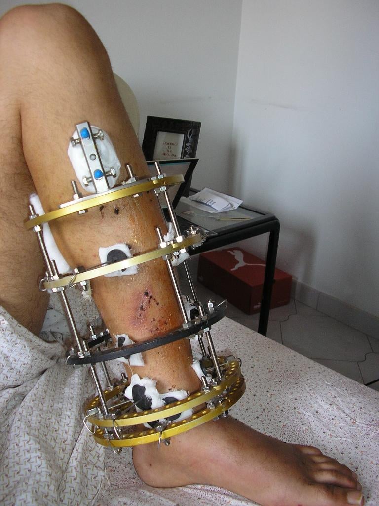
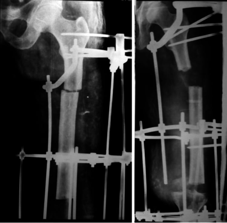
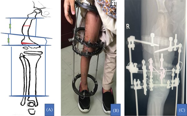
The frame. A circular Ilizarov construct uses stacked rings, threaded rods and tensioned transosseous wires to provide stable fixation while allowing measured adjustment. It permits progressive correction or lengthening while preserving access for pin-site care and joint movement.
The four phases. Treatment runs through latency, distraction, consolidation and remodelling, taken in turn under Clinical Application and Technique below.
Mechanisms and Biology
The Tension-Stress Effect
Gradual tension stimulates cellular proliferation, matrix synthesis and differentiation in several tissue types at once. In the distraction gap the response shows as:
- Osteoblast proliferation, increased in the gap
- Stem cell recruitment, with mesenchymal stem cells migrating to the regenerate zone
- Growth factor release, with VEGF, BMPs and FGFs upregulated
- Angiogenesis, the new vessels running parallel to the bone columns
Blood flow. In Aronson's canine study, flow at the distraction site rose to nearly ten times control, peaking at two weeks, then settled at four to five times control through the rest of distraction and two to three times during consolidation.
Each tissue answers gradual tension in its own way:
- Response to Gradual Tension
- Intramembranous ossification along tension lines
- Clinical Significance
- New bone forms predominantly without a cartilage intermediate (see histology)
- Response to Gradual Tension
- Sarcomere addition, hyperplasia
- Clinical Significance
- Maintains strength during lengthening
- Response to Gradual Tension
- Axonal elongation 1-2mm/day tolerated
- Clinical Significance
- Gradual stretch better tolerated than acute
- Response to Gradual Tension
- Angiogenesis and vessel elongation
- Clinical Significance
- Regional blood flow rises (above)
Corticotomy vs Osteotomy
How the bone is divided critically affects the quality of the regenerate. Ilizarov emphasised preserving the endosteal blood supply, which a corticotomy does and a complete osteotomy does not.
The technique. Multiple drill holes are made through the cortex and connected with a thin osteotome, preserving the medullary contents and endosteal vessels. It is a low-energy technique that minimises thermal necrosis, and it is the gold standard.
Why it heals better. The endosteal blood supply, which provides 70-80% of bone blood flow, stays intact, and the medullary stem cells remain available for regeneration; medullary blood flow is critical to regenerate bone formation. A corticotomy heals faster than a traditional osteotomy, with less risk of delayed union or nonunion.
A saw or high-speed burr creates heat necrosis and damages the endosteal blood supply. This delays healing and risks a poor-quality regenerate.
Stability. The frame is applied with wires or half-pins to achieve rigid stability, and a rigid external frame prevents shear at the distraction gap.
Histology of the Regenerate: The Fibrous Interzone and Its Zones
The evidence describes the regenerate as a "physis-like" structure with "parallel bone columns extending from a central growth zone". Its zones run from the centre of the gap outwards.
The fibrous interzone (FIZ). A radiolucent central band of longitudinally oriented type I/III collagen with fibroblast-like and undifferentiated mesenchymal cells, aligned along the tension vector. It is the germinal or growth zone from which new bone forms in both directions: the true "physis-like" layer, though it is neither a physis nor endochondral.
The primary mineralisation fronts. Zones of microcolumn formation flank the FIZ on each side. Osteoblasts lay osteoid on the aligned collagen scaffold, and it mineralises into parallel bone microcolumns (primary spicules) that grow towards the interzone. This is predominantly intramembranous bone with no cartilage intermediate, though small amounts of transchondroid or endochondral bone appear with less-stable fixation.
Remodelling and mature bone. Peripherally, next to the original cortical ends, osteoclast-osteoblast coupling remodels the microcolumns into lamellar bone with a re-forming medullary canal.
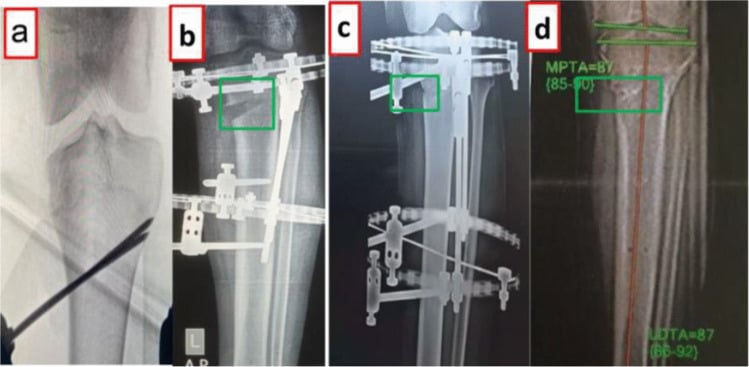
Reading the interzone on film. The width and radiolucency of the interzone on serial films reflect the balance of distraction against mineralisation. A persistently widening lucent interzone signals distraction outrunning bone formation (too fast), while a disappearing interzone signals premature consolidation (too slow). Adequate stability and blood supply keep the interzone organised and the microcolumns parallel.
Differential of a Failing Distraction
A radiolucent or abnormal distraction gap is not one diagnosis. Distinguishing the cause changes management entirely — slowing, pausing, compressing, or revising. The following differential is high-yield for vivas.
- Likely Cause
- Distraction too fast (fibrous regenerate)
- Distinguishing Features
- Gap enlarges faster than bone forms; columns sparse
- Action
- Pause 3-5 days, then resume at 0.5-0.75mm/day
- Likely Cause
- Premature consolidation (rate too slow / latency too long)
- Distinguishing Features
- Resistance to distraction; early bone bridge
- Action
- Increase rate transiently or re-osteotomise if bridged
- Likely Cause
- Inadequate fixation / excess shear
- Distinguishing Features
- Motion at gap, hourglass or cyst formation
- Action
- Improve frame stability; reduce micromotion
- Likely Cause
- Host factors (smoking, diabetes, NSAIDs, malnutrition)
- Distinguishing Features
- Systemically slow healer; otherwise correct setup
- Action
- Optimise host; consider adjuncts; slow rate
- Likely Cause
- Pin-site or deep infection
- Distinguishing Features
- Local signs, pyrexia, raised CRP
- Action
- Treat infection; pin care; debride if deep
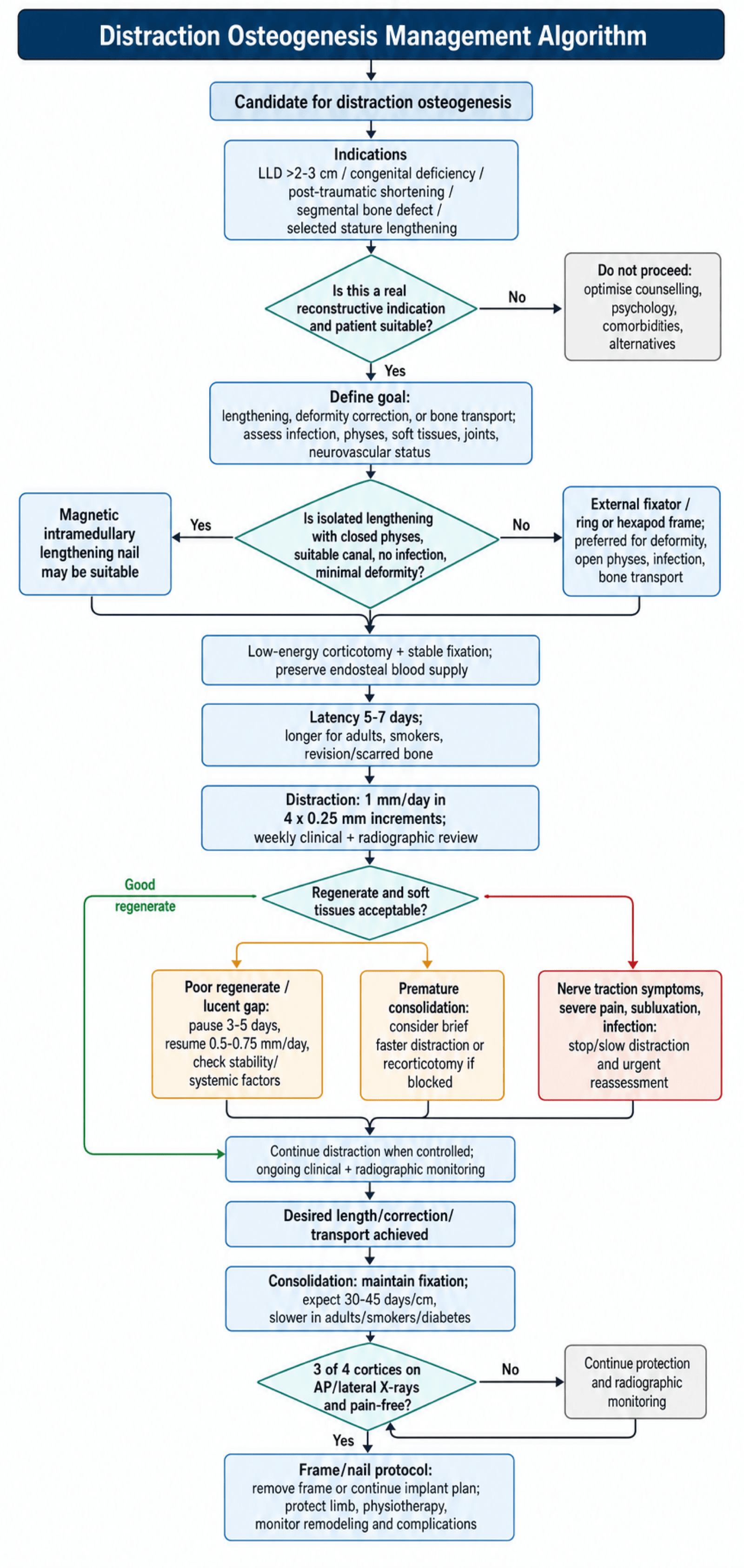
Management Algorithm
Clinical Application and Technique
Phase 1: Latency
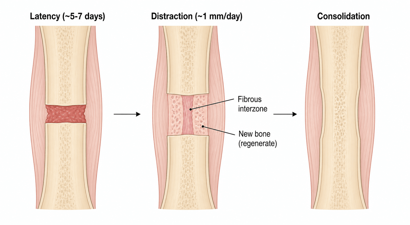
Latency is the wait between corticotomy and the start of distraction, 5-7 days as standard, allowing haematoma formation.
Latency Phase (5-7 Days)
Corticotomy, frame application and soft-tissue closure.
Haematoma forms in the distraction gap and the inflammatory response recruits cells. Mesenchymal stem cells begin to migrate, and the blood clot provides a scaffold.
The fibrin network organises, early fibroblastic proliferation begins and vascular buds form. The tissue is ready for the distraction stimulus.
Adjusting latency. The standard 5-7 days is changed for the patient and the bone:
- Younger children: 5 days (faster healing)
- Older patients or smokers: 7-10 days
- Revision or scarred bone: 10-14 days
Phase 2: Distraction
Rate. The standard is 1 mm per day in total. Faster, and fibrous tissue forms instead of bone, giving a fibrous nonunion; slower, and premature mineralisation blocks the lengthening. One millimetre a day matches the regeneration rate of the bone columns.
Rhythm. The millimetre is given in four increments of 0.25 mm, one every six hours, by slow, controlled turns of the frame. Frequency matters as much as rate: it maintains the biological stimulus, prevents premature consolidation between distractions, gives a better regenerate than once-daily distraction and is less painful than a single large increment.
Changing the rate. The standard is modified when the regenerate or the patient asks for it:
- Poor regenerate on X-ray: slow to 0.5 mm/day
- Premature consolidation: increase to 1.5 mm/day briefly
- Children under 5: can tolerate 1.5 mm/day
- Nerve traction symptoms: slow to 0.75 mm/day or pause 3-5 days
Monitoring During Distraction
Review is weekly, and covers:
- Clinical: pin sites, neurovascular status and pain
- Radiographic: regenerate quality and bone column formation
- Functional: range of motion of the joints above and below
Grading the regenerate. The radiograph is graded on the columns:
- Good: parallel dense bone columns spanning the gap
- Fair: bone columns present but less dense
- Poor: radiolucent gap or minimal bone formation
Warning signs. Any of these must be acted on promptly, on the radiographic appearance and the clinical assessment together:
- Absent bone formation after 2 weeks of distraction
- A widening lucent gap (too fast) or premature consolidation (too slow), read from the interzone as above
- Progressive neurological symptoms
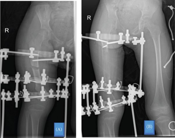
The Two Complications That Arrive During Distraction
Nerve palsy is a distraction-rate problem, and the common peroneal nerve is the one that fails. In tibial lengthening it is tethered at the fibular neck and is the least compliant structure in the segment; in femoral lengthening the sciatic nerve is at risk. Symptoms are ordered, paraesthesia first, then sensory loss, then motor weakness, which is what makes weekly neurological examination during distraction the whole point of follow-up rather than a formality.
Acting on nerve symptoms. The response is graded and immediate: stop distracting, then reverse a few millimetres if symptoms persist, and only then consider decompression. Waiting for a foot drop to declare itself before acting is how a recoverable neurapraxia becomes a permanent deficit. Prophylactic peroneal decompression is used by some units for large planned gains at the proximal tibia; it is a preference, not a proven measure.
Pin-site infection is the commonest complication of any external fixator and the commonest reason a frame is abandoned early. Grade it, because the grade dictates the action:
- Erythema alone: cleansing and oral antibiotics
- Discharge with cellulitis: oral antibiotics and a review of pin mechanics
- A loose or toggling pin: will not settle on antibiotics and must be exchanged or removed, because the infection is being driven by motion at the bone-pin interface
Pain at a pin site with lucency around the pin means loosening until proved otherwise. Deep infection tracking to the regenerate is the feared endpoint and the reason a superficial pin site is never ignored. It is the specific morbidity that magnetic intramedullary nails exist to avoid.
Phase 3: Consolidation
Once the desired length is reached, the frame stays on while the regenerate mineralises and gains strength. The consolidation index is 30-45 days per centimetre lengthened, so a 5 cm lengthening needs 150-225 days of consolidation, and the host moves it:
- Children: 30 days/cm (faster)
- Adults: 40-45 days/cm
- Smokers, diabetes: 50+ days/cm
Consolidation Phase
Woven bone continues forming. Mineralisation begins at the margins and progresses centrally. The regenerate is still mechanically weak and cannot bear full loads without frame support.
Progressive mineralisation is visible on radiographs, and cortices form along the periphery.
Cortical maturation continues and the medullary canal re-establishes.
When the frame comes off. The three-cortices rule: three of the four cortices must be visible on AP and lateral radiographs before frame removal is considered, and removal is safe once those three cortices are mature and the patient is pain-free. The fourth cortex matures after removal.
Getting the timing wrong. Premature removal risks fracture through the regenerate and causes deformity. Delayed removal is unnecessary, wastes time and impacts the patient.
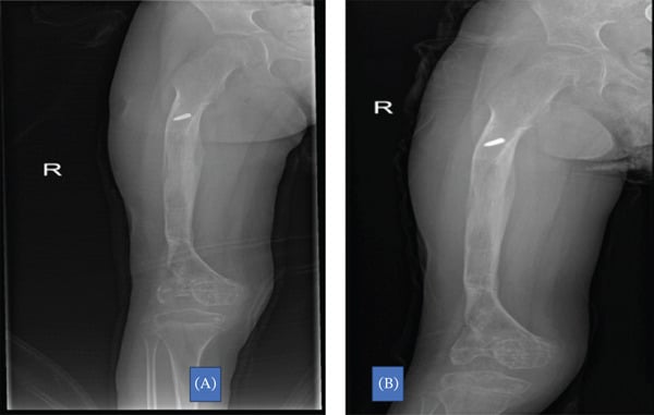
Phase 4: Remodelling
After frame removal the bone continues to remodel for months to years, gradually achieving normal architecture.
- Process
- Cortical thickening
- Radiographic Appearance
- Increased density, defined cortices
- Process
- Medullary canal formation
- Radiographic Appearance
- Central lucency developing
- Process
- Complete remodelling
- Radiographic Appearance
- Normal bone architecture restored
Clinical Relevance and Applications
Indications. The technique enables limb lengthening, deformity correction, bone transport and nonunion treatment:
- Limb length discrepancy greater than 2-3 cm, the most common indication
- Congenital deficiencies (fibular hemimelia, PFFD)
- Post-traumatic shortening
- Bone defects after debridement, or from trauma, infection or tumour resection (bone transport, below)
- Stature lengthening (controversial)
What it offers. No bone graft is required, so there is no donor-site morbidity. The soft tissues lengthen gradually as the bone does, angular deformity can be corrected during the lengthening, the final length is precisely controlled, and weight-bearing is often possible during treatment.
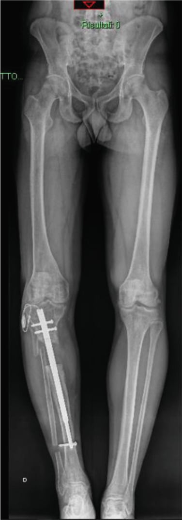
Bone Defects
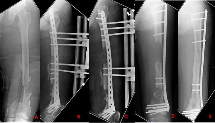
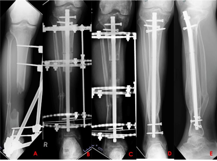
Large bone defects from trauma, infection or tumour resection can be managed with bone transport:
- A corticotomy is performed proximal or distal to the defect
- A bone segment is transported through the defect at 1 mm/day
- Regenerate forms in the transport zone
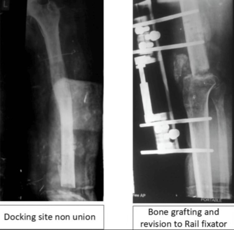
- The segment docks with the opposite end when the defect is filled
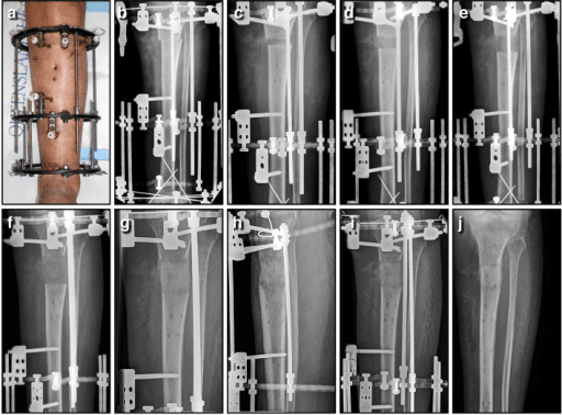
Stature Lengthening
Cosmetic stature lengthening is controversial but increasingly requested, particularly in countries where it is culturally valued.
Cosmetic lengthening in normal individuals raises ethical questions. It carries significant morbidity (9-12 months in frames), complication rates of 30-40% and a risk of permanent nerve injury. Psychological assessment is essential, and most professional societies advise caution.
Only proceed after extensive counselling about risks, timeline and functional impact.
The Distraction, Consolidation and Healing Indices
Distraction index. The time in the distraction (lengthening) phase per centimetre gained, in days/cm. At the standard 1 mm/day it is inherently about 10 days/cm, since a centimetre takes ten 1-mm days, so it is largely fixed by the protocol.
Consolidation index. The time from the end of distraction until the regenerate is mature by the three-cortices rule, per centimetre. This is the variable, biology-dependent index, with the values given under consolidation above.
Healing index (external-fixation index). The total time the frame is on per centimetre gained, distraction and consolidation combined. It is Paley's "lengthening index", classically about one month/cm for a single-level lengthening without deformity, and the single most useful benchmark for counselling and for comparing devices.
Why they matter. They quantify progress and expose problems: a rising consolidation index flags a poor regenerate or adverse host factors. A lower healing index is precisely why implantable magnetic nails (a consolidation index around 25 days/cm in the Nasto series) are attractive against external fixators. Audit outcomes by reporting length gained against goal alongside these indices.
Guidelines, Registries & Global Practice
Global Epidemiology
- Limb length discrepancy is the commonest indication; congenital causes (fibular hemimelia, PFFD, hemihypertrophy), post-traumatic physeal arrest, and post-infective shortening dominate paediatric practice worldwide.
- Bone transport for segmental defects is most prevalent where high-energy trauma and chronic osteomyelitis are common, including many limited-resource settings, making the inexpensive ring fixator a globally important tool.
- Cosmetic stature lengthening clusters in specific cultural and economic contexts and carries the highest medico-legal scrutiny.
Side-by-Side Guidance and Society Positions
- Emphasis
- Ring-fixator deformity correction and bone transport
- Practical Recommendation
- 1mm/day in 4 steps; corticotomy preserving endosteum; three-cortices rule before frame removal
- Emphasis
- Open-fracture and bone-loss reconstruction pathways
- Practical Recommendation
- Manage segmental loss in specialist limb-reconstruction units with combined ortho-plastic input
- Emphasis
- Complication classification and lengthening indices
- Practical Recommendation
- Audit using problems/obstacles/complications; consider magnetic IM nails where deformity is minimal
- Emphasis
- Patient selection and informed consent
- Practical Recommendation
- Caution with cosmetic lengthening; multidisciplinary and psychological assessment
Registry and Outcome Notes
- No dedicated arthroplasty-style registry exists for distraction osteogenesis; evidence is driven by single-centre and multicentre series rather than national implant registries.
- Magnetic intramedullary lengthening nail data (e.g. PRECICE-type devices) report lower pin-site morbidity than external fixators but require post-market surveillance for mechanical and metallosis concerns.
- Outcomes are benchmarked using the distraction index (days/cm during lengthening) and consolidation index (days/cm in frame), with paediatric patients consolidating faster than adults.
High- vs Limited-Resource Practice Variation
- Well-resourced settings: magnetic IM nails, hexapod/computer-assisted frames (Taylor Spatial Frame, TrueLok-Hex), CT-based regenerate assessment, and structured physiotherapy.
- Limited-resource settings: the classic Ilizarov ring fixator remains the workhorse — durable, low-cost, reusable, and effective for transport and deformity correction; patient-led daily distraction and pin-site self-care extend reach where follow-up is sparse.
- Pin-site care principles (regular antiseptic cleansing, early treatment of infection) and VTE awareness during prolonged immobilisation are universal, but specific drug choices follow local antimicrobial stewardship and availability.
Controversies and Areas of Uncertainty
Classic teaching mandates 5-7 days, yet a systematic review of distraction protocols suggests latency may be unnecessary in some models. Optimal latency by age, bone, and device remains debated.
Higher frequency improves regenerate at a given rate, and automated/continuous distraction may be superior in principle, but robust human comparative data versus standard 4x daily manual distraction are limited.
Implantable nails reduce pin-site morbidity, but lack registry-level long-term data and raise device-specific concerns (mechanical failure, metallosis, cost). Best indication boundaries are still being defined.
BMPs, PRP, bisphosphonates, low-intensity ultrasound, and bone-marrow aspirate have all been trialled to accelerate consolidation. Evidence is heterogeneous and none is standard of care.
Ethically contested. High complication rates and prolonged morbidity in otherwise healthy people mean many societies advise caution; thresholds and consent standards vary internationally.
Plain-film three-cortices judgement is subjective. CT, DEXA, and ultrasound are proposed for objective maturity assessment but are not yet universally validated criteria for frame removal.
MCQ Practice Points
Q: What is the standard distraction rate in distraction osteogenesis? A: 1 mm per day in 4 increments of 0.25mm each (every 6 hours). This rate balances bone regeneration capacity with soft tissue tolerance.
Q: What is the consolidation index and typical value? A: 30-45 days per centimeter lengthened - the time required in frame during consolidation phase before safe removal. Children 30 days/cm, adults 40-45 days/cm.
Q: Why is corticotomy preferred over osteotomy in distraction osteogenesis? A: Preserves endosteal blood supply which provides 70-80% of bone blood flow. Multiple drill holes connected with osteotome maintains medullary contents and stem cells critical for regeneration.
Q: What is Ilizarov's tension-stress law? A: Gradual controlled traction on living tissues creates mechanical stress that stimulates and maintains regeneration of bone, soft tissues, nerves, and blood vessels. Foundation principle of distraction osteogenesis.
Q: What radiographic criterion indicates safe frame removal? A: Three of four cortices visible on AP and lateral radiographs, along with consolidation index of 30-45 days/cm. Fourth cortex matures after removal.
Exam Viva Scenarios
Practise clinical reasoning and management decisions out loud
“Examiner asks: Describe the biological principles of distraction osteogenesis and the phases involved.”
“You are lengthening a 12-year-old's femur for limb length discrepancy. At 3 weeks of distraction (21mm gained), radiographs show a widening radiolucent gap with minimal bone formation. What is your assessment and management?”
“A 15-year-old needs 4 cm of femoral lengthening for a post-traumatic discrepancy with no angular or rotational deformity. The family asks whether they really need an external frame. How do you decide, and how do you counsel them?”
Key Principles (Ilizarov)
- Tension-stress law = gradual traction stimulates regeneration
- Stable fixation with rigid external frame
- Corticotomy (not osteotomy) preserves endosteal blood supply
- Low-energy surgical technique respects biology
Four Phases Timeline
- Latency: 5-7 days (hematoma formation)
- Distraction: 1mm/day in 4 increments (bone regeneration)
- Consolidation: 30-45 days/cm (mineralization)
- Remodeling: months-years (cortical maturation)
Critical Parameters
- Rate: 1mm per day total
- Rhythm: 4 increments of 0.25mm (every 6 hours)
- Consolidation index: 30-45 days per cm
- Three cortices visible before frame removal
Troubleshooting
- Too fast distraction = fibrous non-union (radiolucent gap)
- Too slow distraction = premature consolidation
- Poor regenerate = pause 3-5 days, slow to 0.5mm/day
- Nerve symptoms = slow to 0.75mm/day or pause
Biological Responses
- Bone: predominantly intramembranous ossification
- Blood flow: nearly 10x control at the 2-week peak, then 4-5x through distraction
- Nerves: tolerate 1-2mm/day gradual elongation
- Soft tissues: muscle sarcomeres added, vessels elongate
Evidence Base
Tension-Stress Part II: Rate 1mm/day in 4 Steps Optimal
- Canine tibial study of rates (0.5, 1.0, 2.0 mm/day) and frequencies (1, 4, 60 steps/day)
- 0.5 mm/day frequently caused premature consolidation
- 2.0 mm/day produced undesirable changes within elongating tissues
- 1.0 mm/day gave the best regenerate; higher frequency improved outcome at any given rate
- Regenerate forms as a physis-like central growth zone with parallel bone columns
Tension-Stress Part I: Stability and Soft-Tissue Preservation
- Canine tibial experiments varying fixation stability and preservation of periosteum/marrow/medullary blood supply
- Both greater fixator stability and maximal soft-tissue preservation enhanced bone formation
- New bone forms parallel to the tension vector, even with lateral (perpendicular) distraction
- Damage to bone marrow inhibited osteogenesis, confirming the role of marrow elements
- Established the biological rationale for low-energy corticotomy over osteotomy
Blood Flow Surges During Distraction Osteogenesis
- Ten dogs, tibial lengthening, technetium scintigraphy quantifying regional blood flow
- Flow at the distraction site rose to nearly 10x control, peaking at 2 weeks
- Settled to 4-5x control through the rest of distraction, then 2-3x during consolidation
- Distal tibia (away from the gap) showed a similar amplitude and pattern of increased flow
- Supports Ilizarov's idea that distraction can heal hypovascular nonunions and osteomyelitis
Problems, Obstacles and Complications Classification
- 46 patients, 60 limb segments lengthened (mean 5.6 cm, range 1-16 cm)
- Difficulties classified as problems (no surgery), obstacles (surgery to resolve), and true complications
- 27 true complications; original goals achieved in 57 of 60 segments; satisfaction 94%
- Defined complication spectrum: contracture, subluxation, axial deviation, nerve/vessel injury, premature/delayed consolidation, nonunion, pin-site, hardware failure, refracture
- Lengthening index roughly one month/cm for single-level lengthening without deformity
Systematic Review of Optimal Distraction Protocols
- Systematic review of single-variable distraction studies (PubMed 1973-2007)
- 1 mm/day confirmed as the optimal rate (halved in very small animals such as rats)
- A continuous rhythm gave better regenerate than intermittent distraction
- Recommended consolidation period of 6-8 weeks for craniofacial models
- A latency period may not be mandatory in all settings
Magnetic Intramedullary Nail (PRECICE 2) Lengthening
- Multicentre retrospective series, 26 paediatric patients, 26 nails (21 femur, 5 tibia)
- Mean achieved lengthening 44.4 mm versus 49.4 mm goal
- Nail accuracy 91.1% and reliability 88.5%
- Distraction index 11.9 days/cm and consolidation index 25.1 days/cm
- Low complication burden but watch for joint subluxation and mechanical failure