Anaerobic Glycolysis | Matrix Turnover | Mechanotransduction | Catabolic-Anabolic Balance
- Chondrocytes rely on anaerobic glycolysis (95% of ATP) due to avascular nature
- Oxygen tension 1-5% in middle and deep zones; hypoxia induces HIF pathway
- Diffusion from synovial fluid limits cartilage thickness to 1-2mm
- Matrix turnover slow: collagen II half-life 10-15 years, aggrecan 3-24 years
- Mechanical loading essential: cyclic compression enhances metabolism
- “Chondrocytes in OA switch to catabolic phenotype (MMPs up, TIMP down)
- “Glucose transporter GLUT-1 critical for anaerobic metabolism
- “IL-1 and TNF-alpha are major catabolic cytokines
- “Hydrostatic pressure and shear stress activate mechanoreceptors
Overview
Chondrocytes are the sole cell type in articular cartilage, and they synthesise and maintain its extensive extracellular matrix. They make up only 1-2% of the tissue volume, widely separated from one another, yet maintain a matrix 50-100 times their own volume through continuous metabolic activity.
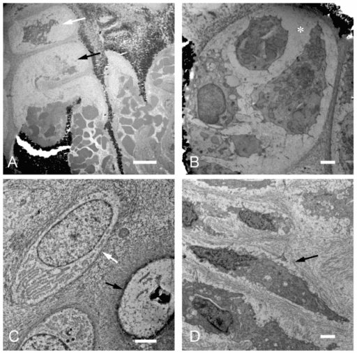
An unusual niche. After skeletal maturity cartilage has no blood supply, and it has no nerves and no lymphatic drainage. Avascularity necessitates anaerobic metabolism, limited diffusion constrains tissue thickness, and the absence of nerves eliminates pain signals from early damage. Chondrocytes have adapted specialised metabolic pathways to function in this hypoxic, avascular, aneural niche.
Understanding chondrocyte metabolism explains why cartilage has limited repair capacity (slow matrix turnover), why injuries are often asymptomatic initially (aneural), and why loading patterns affect joint health (mechanotransduction). It guides treatment strategies including activity modification, viscosupplementation, and emerging biologics.
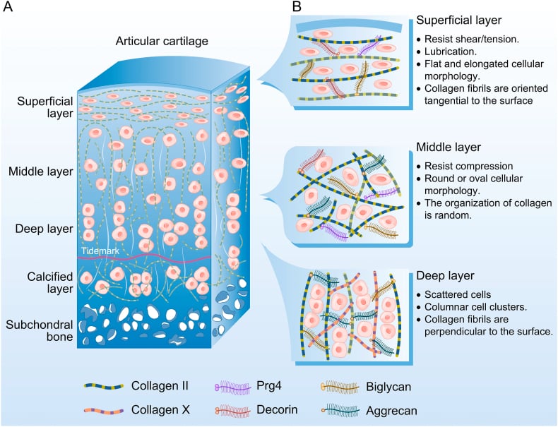
The zones. Cell shape and collagen orientation change with depth, from the joint surface to the bone:
- Superficial: flat cells, collagen parallel to the surface
- Middle: round cells, oblique collagen fibres
- Deep: cells in columns perpendicular to the surface
- Calcified: the interface with subchondral bone
Physiology and Core Concepts
Energy
Anaerobic glycolysis. Chondrocytes derive 95% of their ATP from anaerobic glycolysis (the Embden-Meyerhof pathway) rather than from oxidative phosphorylation. This adaptation reflects the low oxygen environment of cartilage, and the table shows what it costs in yield.
- ATP Yield
- 2 ATP per glucose
- Usage in Chondrocytes
- 95% of energy production
- Advantage in Cartilage
- Functions in hypoxia
- ATP Yield
- 36 ATP per glucose
- Usage in Chondrocytes
- 5% of energy production
- Advantage in Cartilage
- Efficient but needs O2
- ATP Yield
- Variable
- Usage in Chondrocytes
- Emergency energy reserve
- Advantage in Cartilage
- Rapid mobilisation
The oxygen gradient. Oxygen tension falls with depth:
- Synovial fluid and superficial zone: 5-10% O2
- Middle zone: 3-5% O2
- Deep zone: 1-3% O2, hypoxic
- Calcified zone: under 1% O2
Glucose is the primary fuel. Glucose reaches the cell by facilitated diffusion from synovial fluid through the GLUT-1 transporter on the chondrocyte membrane, and uptake is insulin-independent. This transport is the rate-limiting step in energy production, and the reliance on glucose is a hallmark of chondrocyte metabolism.
Diabetes affects cartilage metabolism. Altered glucose homeostasis and advanced glycation end products (AGEs) impair chondrocyte function, and diabetic patients have higher OA rates partly because of this metabolic dysfunction.
Nutrition
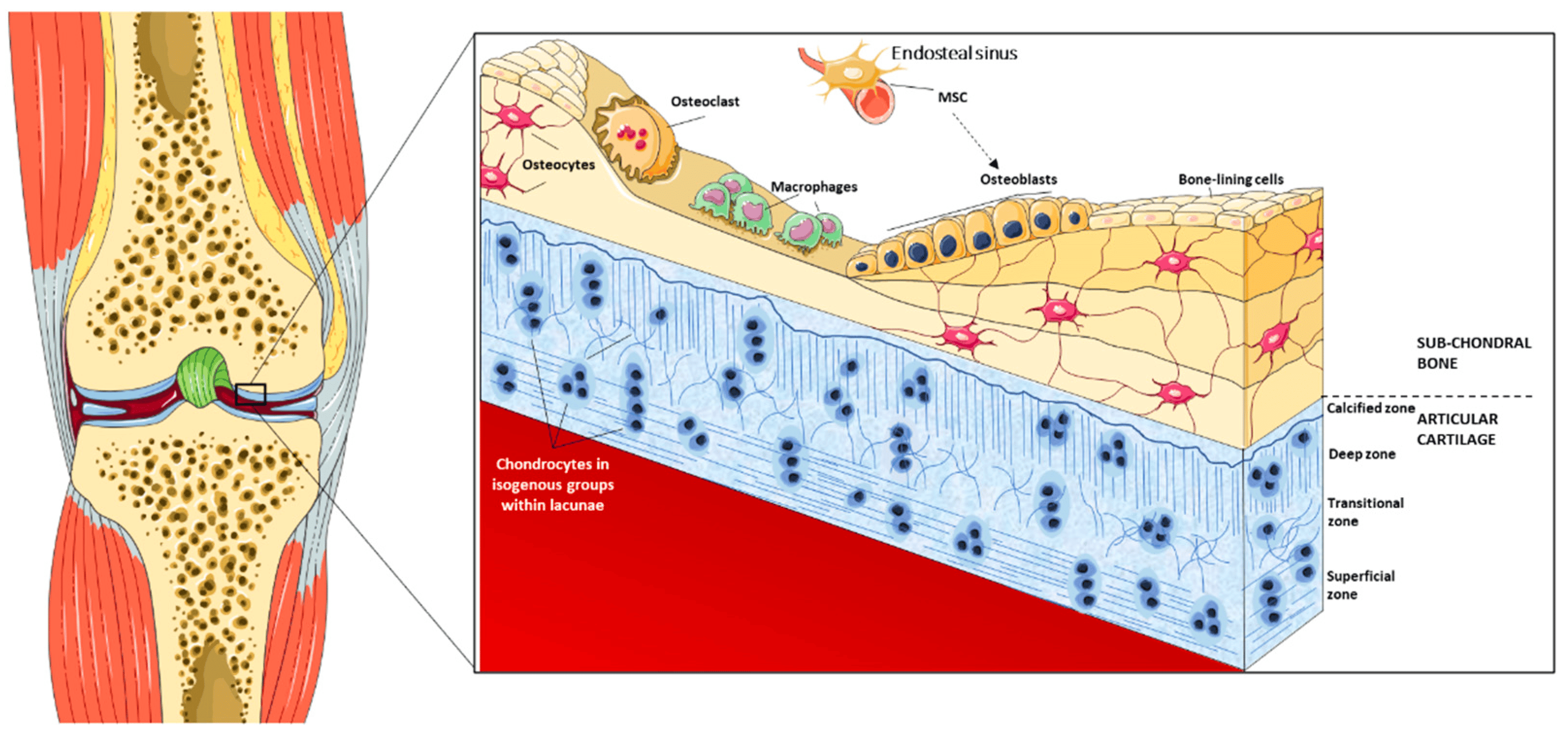
Nutrition by diffusion. Nutrients reach the chondrocyte by diffusion, primarily from the synovial fluid in the joint cavity and secondarily from subchondral bone across the calcified cartilage-bone interface. The maximum diffusion distance of 1-2mm limits adult cartilage to that thickness.
Loading as a pump. Compression pumps nutrients through the matrix, so mechanical loading enhances nutrient transport.
Hypoxia
The HIF pathway. The low oxygen environment activates the transcription factors HIF-1alpha and HIF-2alpha, which regulate chondrocyte gene expression to adapt to hypoxia.
HIF-1alpha keeps the cell alive and making matrix. It upregulates glycolytic enzymes, which maintains ATP in hypoxia, and promotes SOX9, the master transcription factor for chondrogenesis. It also enhances aggrecan and collagen II synthesis and promotes survival in low oxygen, which makes it the primary survival factor.
HIF-2alpha cuts both ways. It regulates cartilage matrix genes but can promote catabolic pathways in excess. Its balance with HIF-1alpha is critical for homeostasis.
Oxidative Stress and Mitochondrial Dysfunction
The redox arm of chondrocyte metabolism is the link between the cell's energy machinery and matrix breakdown.
Reactive oxygen and nitrogen species (ROS/RNS). Even though chondrocytes are mainly glycolytic, they retain functional mitochondria that generate ROS. Inflammatory signalling (IL-1beta, TNF-alpha) induces inducible nitric oxide synthase (iNOS/NOS2), producing large amounts of nitric oxide (NO), and NO combines with superoxide to form peroxynitrite, a potent oxidant.
From signal to damage. At physiological levels ROS act as signalling molecules. In excess they overwhelm the antioxidant defences (superoxide dismutase, catalase, glutathione) and become catabolic.
- Source
- iNOS, induced by IL-1beta/TNF-alpha
- Catabolic effect
- Inhibits aggrecan and collagen II synthesis, activates MMPs, and promotes chondrocyte apoptosis
- Source
- Dysfunctional mitochondria; NADPH oxidases
- Catabolic effect
- Oxidise matrix and proteins, activate catabolic MAPK/NF-kappaB signalling, drive senescence
- Source
- NO + superoxide
- Catabolic effect
- Strong oxidant causing protein nitration and cell death
- Source
- Constitutive
- Catabolic effect
- Protective; their decline with age tips the balance toward catabolism
The mitochondrial link. With ageing and in OA, mitochondrial function declines, with reduced respiratory-chain activity and mitochondrial DNA damage. This raises ROS output, lowers the reserve capacity for oxidative ATP, and promotes chondrocyte senescence and apoptosis.
What the mitochondria are for. They matter less for routine ATP than as a source of damaging ROS and a driver of age-related catabolism. That connects ageing, oxidative stress and the senescent (SASP) phenotype.
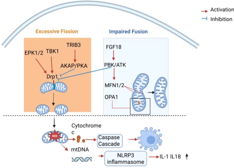
Chondrocytes are glycolytic for ATP but still hold mitochondria. The redox axis is the metabolic bridge between ageing, mitochondrial dysfunction and the catabolic OA phenotype.
Matrix Turnover and Regulation
Turnover is slow. Collagen II has a half-life of 10-15 years. Aggrecan turns over faster, at 3-24 years, but still slowly, and the low metabolic rate limits repair capacity.
Anabolic Pathways
Chondrocytes continuously synthesise extracellular matrix components to maintain cartilage structure and function. The major anabolic factors are:
- Growth factors: IGF-1, TGF-beta, BMPs
- Mechanical signals: cyclic compression, hydrostatic pressure
- Transcription factors: SOX9, RUNX2 (early stages)
- Anti-inflammatory cytokines: IL-4, IL-10, IL-13
What the cell makes. Collagen II is the major fibrillar collagen, 90% of the collagen, and aggrecan is the large aggregating proteoglycan, carrying glycosaminoglycan chains of chondroitin and keratan sulfate. Aggrecan's core protein receives its GAG chains in the Golgi, and it is then secreted and aggregated with hyaluronan.
The minor components. Collagens IX and XI regulate fibril formation and collagen VI sits in the pericellular matrix. Link protein and decorin complete the list.
Collagen II assembly. The molecule is built in stages that run from the nucleus out into the matrix.
Collagen II Synthesis and Assembly
SOX9 activates COL2A1 gene. mRNA transcribed and processed. Exported to rough endoplasmic reticulum.
Pro-alpha chains synthesised on ribosomes. Hydroxylation of proline and lysine residues (requires vitamin C). Glycosylation occurs.
Three pro-alpha1(II) chains align via C-propeptides. Triple helix formation proceeds from C to N terminus. Procollagen secreted.
N and C propeptides cleaved by specific proteinases. Collagen molecules self-assemble into fibrils. Cross-linking by lysyl oxidase stabilises.
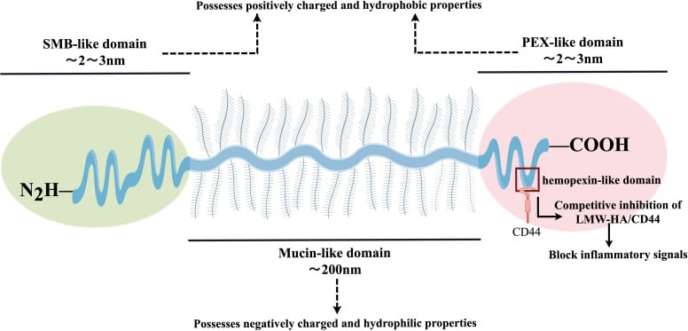
Lubricin. Lubricin (PRG4, superficial zone protein) is a mucinous glycoprotein made almost exclusively by superficial zone chondrocytes and by synoviocytes, and secreted onto the surface rather than into the matrix. Its long, heavily O-glycosylated mucin-like centre is hydrophilic and negatively charged, while compact terminal domains help anchor the molecule at the joint surface.
The boundary lubricant. Lubricin is the boundary lubricant of articular cartilage, working with hyaluronan to keep the coefficient of friction of the loaded surface near 0.001 and to resist cell and protein adhesion.
A product of the same balance. Lubricin production is a zonally restricted synthetic function that is suppressed by IL-1 and TNF and stimulated by TGF-beta, so it moves with the same anabolic-catabolic balance as aggrecan and collagen II. It is lost early, before matrix depletion is visible.
The human proof is monogenic. Loss-of-function mutations in PRG4 cause camptodactyly-arthropathy-coxa vara-pericarditis (CACP) syndrome, in which children develop non-inflammatory joint failure with synovial hyperplasia. No other single cartilage gene product has this clean a demonstration that its absence alone destroys joints, and it establishes that lubrication failure is a mechanism of arthropathy in its own right, not merely a consequence of cartilage loss.
Catabolic Pathways
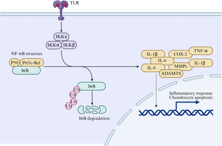
Matrix degradation is normally balanced with synthesis, and imbalance leads to cartilage loss. The drivers are the pro-inflammatory cytokines IL-1beta and TNF-alpha, static compression and excessive load, and the proteases these induce.
The proteases. MMP-13 is the collagenase and ADAMTS-4 and ADAMTS-5 are the aggrecanases. MMP-3 degrades aggrecan and other proteoglycans more broadly, and the TIMPs hold the MMPs in check.
- Substrates
- Collagen II, IX, XI
- Regulation
- IL-1, TNF-alpha induce
- Role in OA
- Primary collagenase in OA
- Substrates
- Aggrecan core protein
- Regulation
- Constitutive + IL-1 induced
- Role in OA
- Aggrecan degradation
- Substrates
- Aggrecan, proteoglycans
- Regulation
- Cytokine-induced
- Role in OA
- Broad matrix degradation
- Substrates
- Inhibit MMPs
- Regulation
- Constitutive expression
- Role in OA
- Protective, reduced in OA
The cytokine cascade. IL-1beta binds the IL-1R1 receptor and activates the NF-kappaB and MAPK pathways. These upregulate MMPs, ADAMTS, iNOS and COX-2 and downregulate aggrecan and collagen II synthesis, and the net effect is a catabolic phenotype with net tissue loss.
Chondrocyte Hypertrophy and the Endochondral Programme in OA
Hypertrophy is a favourite basic-science link between development and disease.
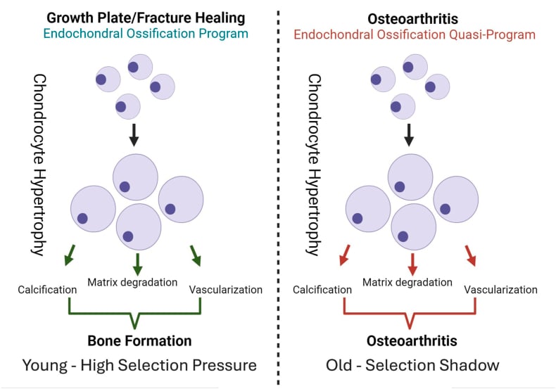
The hypertrophic programme. Terminal (hypertrophic) differentiation is the normal end-point of the growth-plate chondrocyte, driven by a switch in transcription factors away from SOX9 and toward RUNX2 (and MEF2C). The hypertrophic chondrocyte enlarges and switches its matrix output.
- Resting / anabolic chondrocyte
- SOX9 (with SOX5/6)
- Hypertrophic chondrocyte
- RUNX2, MEF2C; SOX9 falls
- Resting / anabolic chondrocyte
- Collagen II
- Hypertrophic chondrocyte
- Collagen X (a hypertrophy marker)
- Resting / anabolic chondrocyte
- Aggrecan, collagen II
- Hypertrophic chondrocyte
- Alkaline phosphatase, MMP-13, VEGF
- Resting / anabolic chondrocyte
- Maintained, resilient
- Hypertrophic chondrocyte
- Mineralised; vascular invasion and replacement by bone
Why it matters in OA. Healthy articular chondrocytes are normally held in a stable, non-hypertrophic state and resist this terminal programme for life. In osteoarthritis they abnormally re-activate it: RUNX2 rises, collagen X and MMP-13 appear, and alkaline phosphatase and VEGF promote calcification and vascular invasion.
The consequences are tidemark duplication and advance, thickening of the calcified-cartilage layer, and osteophyte formation. The same process that builds the growth plate now drives joint destruction. The developmental detail of the growth plate itself is covered in physis-growth-plate-anatomy and ossification-intramembranous-endochondral; here the focus is the metabolic phenotype switch.
"OA chondrocytes recapitulate endochondral ossification" is the one-line answer.
Mechanotransduction
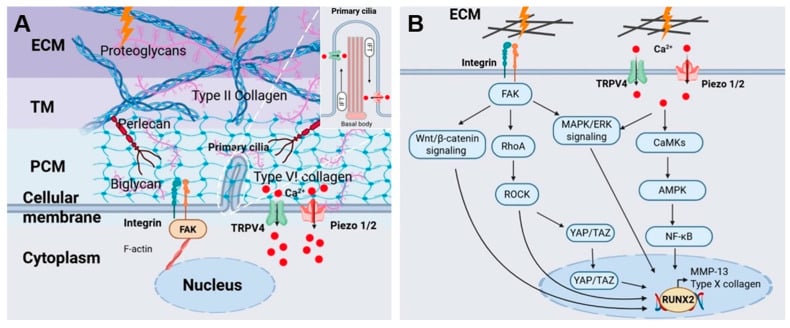
Mechanical loading is a critical regulator of chondrocyte metabolism. Physiological loading maintains cartilage health, and abnormal loading contributes to degeneration.
Mechanosensors. Chondrocytes detect mechanical stimuli through several mechanisms:
- Primary cilium: a solitary non-motile organelle projecting from the cell surface, which bends with matrix deformation
- Integrins: link the cytoskeleton to the matrix, alpha5-beta1 (fibronectin) and alpha10-beta1 (collagen II)
- Ion channels: the mechanosensitive calcium channels Piezo1 and Piezo2
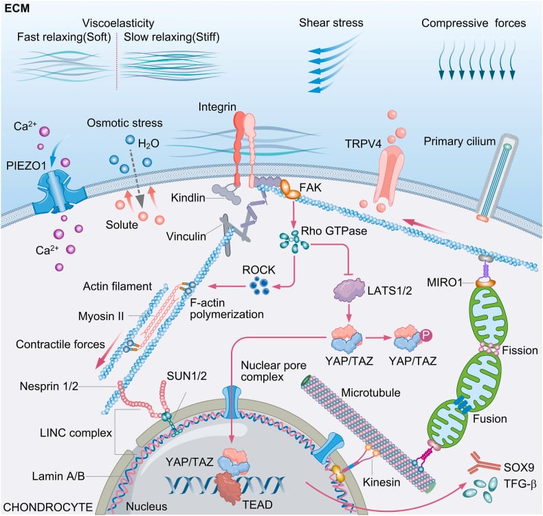
Load-dependent responses. Each loading pattern has its own metabolic effect and matrix response.
- Metabolic Effect
- Anabolic
- Matrix Response
- Increased synthesis
- Clinical Example
- Normal daily activity
- Metabolic Effect
- Anabolic
- Matrix Response
- Enhanced proteoglycan
- Clinical Example
- Swimming, water therapy
- Metabolic Effect
- Catabolic
- Matrix Response
- Increased MMPs
- Clinical Example
- Prolonged standing, obesity
- Metabolic Effect
- Catabolic
- Matrix Response
- Matrix breakdown
- Clinical Example
- Running on concrete, trauma
- Metabolic Effect
- Catabolic
- Matrix Response
- Atrophy
- Clinical Example
- Casting, bed rest
Optimal loading. Moderate cyclic compression at 10-15% strain and a frequency of 0.5-1 Hz, which approximates walking, enhances matrix synthesis and nutrient transport and activates anabolic signalling pathways. This explains why moderate exercise is protective for cartilage while both excessive loading and immobilisation are detrimental.
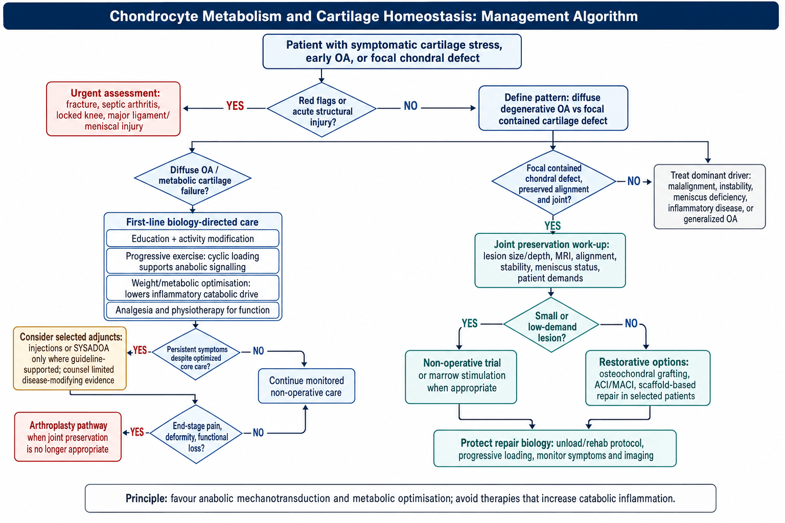
Management Algorithm
Clinical Relevance
Osteoarthritis Pathophysiology
A catabolic shift. OA represents a shift toward catabolic metabolism with failed attempts at repair. MMP-13 and ADAMTS expression increase, the TIMPs (protease inhibitors) decrease, and IL-1beta and TNF-alpha are elevated.
The failed repair. The chondrocytes mount an attempted anabolic response, seen as clusters of cells, before the disease progresses to chondrocyte apoptosis.
The vicious cycle of OA: mechanical injury → cell damage → IL-1beta release → MMP upregulation → matrix degradation → abnormal loading → more injury. Breaking this cycle is the goal of disease-modifying OA drugs.
Therapeutic Targets
Understanding chondrocyte metabolism guides therapeutic strategy:
- Viscosupplementation: hyaluronic acid injections to improve lubrication and potentially stimulate endogenous production
- Corticosteroids: potent anti-inflammatory effect, but can inhibit chondrocyte metabolism if used frequently
- Platelet-rich plasma (PRP): delivers anabolic growth factors (TGF-beta, IGF-1) to shift the balance
- Future targets: senolytics (removing senescent cells), Wnt pathway inhibitors, aggrecanase (ADAMTS5) inhibitors
Rationale and recommendation. The biology behind an injection does not settle its use. AAOS does not recommend hyaluronic acid injections for routine knee OA and NICE advises against intra-articular hyaluronic acid, as the Guidelines section sets out.
Distinguishing Chondrocyte Metabolic States
Separating the chondrocyte phenotypes that dominate in health, ageing and disease is a common viva and MCQ task. Each has a characteristic transcription factor profile, matrix output and clinical correlate.
- Key Drivers
- SOX9, IGF-1, balanced load
- Matrix Effect
- Synthesis = degradation
- Markers
- Collagen II, aggrecan
- Clinical Correlate
- Healthy adult cartilage
- Key Drivers
- TGF-beta, BMP, cyclic load
- Matrix Effect
- Synthesis greater than degradation
- Markers
- Increased proteoglycan
- Clinical Correlate
- Early loading response, immature cartilage
- Key Drivers
- IL-1beta, TNF-alpha, static/impact load
- Matrix Effect
- Degradation greater than synthesis
- Markers
- MMP-13, ADAMTS-5, low TIMP
- Clinical Correlate
- Progressive osteoarthritis
- Key Drivers
- RUNX2, MMP-13, Wnt, low SOX9
- Matrix Effect
- Calcification, collagen X
- Markers
- Collagen X, alkaline phosphatase
- Clinical Correlate
- Growth plate, OA tidemark advance
- Key Drivers
- Ageing, oxidative stress, DNA damage
- Matrix Effect
- Catabolic secretome
- Markers
- p16INK4a, IL-6, MMPs
- Clinical Correlate
- Age-related OA; senolytic target
Guidelines, Registries & Global Practice
The clinical relevance of chondrocyte metabolism is realised through osteoarthritis and cartilage-repair practice. The biology is universal, but how it is translated into guidelines and procedures varies worldwide.
Global Epidemiology
- Osteoarthritis affects an estimated 595 million people worldwide (Global Burden of Disease 2021), making it a leading global cause of disability in older adults.
- The knee is the most commonly affected large joint; prevalence rises sharply with age and obesity.
- Burden is rising fastest in low- and middle-income countries as populations age and obesity increases.
Society Guidance Drawing on Cartilage Biology
- Position on Core OA Biology-Linked Therapies
- Core: exercise, weight loss, education; conditional pharmacology
- Notable Stance
- Strong emphasis on load and metabolic optimisation
- Position on Core OA Biology-Linked Therapies
- Strong evidence for exercise and weight loss; HA injections not recommended for routine knee OA
- Notable Stance
- Skeptical of viscosupplementation
- Position on Core OA Biology-Linked Therapies
- Therapeutic exercise as first-line; advises against intra-articular HA
- Notable Stance
- Does not endorse hyaluronic acid
- Position on Core OA Biology-Linked Therapies
- Stepwise algorithm; supports symptomatic slow-acting drugs (SYSADOA) in some patients
- Notable Stance
- More permissive of glucosamine/chondroitin
There is broad international agreement that mechanical and metabolic optimisation (exercise, weight loss) is first-line because it directly targets the anabolic loading biology described above. Divergence centres on injectables and symptomatic slow-acting agents.
Registries & Cartilage-Repair Practice
- No major registry tracks chondrocyte biology directly, but joint replacement registries (NJR, AJRR, AOANJRR, SHAR, NZJR) record the end-stage of failed cartilage homeostasis and benchmark arthroplasty outcomes.
- Cartilage-repair registries (e.g. the German cartilage registry KnorpelRegister DGOU) capture outcomes of biologically driven procedures such as autologous chondrocyte implantation (ACI) and matrix-assisted ACI (MACI).
High- vs Limited-Resource Practice Variation
- High-resource settings: access to cell-based cartilage repair (ACI/MACI), osteochondral grafting and PRP; structured weight-loss and exercise programmes.
- Limited-resource settings: management is dominated by activity modification, analgesia and physiotherapy, with earlier reliance on arthroplasty once available because biologic repair options are scarce or unaffordable.
- Universal, low-cost interventions (exercise, weight management) carry the strongest evidence and are equitable globally.
Controversies & Areas of Uncertainty
Several long-standing debates in cartilage biology remain unresolved. They are favourite examiner territory because they reward candidates who can argue both sides.
Do chondrocytes use any oxidative phosphorylation? The textbook "95% glycolytic" figure is a simplification. Chondrocytes possess functional mitochondria and basal oxidative phosphorylation persists, but ATP yield is dominated by glycolysis. Mitochondrial dysfunction is increasingly implicated in OA, so the picture is more nuanced than "purely anaerobic".
Is OA a metabolic or mechanical disease? Mechanical malalignment and metabolic or inflammatory drivers (obesity, IL-1, AGEs, metabolic syndrome) are not mutually exclusive. The dominant view (Loeser 2012) is that mechanical injury and low-grade inflammation converge on a final common catabolic chondrocyte pathway.
Why have DMOADs repeatedly failed? Despite compelling animal data (e.g. ADAMTS5 knockout), human aggrecanase, MMP and IL-1 inhibitor trials have not delivered approved disease-modifying drugs. Heterogeneous OA phenotypes, late presentation and the slow matrix turnover that blunts measurable response are leading explanations.
Can senescent chondrocytes be safely targeted? Clearing p16-positive senescent cells reduced OA in animal models, but a phase 2 intra-articular senolytic (UBX0101) failed to meet its primary endpoint, tempering early enthusiasm. Whether senescence is cause or consequence remains debated.
MCQ Practice Points
Q: What percentage of chondrocyte ATP comes from anaerobic glycolysis? A: 95% - Chondrocytes rely predominantly on glycolysis due to the avascular, hypoxic environment of cartilage. Only 5% comes from oxidative metabolism.
Q: Which glucose transporter is critical for chondrocyte energy metabolism? A: GLUT-1 - Insulin-independent facilitated diffusion transporter that allows glucose uptake from synovial fluid in the avascular cartilage.
Q: What is the major catabolic cytokine driving cartilage degradation in osteoarthritis? A: Interleukin-1 beta (IL-1beta) - Upregulates MMP-13 and ADAMTS-4/5, downregulates matrix synthesis, and shifts chondrocytes to catabolic phenotype via NF-kappaB pathway.
Q: What is the half-life of collagen type II in articular cartilage? A: 10-15 years - Extremely slow turnover explains limited repair capacity. Aggrecan turnover is faster (3-24 years) but still slow.
Q: Which organelle serves as the primary mechanosensor in chondrocytes? A: Primary Cilium - A solitary, non-motile cilium that projects into the matrix and deflects with load, triggering intracellular signaling.
Q: Which transcription factor is stabilized by the hypoxic environment of cartilage? A: HIF-1alpha - Hypoxia-Inducible Factor 1-alpha plays a critical role in chondrocyte survival and anabolic function under low oxygen conditions.
Exam Viva Scenarios
Practise clinical reasoning and management decisions out loud
“Examiner asks: Explain how chondrocytes generate energy given the avascular nature of cartilage.”
“A patient asks why you recommend moderate exercise for their early knee osteoarthritis, given that loading damages cartilage.”
“An examiner asks you to explain, at the cellular level, why articular cartilage is lost in osteoarthritis and why we still have no disease-modifying drug.”
Energy Metabolism
- Glycolysis: 95% of ATP (Anaerobic)
- Glucose Transport: GLUT-1 (Insulin independent)
- Hypoxia: HIF-1alpha regulates survival
Nutrition
- Source: Synovial fluid diffusion
- Limit: 1-2mm thickness
- Enhancer: Cyclic pumping action
Matrix Regulation
- Anabolic: TGF-beta, IGF-1, SOX9
- Catabolic: IL-1, TNF-alpha, MMP-13
- Mechanosensor: Primary Cilium, Integrins
Evidence Base
Chondrocyte Energy Metabolism Is Glucose-Driven, Not Oxygen-Driven
- Bovine chondrocytes in agarose monitored for glucose/oxygen uptake and lactate output
- Lactate production predictable from glucose alone; oxygen tension did not influence lactate output
- Confirms anaerobic glycolysis is the dominant ATP pathway in chondrocytes
- Glucose availability, not oxygen, is the rate-limiting substrate for metabolism
HIF-1alpha Drives Anaerobic Glycolysis and Matrix Synthesis
- HIF-1alpha-null epiphyseal chondrocytes cannot maintain ATP under hypoxia
- HIF-1alpha is required for glycolysis under both aerobic and anaerobic conditions
- Loss of HIF-1alpha reduces aggrecan and collagen II mRNA and protein under low oxygen
- Links the hypoxic niche directly to extracellular matrix production
References
-
Sengers BG, Heywood HK, Lee DA, et al. Nutrient utilization by bovine articular chondrocytes. J Biomech Eng. 2005. PMID 16248305.
-
Pfander D, Cramer T, Schipani E, Johnson RS. HIF-1alpha controls extracellular matrix synthesis by epiphyseal chondrocytes. J Cell Sci. 2003. PMID 12665562.
-
Grodzinsky AJ, Levenston ME, Jin M, Frank EH. Cartilage tissue remodeling in response to mechanical forces. Annu Rev Biomed Eng. 2000. PMID 11701528.
-
Fitzgerald JB, Jin M, Dean D, Grodzinsky AJ, et al. Mechanical compression of cartilage explants induces multiple time-dependent gene expression patterns. J Biol Chem. 2004. PMID 14960571.
-
Goldring MB. The role of the chondrocyte in osteoarthritis. Arthritis Rheum. 2000. PMID 11014341.
-
Glasson SS, Askew R, Sheppard B, Morris EA, et al. Deletion of active ADAMTS5 prevents cartilage degradation in a murine model of osteoarthritis. Nature. 2005. PMID 15800624.
-
Lee W, Leddy HA, Guilak F, Liedtke W, et al. Synergy between Piezo1 and Piezo2 channels confers high-strain mechanosensitivity to articular cartilage. Proc Natl Acad Sci USA. 2014. PMID 25385580.
-
Loeser RF, Goldring SR, Scanzello CR, Goldring MB. Osteoarthritis: a disease of the joint as an organ. Arthritis Rheum. 2012. PMID 22392533.