The Hypermobility Syndromes
- Hypermobile EDS: Most common. Joint instability, chronic pain.
- Vascular EDS: COL3A1 mutation. Arterial rupture risk.
- Beighton Score: the cut-off is AGE-ADJUSTED, not a single number - 6 or more prepubertal, 5 or more from puberty to 50, 4 or more over 50. Quoting a flat 4/9 over-diagnoses everyone under 50.
- Skin: Hyperextensible, easy bruising, poor healing.
- Surgery Caution: Poor tissue healing.
- “Beighton cut-off is age-adjusted (6 prepubertal / 5 to age 50 / 4 over 50)
- “Vascular EDS is dangerous
- “Surgery has poor healing
- “Chronic pain is common
Overview and Epidemiology
Ehlers-Danlos syndrome (EDS) is a group of inherited connective tissue disorders caused by defects in collagen or in the proteins that interact with collagen. The result is joint hypermobility, skin hyperextensibility and tissue fragility.
Genetics and frequency. Different types have different genes, and most are autosomal dominant. The prevalence is 1 in 5,000, and hypermobile EDS (hEDS) is the most common type.
Pathophysiology and Pathomechanics
The collagen defect. Different types affect different collagen genes, and the result is weak ligaments, skin and blood vessels.
Why the joints are unstable. Lax ligaments cannot stabilise the joints, so they subluxate and dislocate recurrently. Microtrauma and joint damage add chronic pain, although laxity alone does not explain the pain (see Multisystem Comorbidities below).
Classification Systems
The major types compare as follows.
- Gene/Inheritance
- Unknown gene/AD
- Key Features
- Most common, joint instability, pain
- Gene/Inheritance
- COL5A1 or COL5A2/AD
- Key Features
- Skin hyperextensibility, scarring
- Gene/Inheritance
- COL3A1/AD
- Key Features
- Arterial rupture, thin skin
- Gene/Inheritance
- PLOD1/AR
- Key Features
- Severe scoliosis, hypotonia
Hypermobile EDS. The gene is unknown, so the diagnosis is clinical, by the 2017 criteria set out under Investigations. The orthopaedic picture is joint instability and dislocations, with chronic pain and fatigue.
Classical EDS. The skin is hyperextensible, by more than 1.5cm, and heals with atrophic "cigarette-paper" scars. The joints are hypermobile too.
Vascular EDS. Caused by a COL3A1 mutation, it is the most dangerous type. The skin is thin and translucent with visible veins, and the risks are arterial rupture and bowel perforation.
Kyphoscoliotic EDS. A PLOD1 mutation, inherited as an autosomal recessive trait. It brings severe neonatal hypotonia, progressive kyphoscoliosis and joint hypermobility, and scoliosis surgery is undertaken if needed.
Clinical Assessment
History. Ask about dislocations and subluxations, chronic pain and fatigue, easy bruising, problems with wound healing, and the family history.
The Beighton score. Generalised joint hypermobility is scored out of 9:
- Passive dorsiflexion of the fifth metacarpophalangeal joint beyond 90° - one point each side
- Thumb apposition to the flexor surface of the forearm - one point each side
- Elbow hyperextension beyond 10° - one point each side
- Knee hyperextension beyond 10° - one point each side
- Forward trunk flexion with the knees locked and the palms flat on the floor - one point
The cut-off is age-adjusted: 6 or more before puberty, 5 or more from puberty to age 50, and 4 or more over 50.
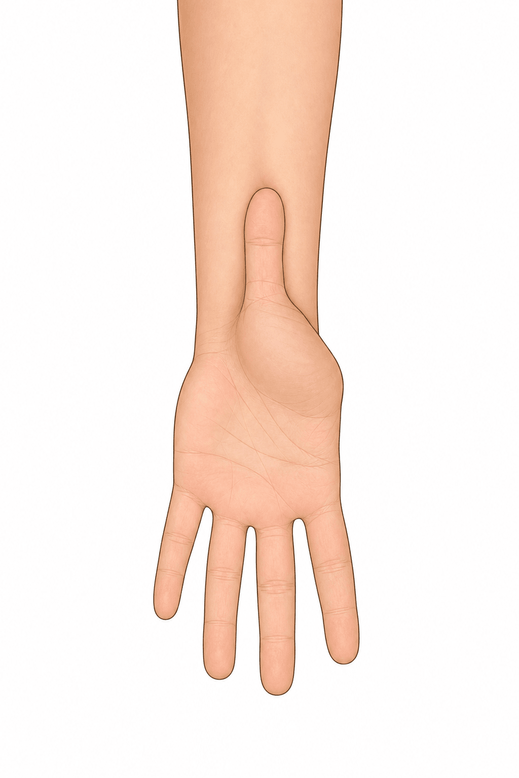
Skin and joints. Examine the skin for hyperextensibility, texture and scars, and the joints for instability and signs of previous dislocations.

Investigations and Diagnosis
Genetic testing. Classical, vascular and other types can be confirmed genetically. hEDS has no genetic test and is diagnosed clinically.
The 2017 hEDS criteria. All three are required:
- Generalised joint hypermobility on the age-adjusted Beighton score. A 5-point self-report questionnaire can substitute one point if the Beighton is one below the cut-off for age.
- Two or more of Features A, B and C:
- Feature A, systemic features of a generalised connective-tissue disorder, five or more of twelve: unusually soft or velvety skin; mild skin hyperextensibility; unexplained striae; bilateral piezogenic heel papules; recurrent or multiple abdominal hernias; atrophic scarring; pelvic-floor, rectal or uterine prolapse; dental crowding with a high narrow palate; arachnodactyly; an arm-span-to-height ratio of 1.05 or more; mitral valve prolapse; aortic root dilatation.
- Feature B, a positive family history: one or more first-degree relatives meeting the criteria.
- Feature C, musculoskeletal, one needed: daily musculoskeletal pain in two or more limbs for at least 3 months; chronic widespread pain for at least 3 months; or recurrent joint dislocations or frank instability without trauma.
- Exclusion of alternatives. Unusual skin fragility is absent (it would suggest another EDS subtype), and other heritable and acquired connective-tissue disorders (including autoimmune disease) and alternative causes of hypermobility or hypotonia (neuromuscular or skeletal dysplasias) are excluded.
Hypermobility spectrum disorder. Symptomatic hypermobility that does not satisfy all three criteria is classified as hypermobility spectrum disorder (HSD), and it is managed identically.
Differential Diagnosis
EDS involves the skin and the joints together, with chronic pain. The other conditions in the differential are distinguished below.
- Key Features
- Joint instability, pain
- Differentiator
- Beighton positive, clinical diagnosis
- Key Features
- Hypermobility only, no pain syndrome
- Differentiator
- No systemic features
- Key Features
- Tall, thin habitus; cardiovascular focus, aortic root
- Differentiator
- FBN1 mutation, lens displaced upwards
- Key Features
- Bifid uvula
- Differentiator
- TGFBR1/2
- Key Features
- Bone fragility primary (fractures)
- Differentiator
- COL1A1/2, blue sclerae
Management
Conservative care comes first. Physiotherapy strengthens the muscles around the joints so that they provide the stability the ligaments cannot. Around it sit multimodal pain management, activity modification that avoids hyperextension and high-impact activities, and bracing for unstable joints.
When to operate. Surgery is for recurrent dislocations that have not responded to conservative treatment. Tissue heals poorly and recurrence is high, so expect a higher failure rate than in the normal population.
Avoid surgery and other invasive procedures if possible, and obtain genetic testing before major surgery when vascular EDS is suspected. Management is arterial surveillance with regular imaging, blood-pressure control and genetic counselling.
Surgical Technique
Joint stabilisation. Recurrent shoulder and patellar dislocations are stabilised with the standard procedures, Bankart repair and MPFL reconstruction among them, but with stronger fixation. The precautions around them follow CARE.
CAREEDS Surgical Precautions
Hook:Take CARE with EDS patients - surgery outcomes are less predictable!
Complications
- Context
- Poor healing
- Management
- Meticulous closure, sutures left in longer
- Context
- Surgical failure
- Management
- Accept the higher failure rate
- Context
- Disease-related
- Management
- Multimodal pain management
Postoperative Care
EDS Postoperative Pathway
Meticulous tension-free closure; leave sutures in LONGER than usual - tissue is fragile and dehiscence risk is high.
Prolonged immobilisation, longer than standard protocols, may help - EDS tissues need more time to heal.
Cautious, graded rehabilitation with proprioceptive retraining; avoid aggressive stretching of hypermobile joints.
Counsel about higher recurrence/failure rates than the general population and the role of ongoing strengthening.
Outcomes and Prognosis
Lifespan. hEDS carries a normal lifespan but chronic symptoms. Vascular EDS reduces life expectancy through arterial rupture: median survival was 48 years in the Pepin 2000 NEJM cohort.
Multisystem Comorbidities & the Pain Mechanism
The comorbidity cluster. hEDS commonly co-occurs with a recognised triad of systemic problems:
- Dysautonomia or orthostatic intolerance, most characteristically postural orthostatic tachycardia syndrome (POTS), with light-headedness, palpitations, presyncope and fatigue on standing
- Functional gastrointestinal disorders and dysmotility: reflux, bloating, early satiety and IBS-type symptoms
- Mast cell activation symptoms: flushing, urticaria, and food or drug sensitivities
Chronic fatigue, anxiety and depression, pelvic-floor dysfunction with urogenital or rectal prolapse, and easy bruising are also frequent.
Why the pain persists. Chronic pain in hEDS is not explained by joint laxity alone. It is increasingly understood as nociplastic pain with central sensitisation, compounded by dysautonomia, deconditioning and impaired proprioception. That is why pain frequently continues even when the joints are mechanically "stable", and why isolated stabilisation surgery rarely resolves it.
The orthopaedic take-home. Treat the orthopaedic complaint as one facet of a systemic disorder. Prioritise graded, proprioceptive physiotherapy and pacing, multimodal and neuromodulatory analgesia, and management of the autonomic and GI comorbidities within a multidisciplinary team, and always screen for the vascular-EDS red flags before any intervention.
Guidelines, Registries & Global Practice
Global epidemiology
- All EDS types combined: roughly 1 in 5,000. Hypermobile EDS / hypermobility spectrum disorders dominate (the large majority of cases).
- Vascular EDS is rare: 1 in 50,000 to 1 in 200,000 (Byers et al, 2017).
- hEDS shows a marked female predominance in clinic populations; rare recessive types (kyphoscoliotic, dermatosparaxis) cluster in consanguineous populations.
Side-by-side guidance
- Position
- Single global standard: 13 subtypes; molecular confirmation for all types except hEDS; revised hEDS criteria
- Position
- Maintains the consortium criteria, the vascular EDS "passport" and emergency-care guidance; international patient registry
- Position
- Confirm COL3A1, manage at centres of excellence, aggressive BP control, annual non-invasive vascular surveillance, minimise surgery
- Position
- Vascular EDS managed within heritable thoracic aortic/connective-tissue disease pathways; celiprolol where available
There is no orthopaedic implant registry signal specific to EDS, but registry-style cohort data (e.g. the Pepin NEJM cohort) underpin natural-history estimates, and the international EDS registry is building genotype-phenotype data.
High- vs limited-resource practice
- Well-resourced: gene-panel/exome testing, MDT (genetics, rheumatology, cardiology, physiotherapy, pain), celiprolol for vascular EDS, annual cross-sectional vascular imaging.
- Limited-resource: diagnosis remains largely clinical (Beighton score, skin and family history); prioritise physiotherapy, activity advice and recognition of vascular EDS red flags for urgent referral when genetic testing and celiprolol are unavailable.
Controversies & Areas of Uncertainty
The hEDS / HSD boundary. The distinction between hEDS and HSD is debated. Many "hEDS" patients meet HSD rather than strict 2017 criteria.
Paediatric diagnosis. A higher Beighton cut-off (the 2023 international paediatric framework proposes 6/9 or more) better identifies true hypermobility in children, and the label hEDS is reserved for biologically mature adolescents.
Surgical decision-making. There is no high-level evidence on whether stabilisation procedures (for example shoulder or patellofemoral) durably outperform sustained rehabilitation in EDS. Higher recurrence is consistently reported, but optimal patient selection is unclear.
Celiprolol generalisability. Benefit was shown mainly in COL3A1-confirmed and clinically diagnosed vascular EDS in a small trial; its value in mutation-negative phenotypes and in non-vascular subtypes is unproven.
MCQ Practice Points
Q: Which EDS type is most common? A: Hypermobile EDS (hEDS).
Q: Which EDS type has the highest mortality? A: Vascular EDS (COL3A1) - arterial rupture.
Q: What score assesses hypermobility? A: Beighton Score, read against an age-adjusted cut-off - 6 or more prepubertal, 5 or more from puberty to age 50, 4 or more over 50.
Q: What is a concern with surgery in EDS? A: Poor tissue healing, higher recurrence rates.
Q: What gene is mutated in vascular EDS? A: COL3A1 (Type III collagen). This causes arterial rupture, bowel perforation, and thin translucent skin.
Q: How is hEDS different from other EDS types for diagnosis? A: hEDS has no genetic test - it is diagnosed clinically. All other EDS types can be confirmed with genetic testing.
Self-Assessment Quiz
Additional Quiz Questions
Viva Scenarios
Practise clinical reasoning and management decisions out loud
“20-year-old female with recurrent anterior shoulder dislocations. Beighton score 8/9. Easy bruising. How do you manage?”
“Same patient mentions her mother died of arterial rupture at age 45. What do you do now?”
“How do you assess the Beighton Score?”
TYPES
- hEDS: Most common
- Classical: Skin
- Vascular: Dangerous
- Kyphoscoliotic: Scoliosis
FEATURES
- Hypermobility
- Skin hyperextensibility
- Dislocations
- Chronic pain
BEIGHTON SCORE
- Pinky, Thumb, Elbow
- Knee, Forward flex
- 9 points total
- cut-off is age-adjusted: 6 / 5 / 4
SURGERY
- Higher failure
- Poor healing
- Avoid in vascular EDS
- Conservative first
VASCULAR EDS
- COL3A1 mutation
- Arterial rupture
- Bowel perforation
- Median survival ~48 years (Pepin 2000)
KEY GENES
- hEDS: Unknown
- Classical: COL5A1
- Vascular: COL3A1
- Kyphoscoliotic: PLOD1
Evidence Base
2017 International Classification of the Ehlers-Danlos Syndromes
- Replaced the 1998 Villefranche Nosology (6 subtypes) with 13 recognised EDS subtypes, each with a set of suggestive clinical criteria.
- Definitive diagnosis of every subtype EXCEPT hypermobile EDS now requires molecular confirmation of a causative variant.
- Revised hEDS criteria were introduced specifically to separate hEDS from other joint hypermobility disorders (HSD).
Clinical and Genetic Features of EDS Type IV (Vascular Type) — Landmark Cohort
- Cohort of 220 index patients plus 199 affected relatives with biochemically confirmed vascular EDS; calculated median survival was 48 years.
- Complications were rare in childhood — 25% had a first major complication by age 20 and over 80% by age 40; most deaths were from arterial rupture.
- Bowel rupture (often sigmoid) caused about a quarter of complications; 12 of 81 women who became pregnant died from pregnancy-related complications.
Celiprolol Prevents Arterial Events in Vascular EDS (BBEST Trial)
- Multicentre randomised, open, blinded-endpoint trial; 53 patients (33 COL3A1-positive) assigned to celiprolol or no treatment.
- Primary arterial events occurred in 5/25 (20%) on celiprolol versus 14/28 (50%) controls (HR 0.36, 95% CI 0.15-0.88, p=0.040); the trial was stopped early for benefit.
- Celiprolol, a beta-1 antagonist with beta-2 agonist action, was well tolerated (fatigue the main adverse effect).
Diagnosis, Natural History and Management of Vascular EDS — International Consensus
- Estimated frequency 1/50,000 to 1/200,000; recommends confirming a COL3A1 variant before applying the diagnosis.
- Advises a vascular EDS 'passport' for emergencies, management at centres of excellence, aggressive blood-pressure control and annual non-invasive vascular surveillance (duplex/CTA/MRA).
- Surgery should be minimised; when unavoidable, tissue handling must be exceptionally gentle.
Musculoskeletal Complaints, Activity and Quality of Life in Hypermobile EDS
- 32 women with hEDS versus 32 matched controls; joint pain was the most frequent and most severe symptom, with significantly more dislocations, cramps, tendinitis and fatigue.
- Habitual physical activity (particularly sport) and all eight RAND-36 quality-of-life domains were significantly reduced in hEDS.
- Demonstrates the heavy musculoskeletal and psychosocial burden that drives the need for structured rehabilitation.