PIP and DIP Joint Injuries | Management Algorithms | Exam Pearls
- Dorsal PIPJ is most common; treated with early motion and buddy taping.
- Volar PIPJ is rare but critical - central slip injury requires 6 weeks extension splint.
- V-sign on lateral X-ray indicates dorsal subluxation and joint instability.
- Digital blocks are required for reduction and comprehensive stability assessment.
- Complications include permanent fusiform thickening and chronic stiffness.
- “Early motion for dorsal; strict extension for volar. Mismanaging volar causes Boutonnière.
- “Assess collateral stability in 30° of flexion after successful reduction.
- “Irreducible dislocations usually implicate volar plate or FDP tendon entrapment.
- “Check for 'Volar Plate Sign' - small avulsion fragment from middle phalanx base.
Finger Dislocations
Crucial Distinction:
- Dorsal: Volar plate avulsion. Treat with early motion (buddy tape) to prevent stiffness.
- Volar: Central slip rupture. Treat with static extension splint (6 weeks) to prevent Boutonnière deformity.
On lateral X-ray, dorsal subluxation creates a V-shaped joint space. This indicates instability and often requires extension block splinting or surgical intervention.
Fusiform swelling and stiffness persist for months. Early mobilization is key for stable (dorsal) dislocations. Always warn patients about permanent finger thickening.
Overview and Epidemiology
Finger dislocations are among the most common hand injuries seen in both primary care and emergency departments. The Proximal Interphalangeal (PIPJ) joint is the most frequently affected, earning its reputation as the "workhorse" joint of the finger.
Mechanism of Injury
- Hyperextension: Most common mechanism for dorsal PIPJ dislocations.
- Axial Loading: Often seen in "jammed finger" sports injuries.
- Rotational Stress: Results in lateral dislocations and collateral ligament disruption.
- Volar Displacement: Occurs with a palm-directed force on a flexed finger, rupturing the central slip.
Anatomy & Joint Stabilisers
The PIPJ is a bicondylar hinge joint stabilised by a three-sided ligamentous "box". Understanding which wall fails dictates the direction of dislocation and the entire treatment pathway.
- Volar: Volar plate (fibrocartilage) — resists hyperextension; thick distal attachment to the middle phalanx base, thin membranous proximal attachment (check-rein ligaments).
- Lateral: Proper and accessory collateral ligaments.
- Dorsal: Extensor mechanism — central slip inserts on the dorsal base of the middle phalanx; lateral bands run laterally.
- Dorsal dislocation → volar plate avulses distally (+/- small bony fragment, the "volar plate sign").
- Volar dislocation → central slip ruptures (obligatory, hence boutonnière risk).
- Lateral dislocation → collateral ligament disruption.
Systematic Review: Treatment of Acute PIPJ Fractures & Fracture-Dislocations
- 37 studies, 471 patients, 480 fingers reviewed (Level III).
- Volar plate arthroplasty achieved the greatest postoperative PIPJ arc (90.6 degrees); dynamic external fixation the lowest (79.7 degrees).
- Recurrent pain and osteoarthritis were highest after extension block pinning (38.5% and 46.2%); ORIF had the highest revision rate (19.7%).
- Closed reduction with percutaneous pinning and volar plate arthroplasty gave good outcomes with the lowest complication rates.
Hemi-Hamate Autograft for Unstable Dorsal PIPJ Fracture-Dislocations
- 13 consecutive patients; mean middle phalangeal volar lip involvement 60% (range 40-80%).
- Size-matched dorsal/distal hamate osteoarticular autograft secured with miniscrews reconstructs the cup-shaped middle phalanx base.
- Mean PIPJ arc 85 degrees; grip 80% of the uninjured side; bony union in all patients.
- Recommended when greater than 50% of the volar base is fractured, or when the joint stays unstable despite a smaller fragment.
Hemi-Hamate Arthroplasty: 10-Year Outcomes
- 12 patients (acute and chronic), mean follow-up 10.7 years.
- Mean active PIPJ arc 76.6 degrees; mean QuickDASH 12.7, mean VAS pain 1; grip and pinch comparable to the uninjured hand.
- Radiographic osteoarthritis in 7 of 12 patients and graft resorption in 3, both associated with reduced motion; union rate 91.6%.
- Despite degenerative change, subjective outcomes and strength remained satisfactory at a decade.
Dynamic (Syringe) External Fixation for Comminuted Intra-Articular Hand Fractures
- 27 patients, 29 MCP/PIP joint injuries treated with a low-cost fixator built from a 1-mL syringe and K-wires.
- Dynamic fixation at the PIPJ gave a mean active arc of 80 degrees (static 64 degrees; static-to-dynamic 66 degrees).
- Low complication profile: 3 pin-site infections and 2 loose pins.
- Ligamentotaxis maintains reduction while permitting early motion in highly comminuted patterns.
Extensor (Zone III) Injuries at the PIPJ: Central Slip & Elson Test
- Acute closed central slip injuries are diagnosed clinically with the Elson test once bony injury is excluded.
- Overlooked central slip injury produces a boutonnière deformity within 1-2 weeks (PIPJ extension lag, DIPJ hyperextension).
- Non-displaced central slip avulsions are treated by extension splinting; displaced or complex injuries are surgical.
- Volar PIPJ dislocation should be managed as an acute central slip injury to prevent fixed deformity.
Volar Plate Arthroplasty (Classification & Technique)
- Described advancement of the volar plate into the articular defect for dorsal PIPJ fracture-dislocations.
- Restores a stable concave gliding surface for the proximal phalanx condyles.
- Best suited to acute or chronic injuries with volar lip involvement up to ~40-50% without a reconstructable fragment.
- Remains a recognised option alongside hemi-hamate reconstruction for non-fixable volar base fractures.
PIPJ Stability Criteria (Articular Surface Rule)
- Volar lip fractures involving less than 30% of the articular surface are usually stable after reduction.
- Fractures involving greater than 50% of the articular surface are usually unstable and subluxate.
- The 30-50% range is a 'grey zone' requiring stress testing/fluoroscopy to define the stable arc.
- Foundation for the modern conservative-versus-surgical treatment algorithm.
Classification
Classification by Direction
- Dorsal: Middle phalanx dorsal to proximal (most common)
- Volar: Middle phalanx volar to proximal (rare, central slip injury)
- Lateral: Collateral ligament disruption (rotatory component)
- Simple dislocation (soft tissue only)
- Fracture-dislocation (bony avulsion)
- Injured Structure
- Volar plate
- Stability
- Usually stable post-reduction
- Injured Structure
- Volar plate + bone
- Stability
- Depends on fragment size
- Injured Structure
- Central slip
- Stability
- Requires extension splinting
- Injured Structure
- Collateral ligament
- Stability
- May have rotatory instability
Clinical Assessment
History
- Hyperextension injury (dorsal dislocation)
- Rotational force (lateral or volar)
- Ball sports most common (basketball, football, cricket)
- Time since injury
- Previous reduction attempts
- Hand dominance and occupation
Examination
- Obvious deformity (dorsally or volarly displaced)
- Swelling and bruising
- Skin integrity (open injuries common in DIPJ)
- Point tenderness
- Neurovascular status (capillary refill, sensation)
- Active ROM after reduction
- Appearance
- Finger shortened, hyperextended posture
- Key Finding
- Volar plate avulsion on X-ray
- Appearance
- PIPJ flexed, DIP extended
- Key Finding
- Cannot actively extend PIPJ
- Appearance
- Ulnar/radial deviation
- Key Finding
- Rotational malalignment
Investigations
Radiographic Assessment
- AP, lateral, and oblique of affected finger
- TRUE lateral is essential for classification
- Confirm dislocation direction
- Identify associated fractures
- Assess joint congruity
- Confirm concentric reduction
- Check for V-sign (dorsal subluxation)
- Assess fracture fragment position
- Significance
- Small fragment common in dorsal dislocation
- Action
- Usually conservative if stable
- Significance
- Dorsal subluxation indicating instability
- Action
- Extension block splinting or surgery
- Significance
- Volar plate interposition
- Action
- Open reduction required


Differential Diagnosis
The swollen, deformed or "jammed" finger has several mimics. The key discriminators are the direction of deformity, active extension capacity, joint congruity on a true lateral film, and whether a mechanical block to reduction is present.
- Key clinical sign
- Hyperextended, shortened finger; reduces easily
- Radiograph
- Middle phalanx dorsal; +/- small volar lip fragment
- Distinguishing feature
- Volar plate avulsion; stable after reduction
- Key clinical sign
- PIPJ flexed, cannot actively extend
- Radiograph
- Middle phalanx volar
- Distinguishing feature
- Obligatory central slip rupture (boutonniere risk)
- Key clinical sign
- Angular/rotational deformity, opens to stress
- Radiograph
- Often congruent; may show condylar avulsion
- Distinguishing feature
- Collateral instability without overt dislocation
- Key clinical sign
- Persistent dorsal subluxation tendency
- Radiograph
- V-sign; volar lip fragment sized on lateral
- Distinguishing feature
- Stability depends on fragment size (30/50 rule)
- Key clinical sign
- Extension lag at PIPJ, no dislocation
- Radiograph
- Normal or small dorsal base avulsion
- Distinguishing feature
- Positive Elson test, joint located
- Key clinical sign
- DIPJ extension lag, droop
- Radiograph
- +/- dorsal distal phalanx fragment
- Distinguishing feature
- DIPJ not dislocated; PIPJ normal
- Key clinical sign
- Skin puckering over palm, MCP hyperextended
- Radiograph
- Sesamoid in joint; parallel surfaces
- Distinguishing feature
- Volar plate buttonholed around metacarpal head
- Key clinical sign
- Focal bony tenderness, deformity
- Radiograph
- Fracture line, joint located
- Distinguishing feature
- No frank dislocation; assess rotation
Two radiographic signs that change your plan:
- V-sign (dorsal V-shaped widening on the true lateral) means dorsal subluxation/instability and triggers extension block splinting or surgery.
- Parallel articular surfaces mean volar plate (or tendon) interposition and an irreducible joint, so proceed to open reduction rather than forcing closed reduction.
Management Algorithm
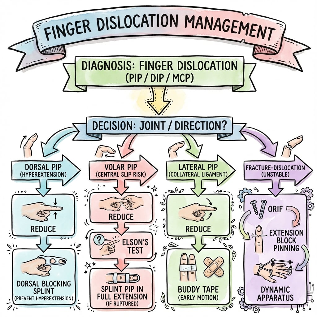
Management
Treatment by Dislocation Type
- Digital block anaesthesia
- Reduction: Longitudinal traction + flexion
- Assess stability through ROM
- If stable: Buddy taping, early motion
- If unstable: Extension block splinting
- Reduction: Traction + extension
- Test active PIPJ extension
- Static extension splint for 6 weeks
- Active DIP flexion exercises during immobilisation
- Usually dorsal, often open injury
- Digital block, irrigate if open
- Reduce and splint for 2-3 weeks
- Stable
- Buddy tape + early motion
- Unstable
- Extension block splint
- Stable
- Extension splint 6 weeks
- Unstable
- Open repair if needed
- Stable
- Extension block if less than 30%
- Unstable
- Surgery if greater than 40%
Surgical Technique
Open Reduction Techniques
- Irreducible dislocation
- Fracture-dislocation greater than 40% articular surface
- Chronic dislocation
- Failed closed treatment
- Volar (Bruner) - preferred for volar plate extraction
- Dorsal - for central slip repair
- Lateral - for collateral ligament repair
- Bruner zigzag incision over PIPJ
- Identify and protect neurovascular bundles
- Retract flexor tendons
- Identify interposed volar plate
- Extract and reduce joint
- Repair volar plate to bone (suture anchors)
- Indication
- Large fragment fracture-dislocation
- Key Points
- Screws, plate, or K-wires
- Indication
- Comminuted volar lip
- Key Points
- Advance volar plate into defect
- Indication
- Chronic defect greater than 50%
- Key Points
- Autograft from hamate
Complications
Common Complications
- Fusiform swelling persists for months
- Warn patients about permanent finger thickening
- Prevention: Early protected motion in dorsal dislocations
- PIPJ flexion with DIPJ hyperextension
- Results from untreated central slip injury
- Common after incorrectly mobilised volar dislocation
- PIPJ hyperextension with DIPJ flexion
- Results from volar plate laxity
- May follow chronic dorsal subluxation
- Cause
- Prolonged immobilisation
- Prevention
- Early motion (dorsal type)
- Cause
- Central slip rupture
- Prevention
- Extension splint 6 weeks (volar)
- Cause
- Volar plate laxity
- Prevention
- Proper treatment of volar plate injury
- Cause
- Inadequate healing
- Prevention
- Appropriate splinting duration
Postoperative Care
Rehabilitation Protocol
- Buddy taping to adjacent finger
- Immediate active motion exercises
- Full ROM expected by 4-6 weeks
- Protect during sports for 6-8 weeks
- Static extension splint (PIPJ in full extension)
- Allow DIP flexion exercises
- Maintain for 6 weeks minimum
- Progressive PIPJ flexion after 6 weeks
- Extension block splinting 2-3 weeks
- Hand therapy referral essential
- Progressive extension by 10°/week
- Full motion by 6-8 weeks
- Dorsal (Stable)
- Buddy tape, active ROM
- Volar
- Extension splint only
- Post-Surgery
- Extension block splint
- Dorsal (Stable)
- Full motion, buddy tape sports
- Volar
- Extension splint, DIP exercises
- Post-Surgery
- Progressive extension
- Dorsal (Stable)
- Return to sport
- Volar
- Begin gentle PIPJ flexion
- Post-Surgery
- Full ROM goals
Outcomes
Expected Outcomes
- Excellent prognosis with early motion
- Greater than 90% achieve functional ROM
- Residual stiffness common but usually mild
- Return to sport within 4-6 weeks
- Good outcomes if splinted correctly
- Higher complication rate than dorsal
- Boutonnière risk with improper treatment
- Full recovery may take 3-6 months
- Outcomes depend on articular involvement
- Less than 30% fragment: good prognosis
- Greater than 50% fragment: guarded prognosis
- Post-traumatic arthritis risk increases with severity
- Good Result Rate
- Greater than 90%
- Main Risk
- Stiffness
- Good Result Rate
- 80-85%
- Main Risk
- Boutonnière deformity
- Good Result Rate
- 60-80%
- Main Risk
- Arthritis, stiffness
DIP Joint Dislocation
The distal interphalangeal (DIP) joint is dislocated far less often than the PIPJ, but when it happens it is almost always dorsal and is frequently open, because the dorsal skin over the joint is thin and closely applied to the extensor mechanism. The usual mechanism is an axial load with hyperextension, classically a ball striking the extended fingertip.
- Open until proven otherwise. Even a small dorsal or volar laceration communicating with the joint makes this an open injury: it needs formal irrigation and debridement, tetanus prophylaxis and antibiotics, not a simple bedside reduction. An unrecognised open DIP dislocation risks septic arthritis and osteomyelitis.
- Screen the terminal tendons after reduction. Test active DIP extension (a lag suggests a terminal extensor / mallet injury, developed in the dedicated mallet-finger topic) and active DIP flexion (loss suggests an FDP avulsion, covered under jersey-finger).
- Nail bed and physis. Look for a nail-bed laceration; in a child a displaced physeal fracture of the distal phalanx (Seymour-type) can masquerade as a DIP dislocation or mallet and is an open physeal injury requiring nail-plate removal and reduction (see phalangeal-fractures).
- Digital block, longitudinal traction with gentle reversal of the deformity; the reduced DIP is usually stable, since it is unusual for both collateral ligaments and the volar plate to fail together.
- Splint the DIP while leaving the PIP free for roughly 2-3 weeks, then move to protected motion. The DIP tolerates short immobilisation, but prolonged splinting causes stiffness.
Irreducible DIP dislocation is rare and, as at the PIPJ, implies soft-tissue interposition, most often the volar plate, occasionally the FDP tendon or a trapped sesamoid, and requires open reduction.
Treat a dorsal DIP dislocation with any breach in the skin as an open joint injury: irrigate, give antibiotic and tetanus cover, and reduce in a clean environment rather than just pulling it back in the cubicle. Always document terminal extensor (mallet) and FDP function after reduction, and in a child actively exclude a Seymour (physeal) injury at the nail fold.
PIPJ Collateral Ligament Injury & Lateral Dislocation
Lateral dislocations and isolated collateral ligament ("sprain") injuries of the PIPJ are common and, because they frequently reduce spontaneously on the field, are easily under-treated. The proper collateral ligament is the primary lateral restraint and is tightest near full extension; the accessory collateral blends with the volar plate. The radial collateral is injured more often than the ulnar.
- Grade I - sprain, ligament in continuity, stable to stress.
- Grade II - partial tear, painful but stable.
- Grade III - complete rupture, demonstrably unstable.
- Apply radial and ulnar deviation stress with the PIPJ in about 20 degrees of flexion (which isolates the proper collateral), always comparing with the same finger on the other hand. Lateral opening of greater than 20 degrees, or a soft/absent endpoint, indicates a complete (Grade III) rupture. A stress radiograph documents both the angulation and any condylar avulsion fragment.
- The overwhelming majority - including reduced lateral dislocations and Grade I-III sprains without a large intra-articular fragment - are stable enough for buddy strapping to the adjacent finger with early motion for 3-6 weeks, and do very well.
- Reserve surgery for a displaced condylar avulsion fragment carrying a meaningful portion of the articular surface, gross instability, or soft-tissue interposition blocking reduction.
- Counsel patients that a lateral PIPJ injury leaves a fusiform, mildly stiff and tender joint for many months, exactly as for dorsal dislocations.
Most PIPJ collateral injuries and reduced lateral dislocations are stable: buddy strap and mobilise early rather than immobilise. Formally stress the collateral at about 20 degrees of flexion against the opposite hand; lateral opening greater than 20 degrees with a soft endpoint signals a complete rupture. A large displaced condylar fragment is the main reason to operate, and a fixed rotational deformity points to an associated phalangeal/condylar fracture (see phalangeal-fractures) rather than a pure ligament injury.
Guidelines, Registries & Global Practice
Global epidemiology:
- Finger and hand injuries are among the most common presentations to emergency and acute care worldwide; the PIPJ is the most frequently dislocated digital joint, with dorsal dislocation by far the commonest pattern.
- Peak incidence is in young, active adults; ball and contact sports (basketball, football/soccer, rugby, cricket, volleyball, handball) dominate the mechanism across regions.
- Most simple dorsal dislocations are reduced and managed by emergency physicians or primary care, with specialist hand referral reserved for volar dislocations, irreducible joints and fracture-dislocations.
- Common ground (AAOS / BOA / AO principles)
- Closed reduction + early protected motion (buddy strapping / dorsal block splint)
- Practical note
- Avoid prolonged rigid immobilisation
- Common ground (AAOS / BOA / AO principles)
- Treat as central slip injury: extension splinting ~6 weeks
- Practical note
- Early flexion mobilisation causes boutonniere
- Common ground (AAOS / BOA / AO principles)
- Assess stable arc + fragment size on true lateral
- Practical note
- No fixed percentage cut-off is universally endorsed
- Common ground (AAOS / BOA / AO principles)
- Digital block with plain lidocaine for reduction
- Practical note
- Document neurovascular status before and after
- Common ground (AAOS / BOA / AO principles)
- Early supervised hand therapy improves motion
- Practical note
- Access varies by health system
Unlike arthroplasty of large joints, finger dislocations are not tracked in national joint registries; the evidence base is institutional case series and systematic reviews rather than registry data, which is itself a recognised limitation.
- High-resource settings: ready access to fluoroscopy, hand surgeons, dedicated hand therapy, and implants (suture anchors, mini-screws, dynamic fixators, hemi-hamate reconstruction).
- Limited-resource settings: emphasis on closed reduction, buddy strapping and improvised dynamic external fixation (for example a syringe-and-K-wire construct) which provides comparable function at minimal cost; early motion and patient education remain the highest-value, lowest-cost interventions everywhere.
Mnemonics
DORALDorsal vs Volar Distinction
Hook:Dorsal dislocations are the 'Typical' type - keep 'em moving!
VOLARVolar Dislocation Management
Hook:Volar = Volatile behavior of the central slip. Splint it!
VSIGNThe V-Sign of Instability
Hook:See the V? Think instability!
MCQ Practice Points
Q: What is the mechanism and treatment of dorsal PIP dislocation?
A: Mechanism: Hyperextension injury with axial load, disrupting the volar plate. Most common finger dislocation. Proximal phalanx displaces dorsally relative to middle phalanx. Reduction: Digital block, longitudinal traction with gentle flexion. Post-reduction: Assess stability through ROM; If stable to 30° flexion: Buddy taping with early mobilization. If unstable: Extension block splinting (blocking last 20-30° of extension) for 2-3 weeks, progressive extension.
Q: What is a volar PIP dislocation and why is it more concerning than dorsal?
A: Volar PIP dislocation: Middle phalanx displaces volarly; Less common but higher complication rate. Mechanism: Rotatory force or direct blow to extended finger. Central slip disruption is common (risk of boutonniere deformity). Reduction often more difficult (may require open reduction). Post-reduction: Splint PIP in full extension for 6 weeks to protect central slip (opposite of dorsal dislocation protocol). Earlier mobilization risks boutonniere deformity.
Q: What is an irreducible PIP dislocation and what causes it?
A: Irreducible dislocation: Cannot achieve closed reduction due to interposed tissue. Causes: Volar plate interposition (flips proximally, blocks reduction); FDP entrapment (tendon wraps around condyle); Lateral band interposition; Button-holing of condyle through volar plate. Clinical clue: Failed gentle reduction attempts, palpable block to reduction. Treatment: Open reduction through volar or dorsal approach, extraction of interposed tissue, volar plate repair.
Q: How do you assess and manage PIP fracture-dislocations?
A: Assess stability: Lateral X-ray - measure percentage of volar articular surface (middle phalanx) fractured. Less than 30%: Usually stable after reduction - extension block splinting. 30-50%: Borderline stable - may require surgical fixation (hemihamate arthroplasty, volar plate arthroplasty, dynamic external fixator). Greater than 50%: Unstable, high subluxation risk - requires surgical stabilization. V-sign (incongruent joint on lateral view) indicates instability requiring intervention.
Q: What is the treatment for MCP joint dislocation and what makes it complex?
A: Dorsal MCP dislocation: Proximal phalanx dorsal to metacarpal head. Simple: Reducible closed with wrist flexion, MCP hyperextension then flexion. Complex (irreducible): Volar plate interposition, often with metacarpal head button-holed through flexor tendons/lumbricals. Clinical sign: Skin puckering over MCP. Complex dislocation contraindication: Repeated forceful reduction attempts (can further tighten noose). Requires open reduction (dorsal or volar approach).
At a Glance
- Mechanism
- Hyperextension
- Stability
- Stable
- Treatment
- Buddy tape / Early motion
- Mechanism
- Rotational / Volar force
- Stability
- Unstable (Central Slip)
- Treatment
- Extension splint 6 weeks
- Mechanism
- Articular greater than 40%
- Stability
- Unstable
- Treatment
- Surgical Fixation
- Mechanism
- Collateral stress
- Stability
- Stable
- Treatment
- Buddy tape 4 weeks
Exam Viva Scenarios
Practise clinical reasoning and management decisions out loud
“A goalkeeper presents with a swollen middle finger. Lateral X-ray shows a dorsal dislocation of the PIPJ with a small avulsion fracture at the base of the middle phalanx. Reduction was easy. How do you treat him?”
“A 28-year-old basketball player presents to the emergency department 2 hours after jamming his index finger. Examination shows the PIPJ held in slight flexion with the fingertip pointing volarly. Lateral X-ray confirms volar dislocation of the middle phalanx relative to the proximal phalanx. The ED resident asks you about the best treatment approach. What do you tell them?”
“A 35-year-old presents with a dorsal PIPJ dislocation of his ring finger sustained 4 hours ago. Multiple attempts at closed reduction in the emergency department have failed. X-rays show the middle phalanx remains dorsally dislocated with the PIPJ joint surfaces parallel to each other rather than overlapping. There is no fracture. What is your assessment and management?”
Direction
- Dorsal: Volar plate injury → Early Motion
- Volar: Central Slip injury → Splint Extension (6w)
Irreducible?
- Volar Plate interposition
- FDP tendon entrapment (rare)
- Condyle buttonholing
Evidence, Controversies & Areas of Uncertainty
The evidence base for PIPJ fracture-dislocations is almost entirely Level III-IV; there are no randomised trials comparing surgical techniques, so management remains opinion- and series-driven. Examiners reward candidates who can articulate these grey areas rather than quote a single dogmatic threshold.
- Argument A
- Operate at greater than 30-40% (subluxation risk)
- Argument B
- Many 40-50% fragments are stable in flexion and do well with extension block
- Pragmatic position
- Decide on dynamic stability (stable arc), not the percentage alone
- Argument A
- Closed methods avoid surgical morbidity
- Argument B
- Open fixation restores articular congruity in large/displaced fragments
- Pragmatic position
- Reserve open surgery for fragments that are reconstructable and unstable through the functional arc
- Argument A
- Hemi-hamate restores bony contour for greater than 50% defects
- Argument B
- Volar plate arthroplasty avoids donor site, good for moderate defects
- Pragmatic position
- Choose by defect size, comminution and reconstructability
- Argument A
- Allows early motion with ligamentotaxis
- Argument B
- Pin-site morbidity and lower final arc in some series
- Pragmatic position
- Useful for comminuted, non-reconstructable patterns
- Argument A
- Early reconstruction limits contracture/arthritis
- Argument B
- Selected chronic cases tolerate delayed reconstruction
- Pragmatic position
- Beyond ~3-6 weeks expect open treatment and counsel on stiffness
- No single articular-surface percentage reliably predicts instability; dynamic testing of the stable arc is more useful than any fixed cut-off.
- Comparative outcome data between hemi-hamate, volar plate arthroplasty, dynamic fixation and ORIF are retrospective and confounded by injury severity selection bias.
- Long-term hemi-hamate series show acceptable function but frequent radiographic osteoarthritis, so "good clinical outcome" does not equal "normal joint".
References
Evidence verified against PubMed.
- Gianakos AL, Yingling J, Athens CM, Barra AE, Capo JT. Treatment for Acute Proximal Interphalangeal Joint Fractures and Fracture-Dislocations: A Systematic Review. J Hand Microsurg. 2020. PMID 33335365. doi:10.1055/s-0040-1713323
- Williams RM, Kiefhaber TR, Sommerkamp TG, Stern PJ. Treatment of unstable dorsal proximal interphalangeal fracture/dislocations using a hemi-hamate autograft. J Hand Surg Am. 2003;28(5):856-65. PMID 14507519. doi:10.1016/s0363-5023(03)00304-6
- Mazhar FN, Noei RR, Zareie B, et al. Long-Term Clinical and Radiological Results of Hemi-Hamate Arthroplasty for PIP Fracture Dislocation. J Hand Surg Am. 2026. PMID 41528296. doi:10.1016/j.jhsa.2025.11.020
- Fleury CM, Yousaf IS, Miles MR, et al. The Syringe External Fixator for Comminuted Intra-Articular Fractures of the Hand. J Hand Surg Am. 2021. PMID 34602335. doi:10.1016/j.jhsa.2021.07.036
- Pillukat T, Windolf J, Schadel-Hopfner M, Fuhrmann RA, van Schoonhoven J. Extensor tendon injuries at the level of the PIP joint. Unfallchirurg. 2021;124(4):265-274. PMID 33616682. doi:10.1007/s00113-021-00984-x
- Eaton RG, Malerich MM. Volar plate arthroplasty of the proximal interphalangeal joint (classic technique). J Hand Surg Am. 1980. (Classic reference)
- Kiefhaber TR, Stern PJ. Fracture-dislocations of the proximal interphalangeal joint: stability criteria. J Hand Surg Am. 1998. (Classic reference)