Salvage Procedure | Functional Limitation | Conversion to THA
- Optimal fusion position: 25-30° flexion, 0-5° adduction, 15-20° external rotation
- Contralateral hip and ipsilateral knee MUST be normal for acceptable function
- Back pain develops in over 50% at 10 years due to increased lumbar stress
- Conversion to THA is technically demanding with unpredictable outcomes
- Leg length discrepancy of 3-5 cm is expected and must be counselled
- “Fusion position determines function: too much flexion impairs standing, too much abduction causes pelvic tilt
- “Ipsilateral knee develops degenerative changes in 30-40% at long-term follow-up
- “Conversion to THA has 40-50% complication rate: instability, leg length issues, abductor deficiency
- “Globally now rare; most fusions are converted to THA within 20 years (mean interval ~27 years in the largest series)
No longer first-line. Hip fusion is a salvage procedure reserved for young patients with failed arthroplasty, eradicated infection, or heavy manual labour requirements where revision THA would fail. Always consider modern revision techniques first.
25-30° flexion, 0-5° adduction, 15-20° external rotation. This position allows sitting, standing, and walking with minimal compensatory pelvic tilt. Too much flexion impairs standing; too much abduction causes pelvic obliquity and gait abnormality.
Adjacent joint disease is inevitable. Lumbar spine develops pain in over 50% at 10 years. Ipsilateral knee degenerates in 30-40%. Contralateral hip experiences increased stress. Patients must understand lifelong functional limitations.
Technically demanding with unpredictable results. Bone loss, leg length discrepancy, abductor deficiency, femoral deformity, and acetabular bone stock issues complicate conversion. Complication rate approaches 40-50% including instability and infection.
- Patient Factors
- Age under 40, normal contralateral hip and knee
- Best Option
- Consider arthrodesis after infection eradication
- Key Pearl
- Counsel about functional limitations and adjacent joint disease
- Patient Factors
- Young patient, high activity demands
- Best Option
- Two-stage: resection arthrodesis then delayed fusion
- Key Pearl
- Ensure infection markers normalized for at least 6 months
- Patient Factors
- Poor bone stock, elderly, low demand
- Best Option
- Resection arthroplasty or modern revision with augments
- Key Pearl
- Arthrodesis rarely indicated in elderly; consider Girdlestone
- Patient Factors
- Patient requesting improved function
- Best Option
- Conversion to THA with extensive preoperative planning
- Key Pearl
- CT for templating, plan for leg length and abductor reconstruction
Overview and Epidemiology
Hip arthrodesis was commonly performed from the 1920s through 1980s for conditions that are now treated with total hip arthroplasty. The development of modern THA, improved infection management, and recognition of long-term sequelae (lumbar spine pain, adjacent joint degeneration) have made fusion a rare salvage procedure. Current indications are limited to young patients with failed arthroplasty or eradicated infection where revision THA has poor prognosis.
- Primary osteoarthritis (now THA)
- Post-traumatic arthritis (now THA)
- Ankylosing spondylitis (now THA)
- Tuberculosis (now medical treatment + THA)
- Primary hip sepsis (rarely fusion now)
These were common fusion indications before modern arthroplasty revolutionized hip reconstruction.
- Recurrent infected THA after multiple failures
- Young manual labourer with destroyed hip
- Massive bone loss precluding revision THA
- Neurologic conditions requiring hip stability
- Failed salvage procedures (Girdlestone with pain)
Fusion is now a last resort when all reconstructive options have been exhausted.
Before considering hip fusion, confirm:
- Contralateral hip is NORMAL (or has well-functioning THA)
- Ipsilateral knee is NORMAL (degenerative changes are a contraindication)
- Lumbar spine is ASYMPTOMATIC (existing back pain will worsen)
- Patient understands functional limitations: no running, limited sitting, eventual conversion likely
- Alternative options (revision THA, resection arthroplasty) have been fully explored
Fusion with pre-existing knee or spine disease leads to early failure and poor outcomes.
Anatomy and Biomechanics
Biomechanical Principles
- Forces distributed through acetabulum and femoral head
- Motion occurs at hip joint
- Lumbar spine compensates minimally
- Forces transmitted directly through fusion mass
- Motion transferred to lumbar spine (flexion/extension)
- Contralateral hip compensates for abduction/adduction
- Ipsilateral knee compensates for rotation
This redistribution is inevitable and drives adjacent joint degeneration.
- Allows sitting without excessive lumbar flexion
- Standing requires lumbar lordosis compensation
- Neutral to slight adduction minimizes pelvic obliquity and functional lengthening
- Abduction is a malposition that causes limping and accelerates ipsilateral knee/back disease
- Permits sitting with knees together
- Allows foot care and shoe donning
Position is determined by functional demands, not by anatomy.
- Normal Hip
- Hip extends 10°, abducts 5°
- Fused Hip
- No hip motion; pelvis tilts posteriorly
- Compensation Strategy
- Lumbar spine hyperextends; contralateral hip abducts more
- Normal Hip
- Hip flexes 30°, clears ground
- Fused Hip
- No hip flexion; leg swings from pelvis
- Compensation Strategy
- Lumbar flexion, pelvic hike, circumduction gait
- Normal Hip
- Hip flexes to 90°
- Fused Hip
- Fusion at 30° flexion position
- Compensation Strategy
- Lumbar spine flexes additional 60°; slouched posture
Stress on Adjacent Joints
- Increased flexion/extension range by 50-100%
- Back pain develops in over 50% at 10-year follow-up
- Disc degeneration accelerates at L4-L5 and L5-S1
- Increased rotational stress
- Symptomatic arthritis in 30-40% at 15-20 years
- May require TKA eventually
- Increased abduction loading
- Accelerated cartilage wear
- Typically remains asymptomatic if initially normal
This is NOT a complication - it is an expected consequence of altered biomechanics.

Classification Systems
Indications Classification
Hip arthrodesis is classified by indication rather than by fracture pattern. Understanding when fusion is appropriate versus when alternative treatments are superior is critical for exam success.
Modern Indications for Hip Arthrodesis
- Specific Scenarios
- Recurrent infected THA after multiple two-stage revisions; poor bone stock
- Prerequisites
- Eradicated infection (CRP/ESR normal 6+ months, negative cultures)
- Alternative Options
- Antibiotic spacer, resection arthroplasty (Girdlestone)
- Specific Scenarios
- Age under 40, high-impact occupation, destroyed hip
- Prerequisites
- Normal contralateral hip and ipsilateral knee; no spine disease
- Alternative Options
- Modern revision THA with trabecular metal, dual mobility
- Specific Scenarios
- Flail hip from polio, spina bifida; requires stability for ambulation
- Prerequisites
- Patient ambulatory with aids; adequate upper extremity function
- Alternative Options
- Hip-knee-ankle-foot orthosis (HKAFO) for stability
- Specific Scenarios
- Pelvic discontinuity, acetabular destruction precluding implant fixation
- Prerequisites
- Young patient where revision THA would fail immediately
- Alternative Options
- Custom triflange acetabular component, pelvic reconstruction cage
Fusion is RARELY superior to modern revision THA techniques. The only clear indication is eradicated infection in a young patient where multiple revision attempts have failed and bone stock is insufficient for further reconstruction. Even then, resection arthroplasty (Girdlestone) may provide acceptable function with less morbidity. Always exhaust revision THA options before offering fusion.
Current indications are extremely narrow and require careful patient selection.
Clinical Assessment and Patient Selection
Preoperative Evaluation
- Pain severity and functional impact
- Previous hip surgeries (number of revisions, complications)
- Infection history (organism, treatment, duration of antibiotics)
- Current mobility (distance, aids required)
- Comorbidities (diabetes, immunosuppression affecting infection risk)
- Smoking status (major nonunion risk factor)
- Occupation and activity level (determines fusion appropriateness)
- Patient expectations (unrealistic expectations are a contraindication)
- "Do you have any back pain?" (lumbar disease will worsen)
- "Do you have any knee pain on the same side?" (knee arthritis will worsen)
- "Can you walk at all currently?" (if non-ambulatory, fusion unlikely to help)
- Range of motion (typically stiff or ankylosed already)
- Pain on motion (indicates active pathology)
- Leg length discrepancy (document baseline)
- Scars from previous surgery (plan incision)
- Full range of motion (must be normal to compensate)
- No pain or crepitus (early arthritis is a contraindication)
- Trendelenburg test (should be negative)
- Full range of motion (any stiffness suggests arthritis)
- No effusion or crepitus (arthritis is contraindication)
- Ligament stability (instability will worsen with fusion stress)
- Range of motion (will need increased lumbar flexion after fusion)
- Tenderness (symptomatic spine disease is contraindication)
- Neurologic exam (rule out radiculopathy)
- Pulses (PVD may worsen with fusion position)
- Sensation and motor function (baseline for sciatic nerve)
This is the most important part of the clinical assessment:
- Obtain standing AP pelvis X-ray (assess BOTH hips)
- Obtain ipsilateral knee X-rays (AP, lateral, skyline)
- If ANY arthritis is present in contralateral hip or ipsilateral knee, arthrodesis is CONTRAINDICATED
- Document thorough examination findings - medicolegal risk is high if fusion proceeds despite diseased adjacent joints
Failure to assess adjacent joints is negligent and will lead to poor outcomes.
Infection Assessment (If Indication Is Post-Septic)
Confirming Eradicated Infection
- CRP and ESR must be NORMAL for at least 6 months after antibiotics stopped
- If elevated, infection is NOT eradicated; do NOT proceed with fusion
- Ultrasound-guided hip aspiration
- Send fluid for culture (aerobic, anaerobic, fungal, mycobacterial)
- Cell count (WBC under 3000, PMN under 80% suggests eradicated infection)
- If positive culture, infection is NOT eradicated; treat with antibiotics
- PET-CT or labeled WBC scan to assess for active infection
- Useful if aspiration not possible (ankylosis) or equivocal results
Proceeding with fusion in the presence of active infection will result in infected nonunion - a devastating complication requiring resection arthroplasty. The infection MUST be eradicated with at least 6 months of normal inflammatory markers after antibiotics stopped. If in doubt, delay fusion and repeat inflammatory markers in 3 months.
Differential Diagnosis: What Else Causes a Stiff, Painful Hip in a Fusion Candidate?
Before committing a young patient to arthrodesis, distinguish the underlying diagnosis - the management diverges sharply, and several mimics are better served by arthroplasty or medical therapy.
- Discriminating Features
- Mechanical pain, prior acetabular fracture, single joint affected
- Key Investigation
- Weight-bearing radiographs; CT for bone stock
- Implication for Fusion
- Classic fusion candidate IF contralateral hip and ipsilateral knee normal
- Discriminating Features
- Sinus, raised CRP/ESR, prior infection history
- Key Investigation
- Aspiration culture, CRP/ESR, alpha-defensin
- Implication for Fusion
- Stage first - eradicate infection before any fusion
- Discriminating Features
- Endemic exposure, indolent course, marked stiffness
- Key Investigation
- Aspirate for AFB/GeneXpert, MRI
- Implication for Fusion
- Treat medically; fusion or THA only after cure
- Discriminating Features
- Bilateral/polyarticular, spine involvement, inflammatory markers
- Key Investigation
- HLA-B27, rheumatology review, axial imaging
- Implication for Fusion
- Contraindicated - spine already stiff; THA strongly preferred
- Discriminating Features
- Pain with spine movement, normal hip rotation, neurogenic features
- Key Investigation
- Lumbar MRI, diagnostic intra-articular hip injection
- Implication for Fusion
- Not a hip problem - fusion will not help and may worsen
- Discriminating Features
- Groin pain, risk factors (steroids, alcohol)
- Key Investigation
- MRI (early), radiographs (collapse)
- Implication for Fusion
- Usually THA; fusion only in young, late, monoarticular cases
A positive response to a diagnostic intra-articular local-anaesthetic hip injection confirms the hip as the pain generator and is invaluable when lumbar or SI pathology coexists. Never fuse a hip for pain that is actually referred from the spine - the most common avoidable error in selection.
Thorough clinical assessment prevents inappropriate patient selection and poor outcomes.
Investigations
Imaging Protocol
Preoperative Imaging Workup
- AP pelvis and lateral hip
- Assess bone stock (acetabulum and proximal femur)
- Identify retained hardware from previous surgeries
- Measure leg length discrepancy (will increase by 3-5 cm)
- AP pelvis includes both hips (assess for arthritis)
- If ANY joint space narrowing, osteophytes, or sclerosis: CONTRAINDICATED
- AP, lateral, and skyline views
- Assess for arthritis (joint space narrowing, osteophytes)
- If ANY arthritis present: CONTRAINDICATED
- AP and lateral lumbar spine
- Assess for degenerative disc disease, spondylolisthesis
- Symptomatic spine disease is a relative contraindication
- Multiple previous surgeries (assess bone stock accurately)
- Pelvic discontinuity or massive acetabular bone loss
- Retained hardware (plan removal strategy)
- Conversion cases (assess fusion mass and plan osteotomy)
- Provides excellent visualization of bone stock
- Helps plan fixation strategy (plate vs screws)
- Essential for conversion THA planning
- Rule out active infection (fluid collections, bone marrow edema)
- Assess soft tissue (muscle atrophy, abductor quality)
- Evaluate lumbar spine if symptomatic (disc herniation, stenosis)
- Active infection: rim-enhancing fluid collection, bone marrow edema
- Chronic changes: muscle atrophy, fatty infiltration of abductors
- High sensitivity for active infection (FDG uptake at fusion site)
- Useful if aspiration not possible or equivocal
- Specific for infection (WBC accumulation at site)
- Combine with bone marrow scan to improve specificity

Laboratory Investigations
- CRP and ESR (must be normal for 6+ months off antibiotics)
- Complete blood count (WBC, differential)
- Hip aspiration culture (if joint accessible)
- CRP over 10 mg/L → likely active infection
- ESR over 30 mm/hr → likely active infection
- Positive culture → active infection; do NOT proceed with fusion
- Full blood count (anemia, infection)
- Renal function (contrast CT, antibiotic dosing)
- Coagulation studies (bleeding risk)
- Blood group and save (large operation, potential blood loss)
- HbA1c if diabetic (glycemic control affects healing)
- Liver function if alcohol history
- ECG and echocardiogram if cardiac disease
Inflammatory markers can be normal despite persistent infection:
- Low-virulence organisms (e.g., Cutibacterium acnes) may not elevate CRP/ESR
- Biofilm infections may have low inflammatory response
- If clinical suspicion for infection, proceed to aspiration or biopsy even if markers normal
The gold standard is culture - not inflammatory markers.
Templating for Fusion Position
Preoperative Planning Steps
- Use AP and lateral radiographs of pelvis
- Mark 25-30° flexion relative to pelvis on lateral view
- Mark 0-5° adduction on AP view (measure pelvic obliquity)
- Plan external rotation 15-20° (measured clinically, not radiographically)
- Measure from ilium to mid-femoral shaft (plate length 20-30 cm)
- Assess bone quality (osteoporosis may require longer plate, more screws)
- Identify screw trajectories (avoid previous screw holes if possible)
- Fusion typically adds 3-5 cm shortening
- Measure current LLD and add 3-5 cm for expected postoperative LLD
- Plan shoe lift requirement (counsel patient preoperatively)
Comprehensive preoperative imaging and planning are essential for successful fusion.
Management Algorithm
Decision Tree for Hip Pathology Requiring Salvage
- Fusion Appropriate?
- YES - if contralateral hip and knee normal
- Alternative Options
- Resection arthroplasty, antibiotic spacer, modern revision THA
- Recommended Approach
- Fusion likely best option; provides stability for high-impact work
- Fusion Appropriate?
- MAYBE - consider alternatives first
- Alternative Options
- Modern revision THA with trabecular metal, dual mobility, antibiotic cement
- Recommended Approach
- Attempt revision THA first; fusion only if multiple revision failures
- Fusion Appropriate?
- NO - resection arthroplasty better
- Alternative Options
- Resection arthroplasty (Girdlestone), antibiotic spacer with chronic suppression
- Recommended Approach
- Resection arthroplasty provides pain relief with less morbidity than fusion
- Fusion Appropriate?
- NO - absolute contraindication
- Alternative Options
- Address knee/spine first, or accept current hip status
- Recommended Approach
- Fusion will worsen knee/spine disease; do NOT proceed
- Fusion Appropriate?
- MAYBE - consider orthotic first
- Alternative Options
- Hip-knee-ankle-foot orthosis (HKAFO) for stability during gait
- Recommended Approach
- Trial orthotic first; fusion if orthotic fails and patient motivated
Hip arthrodesis should be the LAST option considered after all alternatives have been exhausted. The algorithm should be: (1) Attempt modern revision THA with advanced techniques, (2) If infection cannot be eradicated or bone stock insufficient, consider resection arthroplasty, (3) If patient young, high-demand, and resection unacceptable, THEN consider fusion. Jumping straight to fusion without exhausting alternatives is poor practice.
Fusion is rarely the first choice - consider all alternatives first.
Surgical Technique
Patient Selection and Counseling
- Age: Under 40 years
- Occupation: Heavy manual labour
- Activity: High impact demands
- Contralateral hip: Normal
- Ipsilateral knee: Normal
- Spine: Asymptomatic
- Expectations: Realistic about limitations
Even ideal candidates should be counseled about eventual conversion to THA.
- Ipsilateral knee arthritis (will worsen)
- Symptomatic lumbar spine (will worsen)
- Contralateral hip disease (cannot compensate)
- Obesity (poor functional outcomes)
- Elderly patients (resection arthroplasty better)
- Unrealistic expectations (expects normal function)
These are ABSOLUTE contraindications - do not proceed.
Imaging and Templating
Preoperative Assessment
- Assess acetabular bone stock (for fixation)
- Measure leg length discrepancy (will increase by 3-5 cm)
- Evaluate femoral shaft anatomy (for intramedullary fixation)
- Document baseline status of contralateral hip (must be normal)
- Assess ipsilateral knee for early degenerative changes
- If arthritis present, arthrodesis is contraindicated
- Assess for pre-existing degenerative disease
- Symptomatic spine disease is a relative contraindication
- Counsel patient about inevitable progression
- For conversion cases: assess acetabular bone loss
- Identify retained hardware
- Plan fixation strategy (plate vs intramedullary device)
Consent Points
- Cannot run or perform high-impact activities
- Sitting requires lumbar flexion; may be uncomfortable after 30-60 minutes
- Leg length discrepancy of 3-5 cm; requires shoe lift
- Gait abnormality with circumduction
- Back pain develops in over 50% within 10 years
- Ipsilateral knee arthritis in 30-40% at 15-20 years
- May require eventual conversion to THA (challenging procedure)
- Nonunion: 5-10% (requires revision surgery)
- Infection: 2-5% (may require resection)
- Nerve injury: under 1% (sciatic nerve)
- Conversion to THA if fusion fails or function unacceptable
Document this discussion extensively - medicolegal risk is high for this procedure.
Ensure the patient understands that hip fusion provides pain relief and stability at the cost of permanent functional limitation.
Complications
- Incidence
- 5-10% (plate), 10-15% (screws only)
- Risk Factors
- Poor surface preparation, inadequate fixation, smoking, infection
- Management
- Revision arthrodesis with bone grafting and improved fixation; consider conversion to THA if revision fails
- Incidence
- 2-5% (higher if prior infection)
- Risk Factors
- Diabetes, immunosuppression, prolonged operative time, prior septic arthritis
- Management
- Irrigation and debridement with hardware retention (if fusion solid); consider resection arthroplasty if fusion incomplete
- Incidence
- Over 50% at 10 years
- Risk Factors
- Pre-existing spine disease, suboptimal fusion position, young age (longer follow-up)
- Management
- Physiotherapy, NSAIDs, activity modification; may require spinal fusion or conversion to THA if disabling
- Incidence
- 30-40% at 15-20 years
- Risk Factors
- Pre-existing knee disease, excessive fusion flexion (increases knee stress), high activity level
- Management
- Conservative management initially; may require TKA if symptomatic; consider conversion to THA before knee replacement
- Incidence
- 100% (expected outcome)
- Risk Factors
- Shortening of 3-5 cm is typical due to loss of hip motion and fusion position
- Management
- Shoe lift (add 2-3 cm to affected side); counsel preoperatively that this is expected
- Incidence
- Under 1%
- Risk Factors
- Excessive retraction, screw penetration into sciatic notch, positioning injury
- Management
- Exploration if immediate postoperative deficit; nerve recovery unpredictable (may take 12-18 months)
- Incidence
- 10-20% with plate fixation
- Risk Factors
- Thin soft tissue coverage, prominent iliac screws, patient sensitivity
- Management
- Hardware removal after fusion is solid (at least 12-18 months); avoid early removal (nonunion risk)
- Incidence
- 5-10%
- Risk Factors
- Inadequate intraoperative position verification, hardware failure, loss of fixation
- Management
- Revision arthrodesis with osteotomy to correct position if symptomatic and nonunion; very difficult procedure
- Persistent pain at fusion site beyond 6 months
- Radiographic lucency around screws or at fusion interface
- CT scan: lack of bridging bone across acetabulum-femoral head junction
- Revision arthrodesis with bone grafting (autograft from iliac crest or femoral head allograft)
- Upgrade fixation (add compression screws or convert to plate if screws only)
- Address infection (send cultures at revision)
- If revision fails or patient unwilling: consider conversion to THA (challenging but may restore function)
Nonunion is the most common cause of revision surgery after hip arthrodesis.


- Infection: Wound drainage, fever, elevated CRP
- Nerve injury: Sciatic palsy (foot drop)
- DVT/PE: Lower limb swelling, chest pain
- Hardware failure: Loss of fixation (rare if technique sound)
Most complications present within the first 6 weeks and require prompt recognition.
- Nonunion: Persistent pain, motion at fusion site
- Lumbar back pain: Develops gradually over 5-10 years
- Knee arthritis: Progressive over 10-20 years
- Patient dissatisfaction: Functional limitations worse than expected
Late complications are EXPECTED and should be discussed preoperatively.
Long-term complications are inevitable and drive eventual conversion to THA in many patients.
BLANKSComplications After Hip Arthrodesis
Hook:The hip fusion leaves BLANKS - compensatory stresses fill every adjacent joint with degenerative changes!
Postoperative Care and Rehabilitation
Immediate Postoperative Management
- Pain control: Epidural or PCA; transition to oral opioids by day 2
- DVT prophylaxis: LMWH (enoxaparin 40 mg daily) for 35 days (high risk)
- Antibiotic prophylaxis: 24 hours (unless infection history; extend to 48 hours)
- Wound check: Inspect drain output (expect 100-200 mL in first 24 hours)
- Mobilization: Bed rest on day 0; sit in chair on day 1 (if patient tolerates)
- Drain removal: When drainage under 30 mL per 8 hours (typically day 2-3)
- Mobilization: Stand and pivot transfer with physiotherapy
- Gait training: Touch weight-bearing (toe touch only) with crutches or walker
- Hip orthosis: Fit hip abduction orthosis (if not using spica cast)
- Discharge planning: Home with walking aids; ensure safe mobility before discharge
- Wound care: Keep dry; remove staples at 14 days
- Weight-bearing: Touch weight-bearing with crutches (10-20 kg maximum)
- Mobilization: Short walks (50-100 m) multiple times per day
- X-ray: AP pelvis at 2 weeks to check hardware position
- Weight-bearing: Gradually increase to partial (25-50 kg) if no pain
- Radiographs: AP pelvis and lateral hip at 6 weeks
- Goal: Independent mobility with crutches by 6 weeks
If radiographs show early fusion (bridging bone), advance weight-bearing more aggressively.
- Persistent drainage beyond 5 days (concern for deep infection)
- Erythema extending beyond 2 cm from incision (cellulitis vs deep infection)
- Fever over 38.5°C beyond day 3
- Sudden increase in pain (hardware loosening or fracture)
- Inability to bear any weight (fixation failure)
Investigate promptly with inflammatory markers (CRP, ESR) and radiographs.
The first 6 weeks focus on wound healing and protected weight-bearing.
Fracture Through or Around the Arthrodesed Hip
The biomechanics section notes that a fused hip concentrates stress on the proximal femur, and all three imaging examples in this topic show exactly this - subtrochanteric, peri-acetabular and intertrochanteric fractures in previously arthrodesed hips - but their management is never discussed.
- Why they happen. The fused hip transmits load as a single rigid lever from pelvis to femur, concentrating stress at the subtrochanteric/proximal-femoral region and at the fusion mass or acetabulum. The bone is often osteoporotic from disuse and stress-shielded around retained hardware, so a low-energy fall in an elderly patient with an old fusion is the typical mechanism (as in the atlas cases).
- Assessment. Define the fracture level (peri-acetabular/pelvic, through the fusion mass, subtrochanteric, or femoral shaft distal to any implant), the state of the fusion (solid versus nonunion), retained hardware and bone quality. CT with 3D reconstruction is invaluable and is exactly what the imaging examples demonstrate.
- Management principles. A solid fusion with a femoral fracture distal to the mass is treated as a long-bone fracture: restore the mechanical axis with a long lateral plate or a cephalomedullary/retrograde nail that spans the whole femur and bypasses old screw holes and the fusion mass by at least two cortical diameters. A stable fracture with a solid fusion can sometimes be held with percutaneous cannulated screws (as in the atlas). A fracture running through the fusion mass itself is effectively a disrupted/nonunited fusion and needs revision fixation with compression and bone grafting - or, if the patient is symptomatic and the hip is otherwise reconstructable, it becomes an opportunity to convert to THA. Fixation is difficult because of distorted anatomy, sclerotic/osteoporotic bone and retained implants.
Q: An elderly patient with a 30-year-old hip fusion sustains a low-energy subtrochanteric femoral fracture. How do you manage it?
A: Treat it as a long-bone fracture in poor, stress-shielded bone. CT to define the fracture and the state of the fusion, then span the entire femur with a long cephalomedullary nail or lateral plate, bypassing the fusion mass and any old screw holes by at least two cortical diameters. If the fracture runs through the fusion mass (a disrupted, non-united fusion) or the patient wants improved function, consider converting to THA instead. Anticipate osteoporotic bone, distorted anatomy and retained hardware.
Pelvifemoral (Ischiofemoral) Fusion for the Flail or Paralytic Hip
The one-pager lists "pelvifemoral fusion" for the neurologic/flail hip and the FLIP mnemonic names the paralytic indication, but the technique is never described.
- The problem. A flail hip (poliomyelitis, spina bifida, other lower-motor-neuron palsy) has no active muscular stability, so the trunk cannot be balanced over the limb and ambulation - even with aids - is inefficient or impossible. Here the aim of fusion is a stable, painless, load-bearing strut between trunk and leg, not pain relief.
- The construct. Unlike a standard intra-articular hip fusion, a pelvifemoral (ischiofemoral) fusion extends fixation beyond the acetabulum onto the ilium and, classically, incorporates the ischium, so the femur is fused to the whole pelvis as one rigid column (a long cobra/ilio-femoral plate or crossed screws, supplemented by bone graft). This gives greater rotational and coronal stability than an intra-articular fusion alone in a hip that has no muscular protection.
- Position and caveats. Fuse in only slight flexion and neutral-to-slight adduction, matched to the patient's sitting/walking pattern and any orthotic use. Both hips must NEVER be fused - the patient would be unable to sit. A hip-knee-ankle-foot orthosis (HKAFO) is trialled first, and fusion is reserved for the motivated ambulator with adequate upper-limb function in whom bracing has failed.
Q: What is a pelvifemoral fusion and when is it used?
A: A pelvifemoral (ischiofemoral) fusion extends the arthrodesis construct beyond the acetabulum onto the ilium and ischium, so the femur is fused to the whole pelvis as one rigid column. It is used for the flail/paralytic hip (polio, spina bifida) to create a stable weight-bearing strut for ambulation - the goal is stability, not pain relief. Trial an HKAFO first, never fuse both hips (the patient could not sit), and fuse in slight flexion and neutral-to-slight adduction.
Conversion of Hip Arthrodesis to THA
Why Conversion Is Challenging
- Bone loss: Acetabular bone deficiency from prior fusion reaming
- Leg length discrepancy: 3-5 cm shortening; restoring length risks nerve injury
- Abductor deficiency: No functional gluteus medius/minimus (atrophied during fusion)
- Femoral deformity: Proximal femur may have healed in varus or external rotation
- Heterotopic ossification: Bridging bone around fusion site limits motion
40-50% (infection, dislocation, leg length issues, nerve injury).
Preoperative Planning for Conversion
Conversion THA Planning
- Assess acetabular bone stock (often deficient from prior reaming)
- Identify retained hardware (screws may be buried in bone)
- Measure leg length discrepancy (guide for femoral osteotomy)
- Plan cup size and need for augments/structural graft
- Template on non-fused side (mirror image for acetabulum)
- Plan femoral component size (often undersized due to canal narrowing)
- Determine safe amount of leg length restoration (maximum 3-4 cm to avoid nerve injury)
- Plan acetabular reconstruction (jumbo cup, augments, or structural allograft)
- Complications: 40-50% rate (dislocation, infection, nerve injury)
- Leg length: Cannot fully restore (nerve stretch injury risk if over 4 cm lengthening)
- Instability: High dislocation rate (20-30%) due to abductor deficiency
- Reoperation: May require constrained liner or abductor reconstruction
- Alternative: Accept current function (avoid high-risk surgery)
Document extensive discussion - medicolegal risk is very high.
Surgical Technique for Conversion
Approach Selection
- Preferred: Posterior approach (preserves any remaining anterior soft tissue)
- Alternative: Extensile lateral if prior fusion was via lateral approach (utilize old scar)
Exposure
Exposure Steps
- Remove all previous fusion hardware (cobra plate, screws)
- May require extended exposure to access buried screws
- Save removed hardware for reference
- Use oscillating saw to divide fusion mass
- Create plane between femoral head and acetabulum
- Remove bridging bone and heterotopic ossification
- Goal: Separate femur from pelvis to allow dislocation
- Perform femoral neck osteotomy at planned level (based on templating)
- Remove femoral head and any residual acetabular bone
- Assess femoral canal (often narrowed or deformed)
The sciatic nerve is at EXTREME risk during conversion THA:
- Nerve is scarred and adherent from prior surgery
- Leg lengthening over 3-4 cm risks traction injury
- Posterior dissection can directly injure nerve
Protective measures:
- Identify and protect nerve early
- Use intraoperative neuromonitoring
- Limit leg lengthening (accept residual LLD if necessary)
- Consider staged lengthening (femoral osteotomy first, THA later)
Exposure is technically demanding and time-consuming - allow 1-2 hours for this step alone.
Outcomes of Conversion THA
- Expected Result
- 40-50%
- Comparison to Primary THA
- Primary THA: 5-10%
- Notes
- Infection, dislocation, nerve injury, leg length issues
- Expected Result
- 20-30%
- Comparison to Primary THA
- Primary THA: 2-5%
- Notes
- Abductor deficiency is primary cause; dual mobility reduces risk
- Expected Result
- 5-10%
- Comparison to Primary THA
- Primary THA: 1-2%
- Notes
- Higher if prior septic arthritis was indication for fusion
- Expected Result
- 5-10%
- Comparison to Primary THA
- Primary THA: under 1%
- Notes
- Excessive leg lengthening is main cause; monitor intraoperatively
- Expected Result
- 60-70%
- Comparison to Primary THA
- Primary THA: over 90%
- Notes
- Improved function but residual gait abnormality and LLD
- Expected Result
- 30-40%
- Comparison to Primary THA
- Primary THA: 5-10%
- Notes
- For instability, infection, or component loosening
"A patient with a hip arthrodesis from 20 years ago requests conversion to THA to improve function. What do you counsel?"
"Thank you. I would counsel that conversion THA is a high-risk procedure with a complication rate of 40-50%, significantly higher than primary THA. Key risks include instability (20-30% dislocation rate due to abductor deficiency), sciatic nerve injury (5-10% from leg lengthening), and infection (5-10%). I would emphasize that while function typically improves, a residual limp and leg length discrepancy are expected. The reoperation rate is 30-40% within 5 years. I would recommend extensive preoperative planning with CT scan, templating for bone loss, and consideration of dual mobility components. I would also discuss the alternative of accepting current function rather than undergoing high-risk surgery. If the patient proceeds, I would use intraoperative neuromonitoring, limit leg lengthening to 3-4 cm maximum, and employ a dual mobility cup to reduce dislocation risk."
Conversion THA is technically demanding with unpredictable results - thorough preoperative counseling is essential.
FEAROptimal Hip Arthrodesis Position
Hook:FEAR the wrong position - it determines lifelong function! 30° Flexion, 20° External Rotation, neutral-to-slight Adduction is the sweet spot (avoid abduction).
FLIPIndications for Hip Arthrodesis (Historical)
Hook:FLIP the decision tree - arthrodesis is a Last resort after Failed alternatives for young, high-demand patients!
Outcomes and Prognosis
Functional Outcomes
- Typical Outcome
- Unlimited on level ground
- Limiting Factor
- Lumbar fatigue, circumduction gait
- Patient Expectation
- Good - most can walk several kilometers
- Typical Outcome
- 30-60 minutes
- Limiting Factor
- Lumbar flexion requirement, discomfort
- Patient Expectation
- Fair - limited by back pain and position
- Typical Outcome
- Possible with handrail
- Limiting Factor
- Ipsilateral knee stress, leg length discrepancy
- Patient Expectation
- Fair - can manage stairs but requires effort
- Typical Outcome
- Light to moderate work
- Limiting Factor
- Cannot perform high-impact or heavy lifting
- Patient Expectation
- Variable - some return to work, others cannot
- Typical Outcome
- Automatic transmission only
- Limiting Factor
- Cannot operate clutch (requires hip flexion range)
- Patient Expectation
- Good - most can drive automatic vehicles
- Typical Outcome
- Low-impact only (swimming, cycling)
- Limiting Factor
- No running, jumping, or pivoting sports
- Patient Expectation
- Poor - major lifestyle limitation for active patients
Predictors of Poor Outcome
- Young age (longer follow-up → more adjacent joint disease)
- High activity expectations (fusion cannot meet demands)
- Pre-existing knee or spine disease (will worsen)
- Obesity (poor gait mechanics, increased stress on adjacent joints)
- Malposition (excessive flexion or abduction → gait dysfunction)
- Nonunion (persistent pain, instability)
- Leg length discrepancy over 5 cm (severe gait abnormality)
- Manual labor occupation (may not be able to return to work)
- Lack of social support (difficulty with mobility aids)
Identifying these factors preoperatively helps select appropriate candidates.
Long-Term Natural History
Expected Course After Hip Arthrodesis
- Fusion consolidates
- Patient adapts to gait abnormality
- Satisfaction typically high (pain relief achieved)
- Lumbar back pain develops in 30-50%
- Ipsilateral knee symptoms begin (early arthritis)
- Satisfaction declines as functional limitations become apparent
- Lumbar back pain present in over 50%
- Ipsilateral knee arthritis in 30-40%
- Many patients request conversion to THA for improved function
- Majority have been converted to THA
- Those with retained fusion have disabling adjacent joint disease
- Functional decline accelerates with age
Hip arthrodesis is NOT a definitive long-term solution. Most patients eventually require conversion to THA within 15-20 years due to adjacent joint disease or patient dissatisfaction with function. The fusion should be viewed as a TEMPORARY solution that provides pain relief and stability while deferring arthroplasty in young patients or those with contraindications to THA (e.g., active infection).
Outcomes are acceptable in the short to medium term but decline over time due to adjacent joint disease.
Guidelines, Registries & Global Practice
- Frequency: Now under 0.1% of hip reconstructions worldwide; common from the 1920s to 1980s before reliable arthroplasty
- Demographics: Typically young (under 40-45) with monoarticular post-traumatic, post-septic, or failed-arthroplasty disease
- Regional pattern: Still performed more often in regions where revision THA implants and infection-control resources are limited, and for tuberculous or chronic septic hips
- Natural history: Mean interval to conversion THA is around 27 years (Joshi 2002); adjacent-joint pain affects the majority by 20-25 years
- Conversion demand: A substantial proportion of long-standing fusions eventually present requesting conversion for back, knee, or contralateral-hip pain
- AAOS (US): No standalone arthrodesis guideline; fusion framed as salvage for the young high-demand or infected hip when reconstruction will fail
- BOA / BOAST (UK): Emphasise specialist (revision/infection) MDT referral; staged management for prosthetic joint infection before any definitive reconstruction
- AO Foundation: Provides the technical standard - rigid internal fixation (cobra/anterior plate or compression screws) with compression and limb position 25-30 flexion, neutral-to-slight adduction, 15-20 external rotation
- EFORT / European consensus (EBJIS, ICM): Confirm infection eradication (normal markers, negative cultures) before fusion or conversion; favour two-stage strategy
- Convergence: All major societies agree fusion is a last-resort salvage and that modern revision THA is first-line where feasible
- AOANJRR, NJR and AJRR: Across major arthroplasty registry systems, primary arthrodesis is too rare to generate standalone implant-survival curves; it surfaces mainly within complex/revision THA cohorts
- Conversion THA durability: Best single-series data (Joshi 2002, 208 hips) show 96% implant survival at 10 years and 90% at 15 years
- Dislocation signal: Registry and series data show conversion dislocation far exceeds primary THA, driving increasing dual-mobility and large-head use
- Take-home: Where you cannot derive arthrodesis-specific registry numbers, extrapolate from complex-revision and dual-mobility datasets
- High-resource: Modern revision THA (trabecular metal, augments, dual mobility, antibiotic cement) is first-line; fusion reserved for unreconstructable infected/bone-loss cases in the young
- Limited-resource: Arthrodesis retains a wider role - durable, low-maintenance, no implant supply chain, and well suited to heavy manual labour and tuberculous/chronic septic hips
- Implant access: Cobra/arthrodesis plates and dual-mobility cups may be unavailable; compression-screw fusion or non-operative care may dominate
- Decision driver: The choice between fusion and arthroplasty is shaped as much by implant access, infrastructure, and follow-up capacity as by the disease itself
- Functional limitations: no running, limited sitting (30-60 min), gait abnormality, 3-5 cm LLD
- Adjacent joint disease: over 50% develop back pain, 30-40% develop knee arthritis by 15-20 years
- Eventual conversion: most patients request THA within 20 years; conversion is high-risk (40-50% complications)
- Complications: nonunion (5-10%), infection (2-5%), nerve injury (under 1%), hardware prominence
- Alternatives: revision THA with modern techniques (augments, antibiotic cement), resection arthroplasty
- Dislocation risk 20-30% (despite dual mobility cups)
- Nerve injury risk 5-10% from leg lengthening
- Cannot fully restore leg length (residual LLD expected)
- Reoperation rate 30-40% within 5 years
- Alternative: accept current function and manage back pain conservatively
- Record preoperative assessment of contralateral hip, ipsilateral knee, lumbar spine (must be normal)
- Document fusion position planning (goniometer measurements, fluoroscopy images)
- Photograph intraoperative fusion position for medicolegal record
- Document postoperative counseling about weight-bearing and rehabilitation timeline
- Malposition (wrong fusion angle → lifelong gait dysfunction) - PREVENT with meticulous position verification
- Nerve injury in conversion THA (excessive leg lengthening) - PREVENT by limiting lengthening to 3-4 cm
- Failure to warn about functional limitations and adjacent joint disease - PREVENT with extensive documented consent
The medicolegal risk is HIGH for this procedure - document everything.
- Specialist referral: Manage in a revision/infection arthroplasty unit with the relevant implant inventory and expertise
- Implant availability: Cobra/arthrodesis plates and dual-mobility cups are not routinely stocked; confirm and order in advance
- Extended stay: Expect a longer inpatient and rehabilitation course than primary THA, with prolonged protected weight-bearing
- Rehabilitation: Plan early access to physiotherapy and, where needed, inpatient rehabilitation before discharge
Plan logistics in advance - this is not a routine procedure.
Globally, hip fusion is now an uncommon last-resort salvage, and conversion THA is high-risk with unpredictable outcomes - both demand specialist management and thorough consent.
MCQ Practice Points
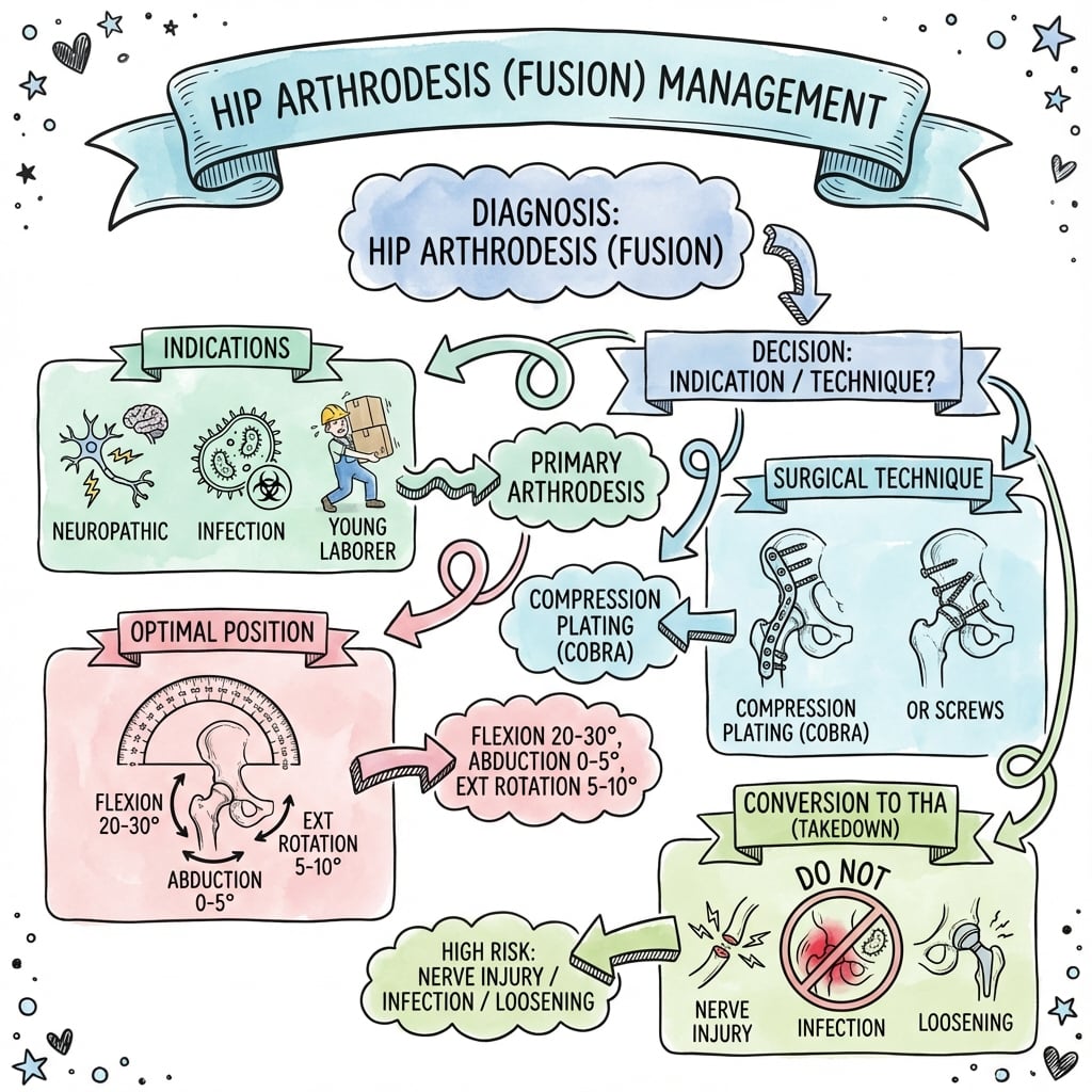
Q: What is the optimal position for hip arthrodesis? A: 25-30° flexion, 0-5° adduction, 15-20° external rotation. This position allows sitting (flexion), standing without excessive lumbar compensation (not too much flexion), walking without pelvic obliquity (neutral to slight adduction, avoiding abduction), and sitting with knees together plus foot hygiene (external rotation). Too much flexion impairs standing; too much abduction causes limping.
Q: What percentage of patients develop lumbar back pain after hip arthrodesis, and why? A: Over 50% develop lumbar back pain within 10 years. The fused hip transfers motion to the lumbar spine, which must compensate for the loss of hip flexion/extension. This increases lumbar range of motion by 50-100%, accelerating disc degeneration at L4-L5 and L5-S1. Back pain is NOT a complication - it is an expected consequence of altered biomechanics.
Q: What is the preferred fixation method for hip arthrodesis, and what fusion rate does it achieve? A: Cobra (arthrodesis) plate fixation is preferred, spanning from the ilium to the mid-femoral shaft with at least 6-8 bicortical screws. This achieves a fusion rate of 90-95%. Screw fixation alone (transfixion screws from ilium through acetabulum to femoral head) achieves only 85-90% fusion and has less rotational stability. Compression across the fusion site is critical regardless of fixation method.
Q: What is the most common complication after conversion of hip arthrodesis to THA, and how can it be reduced? A: Dislocation (20-30% incidence) due to abductor deficiency. The gluteus medius and minimus have atrophied during years of fusion and cannot be restored. Risk can be reduced by using dual mobility cups (reduces dislocation to 10-15%), large femoral heads (36-40 mm), and careful soft tissue repair. Despite these measures, instability remains the most common complication requiring reoperation.
Q: What are absolute contraindications to hip arthrodesis? A: Ipsilateral knee arthritis (will worsen due to compensatory stress), symptomatic lumbar spine disease (will worsen from increased motion demands), and contralateral hip disease (cannot compensate for the fused hip). The contralateral hip and ipsilateral knee MUST be normal, and the lumbar spine should be asymptomatic. If these joints are diseased, hip fusion will fail due to progressive adjacent joint degeneration and patient dissatisfaction.
Q: What is the maximum safe leg lengthening during conversion of hip arthrodesis to THA, and why? A: 3-4 cm maximum. The sciatic nerve is at extreme risk during conversion THA due to scarring from prior surgery and the need to restore leg length (typical LLD after fusion is 3-5 cm). Lengthening over 4 cm causes traction injury to the sciatic nerve, resulting in permanent foot drop and sensory loss. If preoperative LLD is over 5 cm, consider staged lengthening (femoral osteotomy weeks before THA) or accept residual LLD rather than risk nerve injury. Intraoperative neuromonitoring is recommended.
Exam Viva Scenarios
Practise clinical reasoning and management decisions out loud
“A 35-year-old male manual labourer presents with severe hip pain and disability after multiple failed revisions of an infected total hip arthroplasty. The infection has been eradicated with two-stage revision, but he has had recurrent dislocations and poor bone stock precludes further arthroplasty. He asks about hip fusion. What are the indications for hip arthrodesis, and how would you counsel this patient?”
“You have decided to proceed with hip arthrodesis using cobra plate fixation for a young patient with a failed revision THA. Walk me through your surgical technique, focusing on positioning, surface preparation, and fixation.”
“A 55-year-old woman had a hip arthrodesis 20 years ago for post-septic arthritis. She now has disabling lumbar back pain and requests conversion to total hip arthroplasty to improve her function. On examination, she has a 4 cm leg length discrepancy and a Trendelenburg gait (from the contralateral hip compensating). Radiographs show a solid fusion with a cobra plate in situ. How would you approach this case?”
Optimal Fusion Position
- Flexion: 25-30° (allows sitting without excessive lumbar flexion)
- Adduction: 0-5°, neutral (AVOID abduction - Callaghan showed it accelerates knee/back pain)
- External rotation: 15-20° (permits sitting with knees together)
- Verify position with goniometer and fluoroscopy BEFORE draping
Indications (Rare)
- Failed revision THA with eradicated infection and poor bone stock
- Young manual labourer (under 40) with destroyed hip
- Massive bone loss precluding arthroplasty
- Neurologic conditions requiring hip stability (flail hip)
Surgical Technique
- Lateral approach with complete capsulectomy
- Remove ALL cartilage; maximize bone-to-bone contact (70-80% surface area)
- Cobra plate fixation: ilium to femoral shaft, at least 6-8 screws
- Compression across fusion site is critical for union
Long-Term Complications (Expected)
- Lumbar back pain: over 50% at 10 years (increased spinal motion)
- Ipsilateral knee arthritis: 30-40% at 15-20 years (rotational stress)
- Leg length discrepancy: 3-5 cm shortening (counsel preoperatively)
- Nonunion: 5-10% with plate fixation; requires revision with bone graft
Conversion to THA Challenges
- Complication rate: 40-50% (instability, nerve injury, infection)
- Dislocation risk: 20-30% (abductor deficiency); use dual mobility cups
- Limit leg lengthening to 3-4 cm maximum (sciatic nerve stretch injury risk)
- Acetabular reconstruction: often Paprosky IIB-IIIA bone loss; plan for augments
Evidence Base and Key Studies
Hip Arthrodesis: A Long-Term Follow-Up (Landmark Adjacent-Joint Study)
- 28 patients reviewed at a mean of 35 years after arthrodesis (range 17 to 50 years)
- Ipsilateral knee pain in about 60% (mean onset 23 years after fusion)
- Low back pain in about 60% (mean onset 25 years after fusion)
- Contralateral hip pain in about 25% (mean onset 20 years)
- Hips fused in abduction had more ipsilateral knee/back pain and greater knee degeneration; 6 of 28 underwent later THA with marked relief of back pain
Conversion of a Fused Hip to Total Hip Arthroplasty (Largest Conversion Series)
- 187 patients (208 hips) converted from fusion to THA; mean follow-up 9.2 years
- Mean interval from arthrodesis to conversion was 27 years; mean age at conversion 51 years
- 79% pain-free or minimal pain; 83% good-to-excellent function
- 15 nerve palsies and 28 cases of heterotopic ossification; 12 hips required revision
- Implant survival 96.1% at 10 years, 89.9% at 15 years
Hip Fusion Conversion to THA: Systematic Review and Meta-Analysis
- Meta-analysis of 34 pre-post trials of conversion THA after arthrodesis
- Harris Hip Score improved by a weighted mean of 42.3 points (95% CI 38 to 47)
- Leg-length discrepancy decreased by 21 mm (95% CI 19 to 24)
- Greater HHS gain with cementless implants, fusion duration under 12 years, and age under 45 years
- Heterotopic ossification was the most common complication at 14%
Results and Experiences of Conversion of Hip Arthrodesis
- 45 conversions performed; 34 followed for a mean of 77.5 months
- Primary indications were low back pain (21) and ipsilateral knee pain (13)
- 29 of 34 hips (85%) were pain-free or had minimal pain
- Complications included persistent sciatic palsy, 2 superficial infections, 2 periprosthetic fractures and grade IV heterotopic ossification
- Revision arthroplasty required in 4 hips; pronounced limp persisted in half
Hip Arthrodesis: A Procedure for the New Millennium? (Indications and Technique Review)
- Argues arthrodesis remains an excellent option for noninflammatory monoarticular arthritis in the young patient
- Emphasises abductor-sparing technique and avoidance of pelvic deformity
- Limb position is the key determinant of early satisfaction and fusion durability
- Long-term morbidity is driven by pain in contiguous joints (spine, knee, contralateral hip)
- Conversion arthroplasty, when needed, can yield favourable outcomes
Stiff Lumbo-Pelvic Unit and THA Instability (Hip-Spine Relationship)
- Medicare cohort: 14,747 THA patients with prior lumbar fusion vs 839,004 controls
- Dislocation rate 1.55% in controls vs 2.96% with 1-2 level fusion and 4.12% with 3-7 levels
- Risk rose with number of fused levels (3-7 levels: OR 1.60 vs 1-2 levels)
- Loss of compensatory pelvic motion eliminates protective acetabular reorientation when seated
- Effect persisted after age and sex matching