The Clenched Fist Injury | Eikenella corrodens
- A 'Fight Bite' is a Septic Arthritis until proven otherwise.
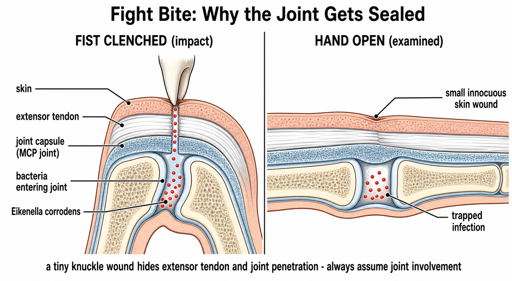
- The tooth penetrates the MCPJ capsule in FLEXION.
- When the finger extends, the tract is sealed by the extensor hood gliding proximally.
- This traps bacteria (Eikenella, Strep, Staph) in the joint.
- Eikenella corrodens is resistant to First Gen Cephalosporins (Keflex) and Clindamycin.
- Treatment is emergent formal arthrotomy and washout.
- “Never trust a 'small cut' over the MCPJ in a young male.
- “Cephalexin alone effectively TREATS the bacteria it covers but SELECTS OUT Eikenella.
- “Augmentin is the drug of choice.
Overview
A "human bite" covers two distinct injuries. The occlusal bite is a direct clamp of the teeth on to a finger, ear or nose. The clenched fist injury (CFI, the "fight bite") is indirect: a punch to the mouth drives a tooth through the skin over a knuckle and inoculates oral flora straight into the metacarpophalangeal (MCP) joint or the extensor apparatus. The clenched fist injury is the dangerous one. Human bites are the third most common mammalian bite, after dog and cat bites.
Why a small wound is a big problem. Human saliva carries up to 10^9 bacteria per mL, a diverse mix of aerobes and anaerobes. The extensor tendon glides over the MCP joint as the fist opens, and that gliding acts as a one-way valve: once the fist relaxes the inoculum is sealed deep within the joint, a closed-loop abscess inside the capsule that destroys cartilage (chondrolysis) within 24-48 hours. Osteomyelitis and amputation are real risks.
- Animal Bite
- Pasteurella / Capnocytophaga
- Human Bite
- Eikenella / Strep Viridans / Staph
- Animal Bite
- Puncture / Crush
- Human Bite
- Inoculation into Joint (CFI)
- Animal Bite
- Augmentin
- Human Bite
- Augmentin (But Ceph/Clinda fail)
- Animal Bite
- Urgent
- Human Bite
- Emergent (if Joint involved)
A laceration over the third, fourth or fifth MCP joint in a young adult is a fight bite until proven otherwise. Treated as a simple laceration and sutured, the septic arthritis proceeds unchecked.
Pathophysiology and Mechanisms
The sliding target. At impact the fist is clenched, the MCP joints are flexed to 90 degrees, and the extensor tendon and hood have moved distally over the metacarpal head. The tooth penetrates skin, tendon and capsule in one aligned track and enters the joint. When the fist opens the MCP joints extend and the extensor tendon glides proximally, so the skin wound now lies proximal to the capsular wound and the two no longer line up. The misalignment prevents drainage and leaves the joint an anaerobic environment.
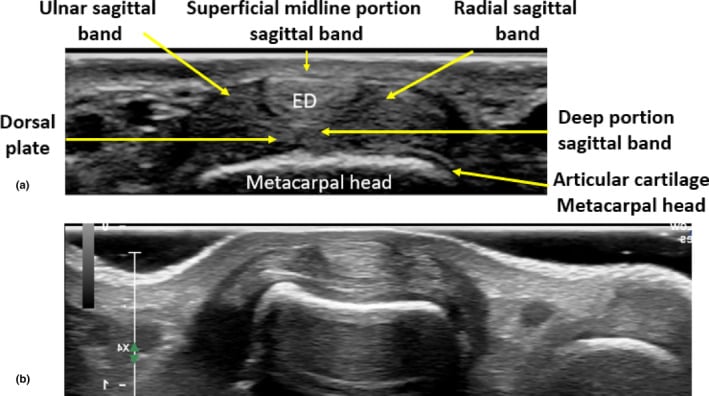
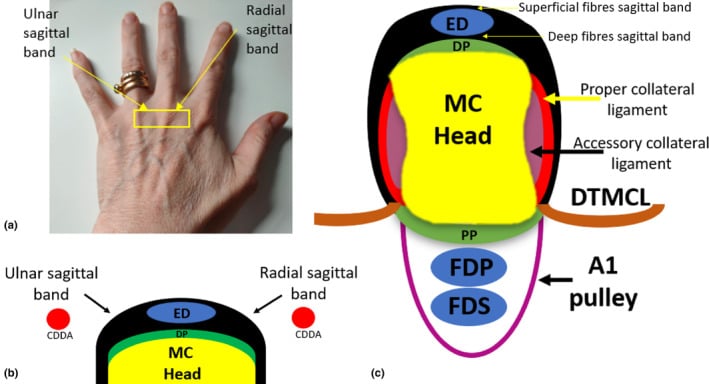
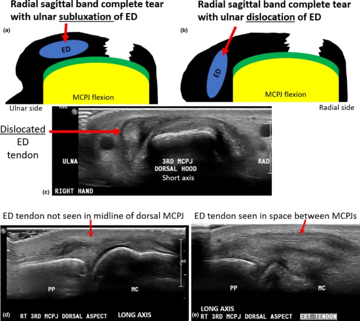
The anatomy that lets it happen. The MCP joint capsule is very thin dorsally and easily penetrated, and the metacarpal head sits just below the tendon, so cartilage damage is common. The sagittal bands centre the extensor tendon over the joint; the tooth can divide one, and a divided band lets the tendon sublux. Once inside, infection spreads readily into the subaponeurotic space dorsal to the tendon or the subfascial space deep to it.
Microbiology
Eikenella corrodens. A Gram-negative facultative anaerobe found in 25-30% of human bite infections, and the organism that makes a human bite different. It acts synergistically with Streptococcus viridans to worsen the infection. It is the E of the HACEK group of fastidious Gram-negatives (Haemophilus, Aggregatibacter, Cardiobacterium, Eikenella and Kingella, the last a cause of septic joints in children), a group associated with endocarditis; the species name records that it corrodes the agar it grows on.
Its resistance profile is the exam point. Sensitive to the penicillins, resistant to the reflex choices:
- Resistant: clindamycin, erythromycin, first-generation cephalosporins (cephalexin), metronidazole (variable)
- Sensitive: penicillin, ampicillin, amoxicillin-clavulanate, ciprofloxacin, ceftriaxone
Cephalexin and clindamycin are the reflex prescriptions for ordinary skin trauma, so treating a human bite with cephalexin (Keflex) is the classic error. It treats the organisms it covers and selects out Eikenella.
The rest of the cocktail. Streptococcus viridans is the most common aerobe (50%) and Staphylococcus aureus is common (30%). The anaerobes are Fusobacterium, Peptostreptococcus and Prevotella (Eubacterium).
Classification Systems
By type. Human bites are grouped by type:
- Occlusal bite - direct clamping, often of a fingertip or soft-tissue area (finger, ear, nose); can cause amputation
- Clenched fist injury - indirect inoculation, with high risk to joint and tendon
- Avulsion - soft-tissue loss, for example ear or nose
- Self-inflicted - nail biting (paronychia), psychogenic
Type sets the urgency. A clenched fist injury needs an urgent arthrotomy; an occlusal bite needs debridement.
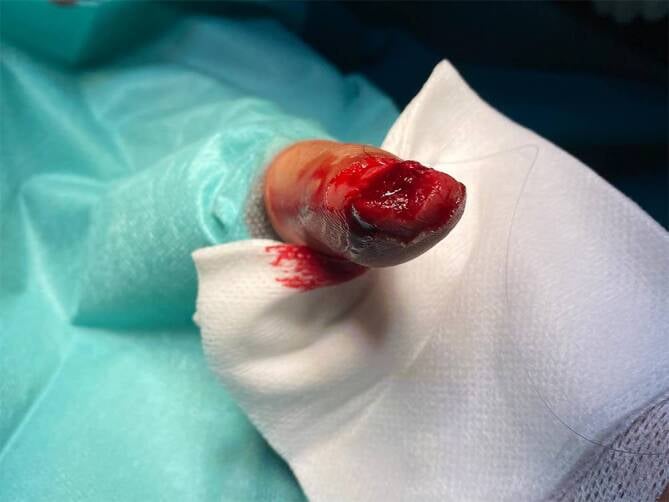
By depth. Beyond the type, it is the depth of penetration of a fight bite that drives prognosis and the operation, and depth is the basis of the large surgical series in the Evidence Base (Chadaev).
- What is involved
- Skin and fat; extensor tendon and capsule intact
- Implication
- Debride and antibiotics; best prognosis
- What is involved
- Tendon laceration or sagittal-band injury, capsule not breached
- Implication
- Explore; watch for tendon subluxation; repair the tendon at a clean second look
- What is involved
- Capsule breached, organisms seeded into the joint
- Implication
- Formal arthrotomy and washout - the classic fight-bite emergency
- What is involved
- Cartilage divots, metacarpal-head erosion, retained tooth fragment
- Implication
- Worst prognosis; thorough debridement, prolonged antibiotics, sometimes joint-spanning distraction
The skin wound is tiny at every depth; what matters is how far the tooth went. In the surgical series outcome tracks directly with depth: results are excellent when only skin or tendon are involved and deteriorate sharply once cartilage or bone is breached. That is why the joint surface must be visualised at operation, and a retained tooth fragment actively hunted for and removed.
Clinical Presentation and Assessment
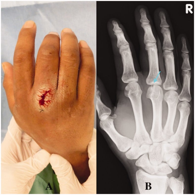
The patient. A young adult man presenting hours to days after a fight, with a deceptively small (3-5 mm) laceration over the dorsum of the third, fourth or fifth MCP joint, most often the third or fourth, whose heads are prominent. The story is frequently concealed or vague ("I cut it on a fence", "cut on metal"), from shame or for legal reasons; ask whether the hand was a fist. Delayed presentation is common; the pain increases overnight. Ask about tetanus and vaccination status.
How it evolves. Three stages, by time since the injury:
- Early (first 24 hours): localised tenderness over the MCP joint, minimal swelling, often no erythema. The wound may already be sealed by gliding of the extensor hood. This benign appearance is the trap.
- Established infection (24-72 hours): increasing dorsal swelling, erythema, throbbing pain, and the cardinal sign of pain on passive movement of the MCP joint, indicating septic arthritis.
- Late or neglected: spreading dorsal cellulitis, purulent discharge, fixed flexion from joint destruction, and systemic features. Crepitus or pain out of proportion raises the spectre of necrotising infection.
Examination. The wound is small and may be sealed. Pain on passive motion of the MCP joint is the sign of septic arthritis. Test the stability of the extensor mechanism for sagittal band injury, examining dynamically for painful snapping or subluxation of the tendon during MCP flexion. Check the digital nerves. Look for gas or crepitus: rare but ominous, the sign of necrotising fasciitis.
Investigations
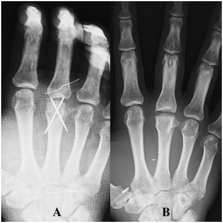
Radiographs are mandatory, and should include oblique views. Four findings are being looked for:
- Fracture of the fifth metacarpal neck (boxer's fracture), which suggests a punch mechanism
- Foreign body: a tooth fragment is radiopaque
- Air in the joint, pathognomonic of penetration
- Osteomyelitis, a late finding: periosteal reaction, osteopenia, erosions
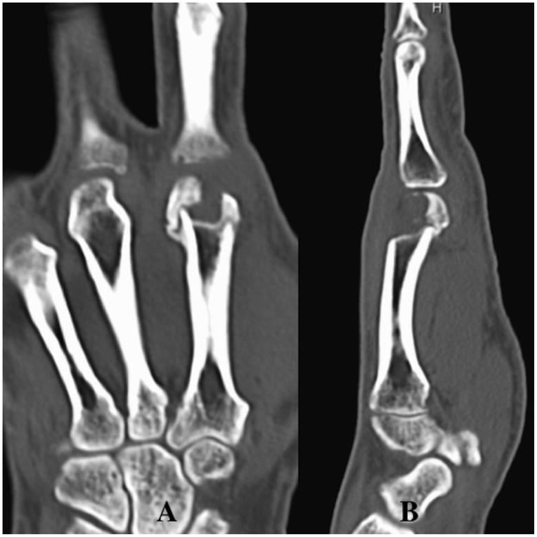
Other imaging. MRI is rarely indicated in the acute setting but is useful for chronic osteomyelitis. Ultrasound can guide aspiration of a joint effusion if the diagnosis is unclear. CT defines the location and extent of metacarpal-head bone loss when late infection or osteomyelitis is suspected, or when reconstruction is being planned.
Differential Diagnosis
A small, infected, painful wound over the dorsum of the hand has several mimics. The clinical task is to decide which patients have a joint or deep-space process that mandates theatre, and which can be managed with antibiotics and observation.
- Distinguishing Features
- Small wound over 3rd-5th MCPJ, young adult, fight history (often concealed), pain on passive MCP motion
- Key Discriminator
- Wound overlies MCPJ; air or tooth fragment on X-ray
- Management Implication
- Emergent formal arthrotomy and washout
- Distinguishing Features
- Diffuse erythema, fluctuance away from joint, no joint-line tenderness, full painless passive ROM
- Key Discriminator
- Joint not penetrated; pain not on passive motion
- Management Implication
- Antibiotics +/- bedside incision and drainage
- Distinguishing Features
- Older patient, prior episodes, no wound, raised urate, monosodium urate crystals on aspirate
- Key Discriminator
- No breach of skin; crystals not organisms
- Management Implication
- NSAIDs/colchicine, not surgery
- Distinguishing Features
- Kanavel signs: fusiform swelling, semiflexed digit, pain on passive EXTENSION, tenderness along flexor sheath (volar)
- Key Discriminator
- Volar not dorsal; flexor sheath not MCPJ
- Management Implication
- Surgical sheath irrigation
- Distinguishing Features
- Vesicles on pulp/digit, healthcare or dental exposure, NO frank pus, viral prodrome
- Key Discriminator
- Vesicular, self-limiting; incision is contraindicated
- Management Implication
- Aciclovir; do NOT incise
- Distinguishing Features
- Pain out of proportion, crepitus, skin necrosis/bullae, systemic toxicity, rapid spread
- Key Discriminator
- Crepitus/gas, systemic sepsis
- Management Implication
- Emergent radical debridement, broad-spectrum IV antibiotics
The two-second discriminator. Pain on passive extension of the digit with tenderness along the volar sheath points to flexor tenosynovitis; pain on passive motion of the MCP joint beneath a dorsal wound points to a fight-bite septic arthritis. Herpetic whitlow is the one infective mimic you must not incise.
Management Strategy
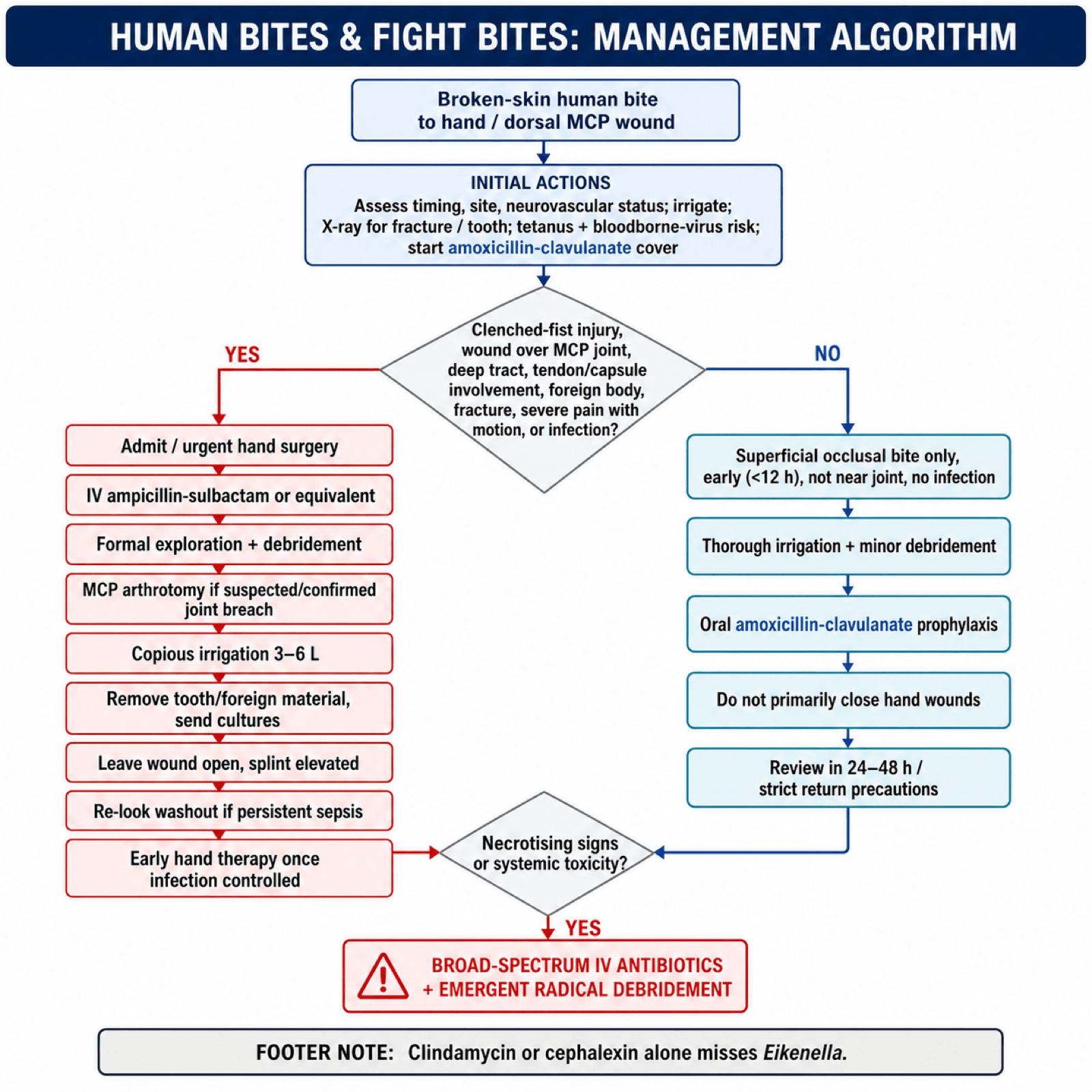
The pathway for a clenched fist injury.
- Admit for intravenous antibiotics
- Explore: surgical exploration is mandatory
- Wash: arthrotomy and irrigation
- Leave open: do not suture
- Rehabilitate: early motion once the infection is controlled
Early mobilisation prevents stiffness and tendon adhesion.
Non-operative management is reserved for the superficial occlusal bite away from a joint, presenting early (less than 12 hours) with no signs of infection. A clenched fist injury is almost never managed non-operatively; the risk of a missed joint penetration is too high. If in doubt, wash it out.
Surgical Technique
Formal arthrotomy. The operation is a fixed sequence:
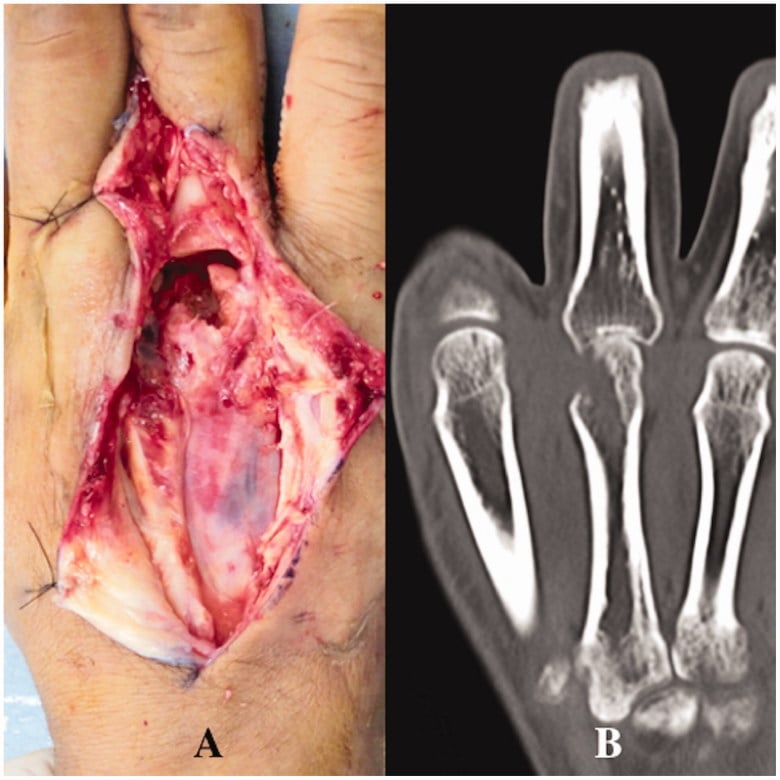
- Incision. Extend the laceration, usually transversely or as a Z-plasty; avoid a longitudinal incision crossing the joint creases.
- Exposure. Expose the extensor tendon and retract it to inspect the capsule. A longitudinal split of the tendon, splitting the sagittal fibres, may be needed to see the joint surface directly.
- Capsulotomy. If a tear is found, open it. If no tear is found but clinical suspicion is high, open it. The joint must be visualised.
- Irrigation. Copious saline, 3-6 litres, with an 18G catheter on a syringe for jet lavage within the joint.
- Staging. Inspect the metacarpal head for divots from the tooth and for cartilage loss.
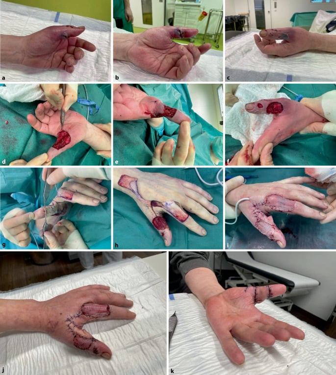
- Closure. Leave the skin open; loose approximation only if the wound is very clean.
A strict no-closure policy is the safest rule for the junior surgeon.
The tooth. A fragment embedded in the metacarpal head must be removed, with a curette or rongeur, and the bone defect it leaves treated as potential osteomyelitis. Cultures of the bone fragment are sent separately.
Antibiotic Protocol
Amoxicillin-clavulanate (Augmentin) is the gold standard, first line for every human bite, because it covers Staph, Strep, the anaerobes and Eikenella in one drug. For a clenched fist injury it is started intravenously at once.
Penicillin allergy is covered with a combination:
- Ciprofloxacin (covers Eikenella) or trimethoprim-sulfamethoxazole
- plus clindamycin or metronidazole for the anaerobes and Staph
Clindamycin alone misses Eikenella. Cephalexin alone misses Eikenella.
Tetanus, Rabies and Bloodborne Viruses
Tetanus. A human bite is a tetanus-prone wound: contaminated, often with devitalised tissue, and often presenting late. Action depends on the immunisation history:
- Fully immunised with a booster within the last 10 years: no further tetanus action needed
- Immunised but the last dose was more than 10 years ago, or the course is incomplete or uncertain: give a tetanus toxoid booster
- Unimmunised or uncertain, or a heavily contaminated wound in an incompletely immunised patient: give toxoid and human tetanus immunoglobulin at separate sites, and complete the vaccination course
Rabies. This is the high-yield discriminator from animal bites: a human bite does not require rabies prophylaxis, because humans are not a rabies reservoir host. Rabies post-exposure prophylaxis is the concern for many animal bites (dog, bat), not human bites.
Bloodborne viruses. The risks quoted are approximately 30% for hepatitis B if the source is positive, 3% for hepatitis C and 0.3% for HIV. Transmission through a clenched fist injury is rare, unlike a needle-stick, but possible. Hepatitis B is the virus to consider for every human bite; HIV and hepatitis C are considered only where there was visible blood.
Complications
Infection-related complications.
- Risk Factors
- MCP joint penetration, delay
- Prevention
- Early I&D, IV antibiotics
- Management
- Joint washout, may need multiple
- Risk Factors
- Metacarpal head involvement, delay
- Prevention
- Early debridement
- Management
- Prolonged IV antibiotics, debridement
- Risk Factors
- Palmar involvement, immunocompromised
- Prevention
- Aggressive exploration
- Management
- I&D of web space, thenar space
- Risk Factors
- Diabetes, delayed presentation
- Prevention
- High index of suspicion
- Management
- Emergent radical debridement
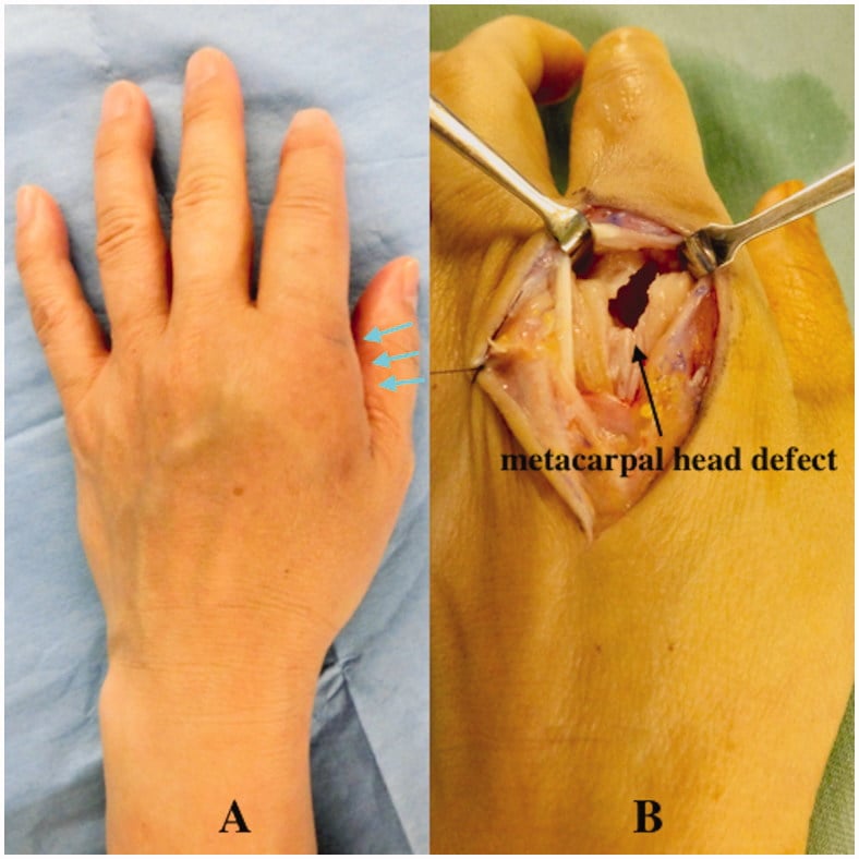
Structural complications. Tendon rupture may follow the initial injury or the infection, the extensor tendons being most vulnerable at the MCP level. Sagittal band disruption allows the extensor tendon to sublux between the metacarpal heads and may need surgical repair. Joint stiffness is common after septic arthritis, which is why early mobilisation once the infection is controlled matters so much. Contractures follow collateral ligament shortening if the joint is immobilised in extension, the reason for splinting in the intrinsic plus position.
Worst-case scenarios. In severely neglected cases, with immunocompromise or a long delay:
- Amputation may be required for uncontrollable infection or extensive tissue loss
- Loss of hand function: permanent grip weakness and finger stiffness
- Systemic sepsis, life-threatening if the infection spreads
Rehabilitation
Splint first, then move as soon as the infection allows.
- Volar splint in intrinsic plus (MCP joints flexed 70, IP joints extended) to prevent collateral ligament shortening
- High elevation in a Bradford sling to reduce oedema
- Saline-soaked gauze dressings to encourage wicking of purulence
- Start active range of motion as soon as the cellulitis resolves and the wound is clean
- Isolated extensor digitorum communis (EDC) gliding exercises
- The wound heals by secondary intention (granulation)
- Scar management and desensitisation once healed
- Grip strengthening once soft-tissue coverage is complete
Prognosis
Time is the prognostic factor. The later the patient presents, the higher the infection rate and the worse the joint does.
- Infection Rate
- 10%
- Joint Outcome
- Good if no joint penetration
- Overall Prognosis
- Excellent with appropriate treatment
- Infection Rate
- 30-40%
- Joint Outcome
- Moderate risk of stiffness
- Overall Prognosis
- Fair with aggressive management
- Infection Rate
- 50%+
- Joint Outcome
- High risk of permanent damage
- Overall Prognosis
- Guarded, multiple surgeries likely
Injury pattern. A simple bite wound has a good prognosis with early antibiotics. A clenched fist injury does worse because of joint penetration and contamination, and once the joint is involved, 20-30% develop some permanent stiffness even with treatment.
What predicts a poor outcome. Negative prognostic factors include:
- Delay in presentation: greater than 24 hours significantly worsens outcomes
- MCP joint penetration, with its risk of septic arthritis and cartilage destruction
- Eikenella corrodens, often resistant to empiric therapy, and may be missed
- Patient comorbidities: diabetes, immunosuppression, alcoholism
- Incomplete debridement, since retained contamination leads to persistent infection
Long-term function. Most patients treated appropriately within 24 hours return to full hand function. Those presenting late or with joint involvement may be left with reduced grip strength (20-40%), limited MCP range of motion, cold intolerance and persistent pain.
Guidelines, Registries & Global Practice
Global Epidemiology
Human bites are the third most common mammalian bite presenting to emergency departments worldwide, after dog and cat bites. They cluster in young adult males, often around interpersonal violence and alcohol, with most injuries to the hand. In the Talan multicentre series, 70% of those involved were young adult men and 56% of injuries were clenched-fist injuries. Under-reporting is common because patients conceal an assault mechanism; delayed presentation (beyond 24 hours) is consistently associated with higher complication rates across published series from Europe, North America, Africa and Asia.
Guidelines Side by Side
- Antibiotic of Choice
- Amoxicillin-clavulanate; ampicillin-sulbactam IV
- Wound Closure (Hand)
- Leave open; arthrotomy for CFI
- Notable Stance
- Fight bite over MCPJ treated as septic arthritis
- Antibiotic of Choice
- Co-amoxiclav; clindamycin + ciprofloxacin if penicillin-allergic
- Wound Closure (Hand)
- Open; debride in theatre
- Notable Stance
- Low threshold for surgical exploration of dorsal MCPJ wounds
- Antibiotic of Choice
- Amoxicillin-clavulanate first-line
- Wound Closure (Hand)
- Open for hand/high-risk; closure acceptable for selected facial wounds
- Notable Stance
- Routine prophylaxis advised for human and hand bites
- Antibiotic of Choice
- Cover Eikenella, Strep, Staph, anaerobes
- Wound Closure (Hand)
- Open, delayed closure
- Notable Stance
- Treat associated metacarpal fracture as open/contaminated
The consistent global message: amoxicillin-clavulanate is first-line, cephalexin and clindamycin monotherapy are inadequate, hand bites are left open, and a wound overlying the MCP joint mandates surgical exploration.
Evidence and Registry Notes
There is no implant registry for bite injuries, but the evidence base is anchored by a Cochrane systematic review (Medeiros and Saconato) confirming antibiotic prophylaxis reduces infection after human and hand bites, the Talan microbiology series defining the organisms, and surgical series (Chadaev) showing outcome tracks with depth of joint and bone involvement.
Bloodborne Virus Assessment (Global Principle)
- Hepatitis B: Assess immunisation status of the person bitten; offer vaccination or immunoglobulin where indicated, as HBV can transmit via mucosal or non-intact-skin exposure.
- HIV / Hepatitis C: Transmission requires visible-blood exposure and is negligible from ordinary (blood-free) saliva; reserve testing and post-exposure prophylaxis discussion for bloody-saliva exposures.
- Consent and follow-up: Source and recipient testing requires informed consent; baseline and repeat serology where risk is identified.
High-Resource vs Limited-Resource Practice
- High-resource: 24-hour theatre access, formal arthrotomy under regional/general anaesthesia, IV co-amoxiclav, hand-therapy-led rehabilitation, and structured bloodborne-virus pathways.
- Limited-resource: Emphasis on meticulous bedside debridement and irrigation, oral amoxicillin-clavulanate (oral shown equivalent to IV for uncomplicated early bites in the Zubowicz trial), early referral of any joint-penetrating injury, and tetanus cover, which remains a priority where immunisation coverage is incomplete.
Documentation and Prevention
These injuries carry medicolegal weight: record the time and mechanism, clinical findings (with photographs where consented), treatment given, and the prognosis discussed. Prevention centres on reducing alcohol-related violence and on public awareness that a punch to the mouth is a high-risk inoculation injury, not a trivial graze.
Controversies and Areas of Uncertainty
Much of the surgical dogma for fight bites rests on small retrospective series and a single randomised trial that deliberately excluded joint and tendon injuries. The high-yield exam answers are well established; several practical questions remain genuinely debated.
Routine arthrotomy or selective exploration. Some authors advocate formal arthrotomy for every wound overlying the MCP joint; others reserve it for a proven capsular breach, air on imaging, established septic arthritis or failure of antibiotics. The safe exam position is unchanged: if the joint may be involved, explore it.
Antibiotic duration. Prophylaxis after a clean early bite (commonly 3-5 days) is a different question from treatment of established septic arthritis or osteomyelitis (often weeks, intravenous then oral). No high-quality trial defines the optimal duration for the established-infection group.
Imaging the clean wound. Plain radiographs are universally recommended to exclude fracture, tooth fragment and intra-articular air. The role of early MRI or ultrasound in the equivocal, apparently clean wound is undefined and rarely changes acute management.
Negative-pressure wound therapy and distraction. Case reports and small series describe NPWT and joint-spanning distraction for severe established infection. They are adjuncts, not first-line, and lack comparative evidence.
Bloodborne-virus testing thresholds. Whether to test asymptomatic recipients of blood-free bites is contested. The current synthesis favours universal hepatitis B follow-up and selective HIV and hepatitis C testing based on visible-blood exposure.
MCQ Practice Points
Q: Which bacteria is characteristically resistant to Clindamycin and Cephalexin in human bites? A: Eikenella corrodens.
Q: In a Clenched Fist Injury, the bacterial inoculum is trapped because: A: The extensor tendon glides proximally upon finger extension, sealing the tract.
Q: What is the first-line oral antibiotic for a human bite? A: Amoxicillin + Clavulanate (Augmentin).
Q: What is a 'Honeymoon Period' in flexor tenosynovitis? A: The period (12-24h) where the bacteria are proliferating but signs are subtle, before rapid escalation.
Q: What is the mandatory surgical approach for a confirmed clenched fist injury? A: Formal arthrotomy with copious irrigation (3-6L saline) and the wound left open.
Q: What complication should you suspect if a fight bite patient presents with crepitus? A: Necrotizing fasciitis - requires emergent radical debridement.
Viva Scenarios
Practise clinical reasoning and management decisions out loud
“A 24-year-old male presents with a 4mm laceration over his right long finger MCPJ. He says he cut it on a tooth during a fight 2 days ago. It is red, swollen, and he cannot extend the finger due to pain. He has been taking Keflex from his GP.”
“X-ray shows a Boxer's fracture (5th MC Neck) with volar angulation. On clinical exam, there is a small scab over the 5th MCPJ. Patient denies a bite.”
“Why is Eikenella corrodens significant in human bites? What is its unique resistance profile?”
“A 40-year-old carer presents 6 hours after being bitten on the dorsum of the hand by a patient with intellectual disability. The skin is broken but there is no erythema, no swelling, the joint is not involved and passive movement is painless. She asks whether she really needs treatment for 'such a tiny bite'.”
Diagnosis
- Small wound over MCPJ = Fight Bite
- Boxer's Fracture + Wound = Open/Infected
- Pain on passive ROM = Septic Arthritis
- X-ray: Look for Air and Tooth
Microbiology
- Polymicrobial
- Eikenella corrodens (Gram Neg Anaerobe)
- Strep viridans
- Staph aureus
Management
- Admit + IV Augmentin
- Formal Arthrotomy + Washout
- Leaves Wounds Open
- Splint in Intrinsic Plus
Evidence Base
Microbiology of Infected Human Bites (Landmark)
- Multicentre prospective study of 50 patients with infected human bites; 56% were clenched-fist injuries, 44% occlusal
- Median 4 isolates per wound (3 aerobes, 1 anaerobe); polymicrobial in the majority
- Key isolates: Streptococcus anginosus 52%, Staphylococcus aureus 30%, Eikenella corrodens 30%, Fusobacterium nucleatum 32%, Prevotella melaninogenica 22%
- Amoxicillin-clavulanate and moxifloxacin showed excellent in-vitro activity against common isolates
Antibiotic Prophylaxis for Mammalian Bites (Cochrane)
- Systematic review of 8 randomised controlled trials of prophylactic antibiotics after mammalian bites
- Prophylactic antibiotics significantly reduced infection after human bites (unlike cat or dog bites)
- For bites of the hand, prophylaxis reduced infection (OR 0.10, 95% CI 0.01 to 0.86; NNT = 4)
Antibiotic Prophylaxis in Early Hand Bites (RCT)
- Prospective randomised trial of 48 patients with early (under 24h) hand bites without joint or tendon involvement
- Placebo (mechanical wound care alone): 7 of 15 (46.7%) became infected
- Oral and parenteral antibiotic groups: zero infections
- Oral antibiotics were equivalent to intravenous for prophylaxis of uncomplicated early bites