Osteomyelitis, Septic Arthritis & Prosthetic Joint Infection
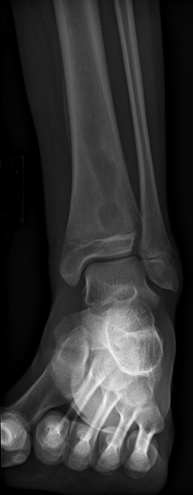
Radiography: First-line screening — normal early; periosteal reaction, lysis at 10-14 days
Ultrasound: Best for joint effusion detection and aspiration guidance for septic arthritis
MRI with contrast: Gold standard for osteomyelitis, spinal infection, soft tissue abscess
Bone Scan (Triple-phase): Sensitive but non-specific; useful when MRI unavailable
Labelled WBC Scan: Best nuclear medicine test for PJI and chronic osteomyelitis
FDG-PET: High sensitivity for chronic osteomyelitis; limited specificity around metalwork
Key: Normal radiographs DO NOT exclude infection — MRI is needed for early and accurate diagnosis
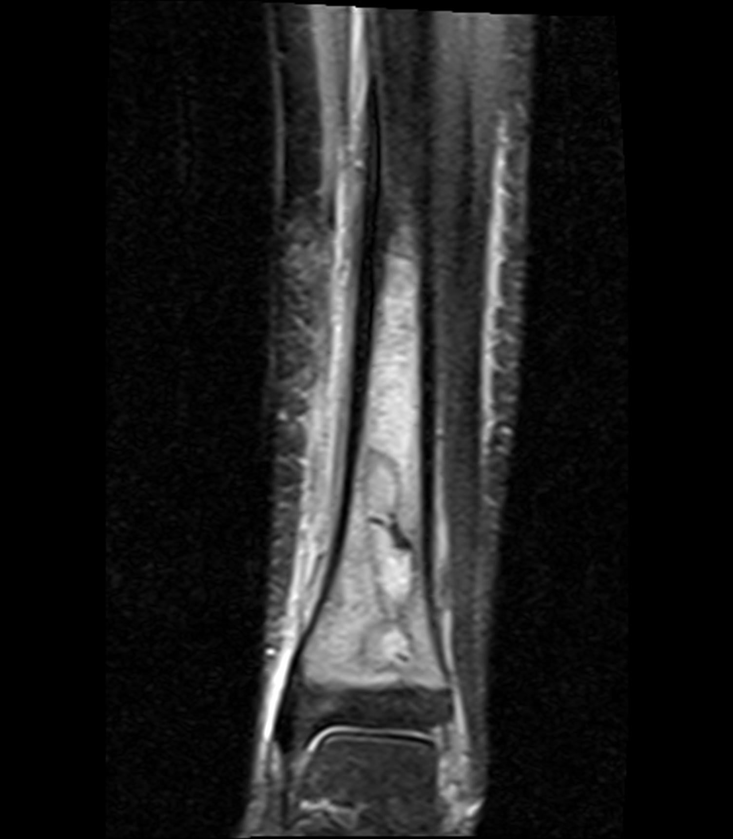
- MRI with contrast is the gold standard imaging modality for osteomyelitis — sensitivity 95%, specificity 88%.
- Radiographic changes of osteomyelitis (periosteal reaction, lytic destruction) lag 10-14 days behind clinical onset — NORMAL RADIOGRAPHS DO NOT EXCLUDE OSTEOMYELITIS.
- Ultrasound is the first-line imaging modality for suspected septic arthritis — it detects effusion and guides aspiration for diagnostic confirmation.
- For PJI: labelled WBC scan (combined with sulphur colloid) has the best accuracy among nuclear medicine modalities.
- Spinal infection (discitis/osteomyelitis): MRI shows disc signal change, endplate destruction, and paraspinal/epidural abscess — gadolinium helps differentiate abscess from phlegmon.
- “Penumbra sign on MRI: a thin rim of T1 hyperintensity surrounding an intraosseous abscess = SPECIFIC for infection (Brodie abscess). Distinguishes from tumour.
- “In children under 18 months, osteomyelitis can cross the growth plate (transphyseal vessels still patent) — this causes concurrent septic arthritis in adjacent joints.
- “The Cierny-Mader classification combines anatomical type (medullary, superficial, localised, diffuse) with host status (A: healthy, B: compromised, C: treatment worse than disease).
- “FDG-PET has high sensitivity for chronic osteomyelitis (96%) but limited specificity around metalwork.
- “MRI features that distinguish osteomyelitis from neuropathic (Charcot) arthropathy: geographic marrow signal change, sinus tract, soft tissue abscess, and cortical destruction.
Infection imaging is a high-yield examination topic. You must know: the temporal evolution of radiographic changes (10-14 day lag), the superiority of MRI for early osteomyelitis detection, the penumbra sign (Brodie abscess), the approach to septic arthritis (USS-guided aspiration), imaging of PJI (labelled WBC scan), and spinal infection imaging (MRI with contrast). Classic traps: excluding osteomyelitis based on normal radiographs and not knowing the difference between abscess (ring-enhancing) and phlegmon (diffuse enhancement) on contrast MRI.
DRIPSRadiographic Signs of Osteomyelitis
Hook:DRIPS: the five classic radiographic signs of osteomyelitis — remember they take 10-14 days to appear.
PACESMRI Features of Osteomyelitis
Hook:PACES: the five key MRI features of osteomyelitis — the Penumbra sign is the most specific.
RUMAImaging Algorithm for Suspected MSK Infection
Hook:RUMA: Radiograph, Ultrasound, MRI, Alternative nuclear medicine — the stepwise infection imaging algorithm.
Overview
Imaging of musculoskeletal infection requires an understanding of the temporal evolution of radiographic changes, the strengths and limitations of each imaging modality, and the specific clinical scenarios that determine imaging selection. The fundamental principle is that normal radiographs do NOT exclude infection — radiographic changes lag 10-14 days behind clinical onset, and by the time changes are visible, significant bone destruction has occurred.
Day 0-3: Radiographs normal. MRI may show soft tissue oedema. Bone scan becomes positive (increased vascularity). Day 3-7: Radiographs may show soft tissue swelling. MRI shows bone marrow oedema. Triple-phase bone scan positive on all three phases. Day 7-14: Radiographs begin showing periosteal reaction and rarefaction. MRI shows established marrow changes with possible abscess formation. Day 14+: Radiographs show lytic destruction, sequestrum/involucrum in chronic cases. MRI delineates the full extent of infection and guides surgical planning.
Aspiration is the gold standard for confirming infection — imaging supports but does not replace microbiological diagnosis. Joint aspiration for septic arthritis: WBC more than 50,000 cells/microlitre (more than 75% neutrophils) is highly suggestive. Crystals may coexist with infection (gout and septic arthritis can occur simultaneously). Bone biopsy for osteomyelitis: image-guided biopsy for culture and histology when the organism is unknown. Blood cultures are positive in only 50% of osteomyelitis cases.
Systematic Approach
Infection Imaging Selection Guide
- First-Line Imaging
- AP + lateral radiograph (often normal). Bloods (CRP, ESR, WCC)
- Advanced Imaging
- Urgent MRI with contrast: marrow oedema, subperiosteal abscess, extent. Bone scan if MRI delayed
- First-Line Imaging
- Radiograph (sequestrum, involucrum, cortical thickening)
- Advanced Imaging
- MRI with contrast for extent and abscess delineation. CT for sequestrum identification and surgical planning
- First-Line Imaging
- Ultrasound: effusion detection + aspiration guidance
- Advanced Imaging
- MRI with contrast if bone involvement suspected (adjacent osteomyelitis). USS is usually sufficient for diagnosis
- First-Line Imaging
- Radiograph (loosening, osteolysis — often non-specific)
- Advanced Imaging
- Combined WBC/sulphur colloid scan or FDG-PET. MRI limited by metalwork artefact. Joint aspirate is gold standard
- First-Line Imaging
- Radiograph (disc space narrowing, endplate irregularity — may be delayed)
- Advanced Imaging
- MRI with contrast: gold standard. Shows disc destruction, endplate oedema, paraspinal/epidural abscess. CT-guided biopsy
- First-Line Imaging
- Radiograph (cortical erosion, periosteal reaction)
- Advanced Imaging
- MRI with contrast: distinguishes osteomyelitis from neuropathic (Charcot) changes. Probe-to-bone test correlates
Imaging Differential Diagnosis: Mimics of Infection
Marrow oedema and aggressive periosteal reaction are non-specific — several entities mimic osteomyelitis on MRI. Knowing the discriminators avoids both over- and under-treatment.
- Key Overlapping Feature
- Marrow oedema, periosteal reaction, soft-tissue enhancement
- Discriminating Feature(s)
- Sinus tract, rim-enhancing abscess, penumbra sign, raised CRP/ESR with clinical sepsis; geographic confluent T1 marrow replacement
- Key Overlapping Feature
- Marrow oedema, bone destruction, joint disorganisation
- Discriminating Feature(s)
- Periarticular, midfoot/Lisfranc distribution, subchondral cysts and intra-articular bodies, NO sinus tract or abscess (Ahmadi)
- Key Overlapping Feature
- Aggressive periosteal reaction, marrow signal change
- Discriminating Feature(s)
- Solid enhancing mass, cortical destruction with soft-tissue mass, ABSENCE of penumbra sign; biopsy decisive
- Key Overlapping Feature
- Marrow oedema, periosteal reaction
- Discriminating Feature(s)
- Linear low-signal fracture line, no abscess or sinus tract, mechanical history
- Key Overlapping Feature
- Marrow signal change, double-line sign
- Discriminating Feature(s)
- Serpiginous geographic margin, double-line sign on T2 (not penumbra), no soft-tissue collection
- Key Overlapping Feature
- Marrow oedema, periosteal reaction, lytic-sclerotic lesions — looks infective
- Discriminating Feature(s)
- Children/adolescents, MULTIFOCAL and recurrent, culture-NEGATIVE, classic medial-clavicle/metaphyseal/pelvic/spinal distribution; whole-body MRI maps lesions; SAPHO adds synovitis/acne/pustulosis/hyperostosis in adults — diagnosis of exclusion
- Key Overlapping Feature
- Synovitis, erosions, effusion
- Discriminating Feature(s)
- Tophi (gout), symmetrical erosive distribution (RA), crystals on aspirate — but remember crystals and sepsis can coexist
Crystal arthropathy and septic arthritis can occur SIMULTANEOUSLY. The presence of urate or pyrophosphate crystals on synovial fluid analysis does NOT exclude infection — always send fluid for Gram stain and culture and interpret in the context of the cell count and clinical picture.
Clinical Applications
MRI Assessment of Osteomyelitis
MRI with gadolinium contrast is the gold standard imaging modality for osteomyelitis, with sensitivity of 95% and specificity of 88%. The key MRI findings (PACES mnemonic):
Bone marrow oedema: The most sensitive but least specific finding. On T1: the normal bright fatty marrow is replaced by dark signal. On STIR/T2 fat-sat: marrow shows bright signal indicating oedema. This pattern is also seen in tumour, stress fracture, and contusion — clinical correlation is essential.
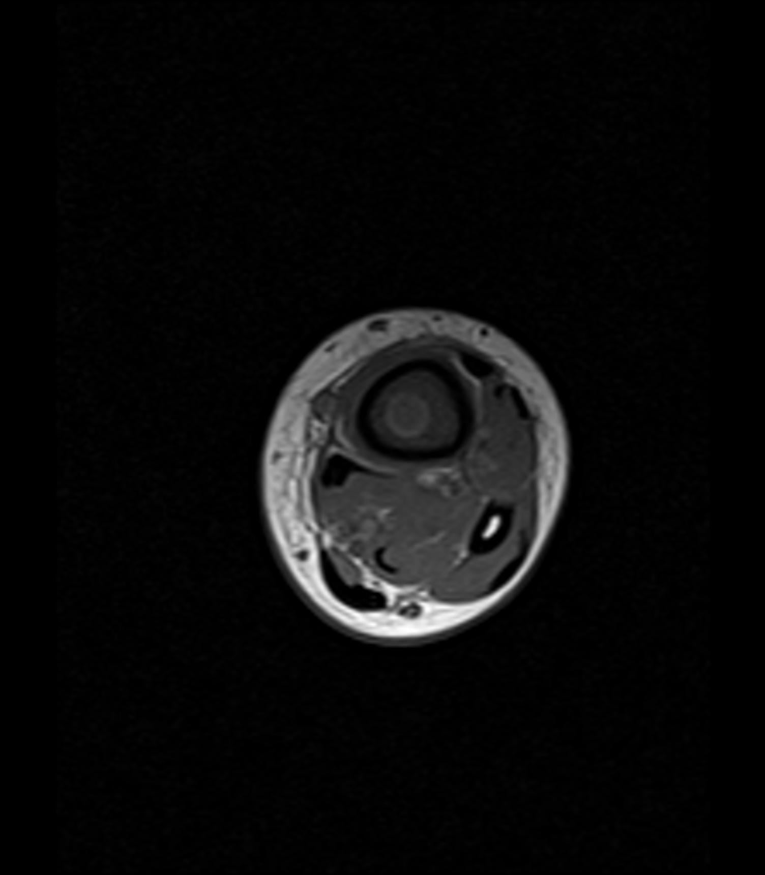
Abscess formation: On contrast-enhanced T1 fat-sat images, an abscess appears as a collection with ring enhancement (wall enhances, centre does not). This distinguishes it from phlegmon, which shows diffuse, homogeneous enhancement. Abscess identification is critical for surgical planning — abscesses typically require drainage, while phlegmon may respond to antibiotics alone.
Penumbra sign: A thin rim of T1 hyperintensity (1-2mm) immediately surrounding an intraosseous abscess (Brodie abscess). This finding is SPECIFIC for infection and helps distinguish a Brodie abscess from a bone tumour. The hyperintense rim represents granulation tissue at the interface between abscess and viable bone.
Cortical erosion and sinus tract: Cortical destruction is best assessed on contrast-enhanced images. A sinus tract appears as a linear enhancing track extending from the infected bone through soft tissues to the skin surface — this is specific for chronic osteomyelitis.
Paediatric considerations: In children under 18 months, the growth plate has transphyseal blood vessels that allow infection to cross from the metaphysis to the epiphysis, potentially causing concurrent osteomyelitis and septic arthritis. After 18 months, the growth plate acts as a barrier to infection spread.
Guidelines, Registries & Global Practice
Musculoskeletal infection is a global problem with rising incidence driven by an ageing population, increasing arthroplasty volumes, diabetes and intravenous drug use. Periprosthetic joint infection complicates roughly 1-2% of primary hip and knee arthroplasties and a higher proportion of revisions, and native vertebral osteomyelitis has an incidence of approximately 2.4 per 100,000 person-years that rises sharply with age. Imaging recommendations are broadly consistent across major societies, with differences mainly in the role of nuclear medicine versus MRI when the diagnosis is uncertain.
- Osteomyelitis & Spinal Infection
- Gadolinium-enhanced MRI is the modality of choice for native vertebral osteomyelitis; gallium/bone scan or FDG-PET/CT when MRI is contraindicated
- PJI / Periprosthetic
- Radiographs in all; arthrocentesis is the cornerstone; advanced nuclear-medicine imaging only when diagnosis remains uncertain
- Osteomyelitis & Spinal Infection
- MRI with contrast first-line for osteomyelitis and discitis; urgent imaging for suspected spinal epidural abscess
- PJI / Periprosthetic
- Aligns with MSIS/EBJIS criteria; aspiration and synovial biomarkers prioritised; nuclear medicine as adjunct
- Osteomyelitis & Spinal Infection
- MRI for soft-tissue and marrow extent; CT for sequestrum and surgical planning
- PJI / Periprosthetic
- EBJIS 2021 definition integrates clinical, synovial, microbiological and histological criteria; imaging supportive
- Osteomyelitis & Spinal Infection
- Not the primary remit (focus on PJI definitions)
- PJI / Periprosthetic
- 2018 ICM scoring: serum and synovial markers, culture and histology define PJI; imaging is a minor/adjunct criterion
- Osteomyelitis & Spinal Infection
- Confirmatory FRI criteria include fistula/sinus, purulent drainage and positive cultures; imaging (radiograph, CT, MRI, WBC-SPECT/CT) is suggestive
- PJI / Periprosthetic
- Hardware infection assessed with WBC scintigraphy/SPECT-CT and FDG-PET where MRI is degraded by metal
Registry and outcome data. National arthroplasty registries (NJR for England/Wales, AJRR in the US, AOANJRR in Australia, the Swedish and Norwegian registries, and the NZJR) consistently identify infection as a leading cause of early revision after hip and knee arthroplasty, underpinning the high prior probability that drives PJI imaging and aspiration pathways. Registry follow-up of revision-for-infection also informs the choice between debridement-and-implant-retention and one- or two-stage revision once infection is confirmed.
High- vs limited-resource practice variation. Where MRI and radiopharmaceuticals are readily available, gadolinium-enhanced MRI is the default for osteomyelitis and spinal infection, with FDG-PET/CT or WBC/marrow imaging reserved for metal-laden or equivocal cases. In limited-resource settings, plain radiographs, ultrasound (for effusion detection and aspiration guidance) and image-guided or clinical aspiration carry a greater diagnostic burden; triple-phase bone scintigraphy may substitute when MRI is unavailable. Across all settings the unifying principle is unchanged: imaging localises and stages infection, but microbiological confirmation by aspiration or biopsy remains decisive.
Controversies & Areas of Uncertainty
FDG-PET is attractive (single visit, no cell labelling) and highly sensitive, but specificity around metalwork is limited because inflammatory periprosthetic uptake overlaps with infection. Combined labelled-leukocyte/marrow imaging remains more specific (Love et al), yet is labour-intensive and operator-dependent. Standardised FDG-PET interpretation criteria are still evolving, so practice varies between centres.
The major PJI definitions (MSIS, 2018 ICM, EBJIS 2021) are built on serum and synovial biomarkers, culture and histology — imaging contributes little to the formal score. This reflects the non-specificity of cross-sectional imaging around implants, and means a normal scan never excludes PJI when clinical and laboratory suspicion is high.
Gadolinium improves detection of epidural and paraspinal abscess and differentiates abscess from phlegmon, but non-contrast MRI already shows the cardinal features (disc and endplate signal change). In renal impairment or gadolinium contraindication, diffusion-weighted imaging and careful non-contrast assessment are used, with FDG-PET/CT as a problem-solving adjunct.
Superimposed osteomyelitis on a Charcot foot remains one of the hardest calls in MSK radiology. Secondary signs (sinus tract, soft-tissue collection, ulcer adjacent to bone) carry the most weight, but no single feature is perfect, and image-guided bone biopsy is often required for a definitive answer before prolonged antibiotics or amputation.

Clinical Decision Scenarios
Practise clinical reasoning and management decisions out loud
“A 5-year-old boy presents with a 3-day history of refusal to walk, fever (39 degrees Celsius), and tenderness over the proximal tibia. His radiograph is normal. Blood tests show CRP 85 and WCC 16,000.”
“A 72-year-old man with a total knee replacement performed 2 years ago presents with persistent knee pain, warmth, and a CRP of 45. The radiograph shows no obvious periprosthetic fracture.”
“An examiner asks you to explain how MRI differentiates osteomyelitis from neuropathic (Charcot) arthropathy in a diabetic foot.”
Imaging Timeline
- Day 0-3: Radiographs NORMAL. MRI shows marrow oedema. Bone scan positive
- Day 7-14: Radiographs show periosteal reaction and rarefaction
- Day 14+: Radiographs show lytic destruction, sequestrum/involucrum
- KEY: Normal radiographs do NOT exclude osteomyelitis
MRI Features (PACES)
- Penumbra sign: T1 hyperintense rim around Brodie abscess (SPECIFIC for infection)
- Abscess: ring enhancement on Gd+. Phlegmon: diffuse enhancement
- Cortical breach: interrupted cortex with surrounding oedema
- Elevated marrow signal on STIR: sensitive but NOT specific
- Sinus tract: linear enhancing tract to skin (SPECIFIC for chronic OM)
PJI Imaging
- Joint aspiration is GOLD STANDARD (WBC count, culture, alpha-defensin)
- Combined WBC + sulphur colloid scan: best nuclear medicine test (sensitivity 91%)
- Triple-phase bone scan: sensitive but NOT specific (positive for 12-18 months post-op)
- FDG-PET: high sensitivity (96%) but limited specificity around metalwork
Osteomyelitis vs Charcot
- OM: geographic marrow oedema, sinus tract, abscess, ulcer-bone proximity
- Charcot: diffuse marrow oedema, joint destruction, NO abscess
- Sinus tract = 100% specific for osteomyelitis
- Both can coexist (superimposed infection on Charcot)
Evidence Base
MRI Accuracy for Foot Osteomyelitis (Meta-Analysis)
- Across 16 studies, the diagnostic odds ratio for MRI in foot osteomyelitis was 42.1 (95% CI 14.8-119.9), with specificity 82.5% at a 90% sensitivity cut point.
- MRI was markedly superior to technetium-99m bone scanning (DOR 149.9 vs 3.6 in head-to-head studies), plain radiography (81.5 vs 3.3), and white-cell studies (120.3 vs 3.4).
- MRI performance allowed the diagnosis of foot osteomyelitis to be both ruled in and ruled out.
Penumbra Sign in Subacute Osteomyelitis
- In 32 patients referred to an orthopaedic oncology service but proven to have osteomyelitis, the penumbra sign (T1 hyperintense rim around an intraosseous abscess) was present in 24 (75%).
- The penumbra sign was more sensitive than the T2/STIR double-line sign, which was seen in only 29% of cases.
- On histology the rim was composed of highly vascularised granulation tissue with thick-walled arterioles.
MRI evidence supports its role as the gold standard for osteomyelitis diagnosis.