Navicular Osteochondrosis | Self-Limiting | Boys 3-7 Years | Excellent Prognosis
- Self-limiting condition with excellent prognosis - no surgery required
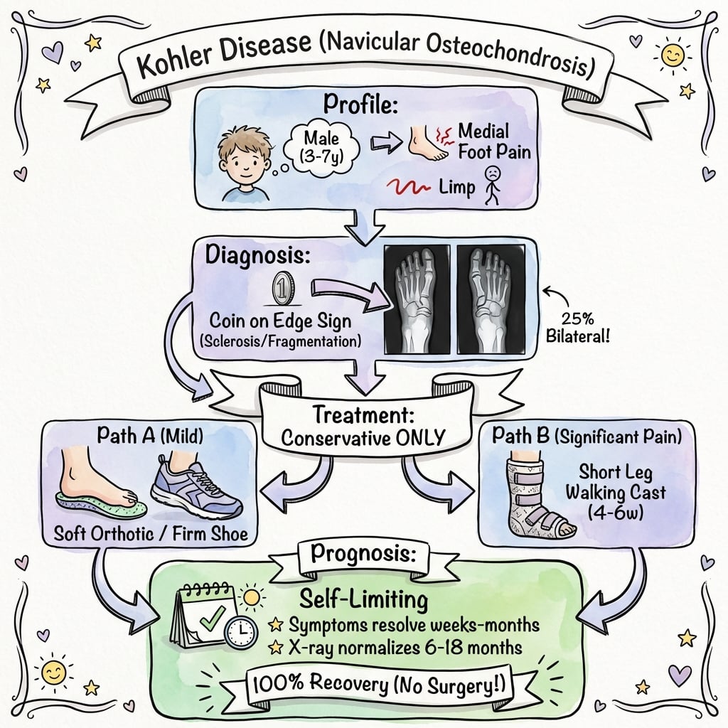
- Boys aged 3-7 years predominantly affected (5:1 male predominance)
- Navicular is last tarsal bone to ossify making it vulnerable to AVN
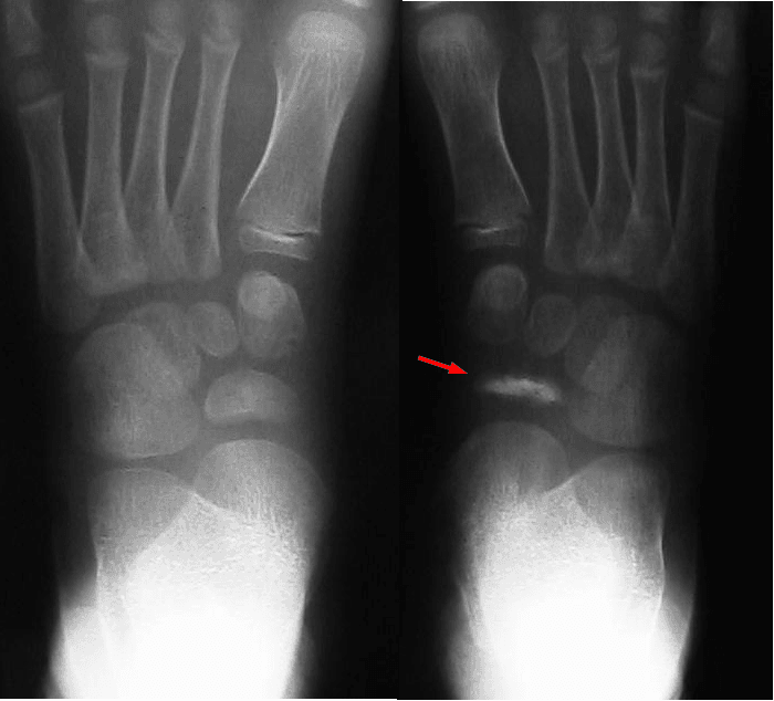
- Coin on edge appearance on lateral radiograph is classic finding
- Complete radiographic reconstitution occurs within 6-18 months
- “Always bilateral X-rays as 25% are bilateral
- “Conservative treatment only - cast or firm-soled shoe
- “Do not confuse with accessory navicular (older children, different location)
- “Natural history is spontaneous resolution regardless of treatment
Boys aged 3-7 years most commonly affected with 5:1 male predominance. The navicular is the last tarsal bone to ossify (ages 2-4 years) making it vulnerable to vascular compromise during this critical period.
Delayed ossification combined with mechanical compression causes avascular necrosis. The navicular receives its blood supply from plantar and dorsal vessels that are vulnerable during the ossification period when demands are high.
Coin on edge appearance - navicular appears sclerotic, flattened, and fragmented on lateral radiograph. AP view shows decreased navicular height with irregular margins. Always image both feet as 25% are bilateral.
Excellent prognosis - symptoms resolve within weeks to months with conservative treatment. Complete radiographic reconstitution within 6-18 months regardless of treatment. No surgery ever indicated.
Overview and Epidemiology
Kohler disease (also known as navicular osteochondrosis or Kohler I disease) is a self-limiting avascular necrosis affecting the tarsal navicular in young children. First described by Alban Kohler in 1908, it represents a temporary disruption of blood supply to the developing navicular bone during a critical period of ossification.
Epidemiology
The condition predominantly affects boys aged 3-7 years with a peak incidence at 4-5 years. The male-to-female ratio is approximately 5:1. The navicular is the last tarsal bone to ossify (typically between ages 2-4 years), which is believed to be a key factor in its susceptibility. Bilateral involvement occurs in approximately 25% of cases, though often asymmetrically. There is no clear racial or ethnic predisposition, and the condition is relatively uncommon compared to other pediatric osteochondroses.
Pathophysiology
The navicular bone occupies a central position in the medial longitudinal arch, making it subject to significant compressive forces during weight-bearing. The etiology involves a combination of factors including delayed ossification, mechanical compression, and vascular insufficiency. During the ossification period (ages 2-4 years), the navicular is particularly vulnerable as its blood supply is tenuous. The developing bone must support the mechanical loads of walking while its vascular supply is establishing. This mismatch between mechanical demand and vascular capacity leads to ischemia, osteonecrosis, and the characteristic radiographic changes. The condition is self-limiting because once ossification completes and vascular supply matures, normal bone architecture is restored.
Clinical Presentation
History
Children present with an insidious onset of limping and medial midfoot pain. Parents typically notice their child has developed a limp over days to weeks. The child may refuse to walk long distances or complain of foot pain with activity. Pain is localized to the medial midfoot and worsens with weight-bearing. Unlike septic arthritis or acute fractures, there is no history of trauma and the child remains systemically well. Some children may walk on the lateral border of their foot to offload the painful navicular.
Examination
Inspection: The child demonstrates an antalgic gait pattern, often favoring the lateral border of the foot. There may be mild swelling over the dorsomedial midfoot, though this is often subtle or absent.
Palpation: Point tenderness is present over the navicular bone on the medial aspect of the midfoot. The navicular is located approximately 2-3 cm distal to the medial malleolus.
Range of Motion: Midfoot motion is typically preserved but may be painful at extremes. Subtalar and ankle motion should be normal.
Gait: Classic antalgic gait with shortened stance phase on the affected side. Some children adopt a supinated foot posture to reduce pressure on the navicular.
Neurovascular: Normal - any neurovascular compromise should prompt consideration of alternative diagnoses.
Imaging and Diagnosis
Radiographic Findings
Standing AP and lateral views of both feet are the primary imaging modality. Always image both feet as 25% of cases are bilateral.
Classic findings include:
- Sclerosis: Increased bone density of the navicular
- Fragmentation: Navicular appears to have multiple fragments
- Flattening: Loss of normal navicular height
- Coin on edge appearance: On lateral view, the navicular appears thin and dense like a coin viewed edge-on
- Irregular margins: Loss of smooth cortical outline
Progression: Over 6-18 months, radiographs show progressive reconstitution with return to normal navicular morphology. The radiographic appearance may lag behind clinical improvement - children often become asymptomatic while radiographic changes persist.
Key Differentials
1. Accessory Navicular (Os Tibiale Externum)
This occurs in older children and adolescents (typically age 10 and above). There is a medial prominence at navicular with pain at posterior tibial tendon insertion. X-ray shows accessory ossicle separate from main navicular. Treatment differs - may require excision if symptomatic.
2. Tarsal Coalition
Calcaneonavicular or talocalcaneal coalition presents with rigid flatfoot and limited subtalar motion. Pain is often in sinus tarsi region. Oblique radiographs or CT confirm diagnosis.
3. Navicular Stress Fracture
This occurs in older children and adolescent athletes with acute onset and specific injury mechanism. Tenderness at navicular is more focal. MRI shows linear fracture line.
4. Infection or Osteomyelitis
Patients have systemic symptoms (fever, malaise) and elevated inflammatory markers. Soft tissue swelling is more prominent. MRI shows bone marrow edema with soft tissue involvement.
The key distinguishing feature of Kohler disease is the characteristic age group (3-7 years), typical radiographic appearance, and self-limiting course.
Management
Non-Operative Management
All cases of Kohler disease are managed conservatively - there is no role for surgery. The goal of treatment is symptom relief while awaiting natural resolution.
Treatment Options include:
1. Short Leg Walking Cast (4-8 weeks): Provides excellent pain relief, allows continued ambulation, may accelerate symptomatic recovery, and is reserved for more symptomatic children.
2. Firm-Soled Shoe or Walking Boot: Reduces stress on midfoot, allows some activity modification, and is appropriate for milder symptoms.
3. Activity Modification and NSAIDs: Avoid high-impact activities, use analgesia for comfort, and is suitable for minimally symptomatic cases.
4. Medial Arch Support: May provide symptomatic relief and is useful during recovery phase.
Expected Timeline: Symptom resolution occurs within weeks to months. Radiographic reconstitution takes 6-18 months. Long-term outcome is complete recovery expected.
Complications and Prognosis
Kohler disease is fundamentally a benign, self-limiting condition, and true complications are rare. The principal risks are iatrogenic and diagnostic rather than from the disease itself.
- Excellent natural history. Long-term series report a normal foot in adulthood in essentially all patients, with complete radiographic reconstitution within 6-18 months irrespective of treatment.
- Persistent or recurrent symptoms. Genuine ongoing pain after reconstitution should prompt a search for a coexisting diagnosis (talocalcaneal coalition, large or symptomatic accessory navicular, or stress injury), as documented in long-term follow-up.
- Iatrogenic harm from overtreatment. Unnecessary surgery, prolonged non-weight-bearing immobilisation, or repeated imaging (including MRI requiring sedation in young children) are avoidable harms driven by misdiagnosis or failure to reassure.
- Misdiagnosis. Mislabelling an incidental irregular ossification variant, an accessory navicular, or adult-type Mueller-Weiss osteonecrosis as Kohler disease leads to inappropriate management.
There is no recognised association with later degenerative arthritis, deformity, or arch collapse attributable to Kohler disease itself.
Kohler I versus Kohler's Second Disease (the Eponym)
The topic calls this 'Kohler I disease' but never explains what the 'I' distinguishes.
- Kohler I is this condition - osteochondrosis of the tarsal navicular (Alban Kohler, 1908).
- Kohler's second disease ('Kohler II') is an older name for osteochondrosis of the second (or third) metatarsal head - the entity now usually called Freiberg infraction (Freiberg, 1914, is the preferred eponym; see the dedicated Freiberg topic).
- The two are almost opposite in demographics: Kohler I affects young boys (~3-7 years) and is benign and self-limiting; Freiberg / Kohler II affects adolescent girls and, unlike Kohler I, can flatten the metatarsal head and require surgery. Keeping the eponyms straight is a classic exam trap.
Q: What is 'Kohler's second disease', and how does it differ from Kohler I?
A: 'Kohler's second disease' (Kohler II) is an older name for osteochondrosis of the 2nd (or 3rd) metatarsal head - now usually called Freiberg infraction (Freiberg, 1914, is the preferred eponym). Kohler I = the tarsal navicular in young boys (~3-7y), benign and self-limiting; Freiberg / Kohler II = the 2nd metatarsal head in adolescent girls and can require surgery. Do not conflate them.
Guidelines, Registries & Global Practice
Global Epidemiology
Kohler disease is an uncommon paediatric osteochondrosis reported worldwide with a consistent demographic profile: boys aged roughly 2-10 years (peak 4-5), male predominance of about 5:1, and bilateral involvement in around a quarter of cases. There is no recognised racial or geographic predilection, and incidence figures mirror each other across published series from North America, Europe and Asia. Because the condition is benign and self-limiting, it is not tracked by any arthroplasty or trauma registry; the evidence base is built from single-centre case series rather than registry or trial data.
Guideline and Society Positions
- Position on Kohler disease
- Clinical and radiographic diagnosis; no operative role; short-leg cast or supportive footwear for symptom control
- Position on Kohler disease
- Conservative management within "limping child" pathways; image to exclude mimics, reassure regarding natural history
- Position on Kohler disease
- Same conservative consensus; MRI reserved for atypical or persistent cases
- Position on Kohler disease
- Self-limiting AVN of the navicular; cast shortens symptoms but does not alter outcome
No named society publishes a dedicated Kohler disease guideline because management is uncontroversial and uniformly non-operative; recommendations are extrapolated from the limping-child and paediatric foot pain literature.
High- vs Limited-Resource Practice
In well-resourced settings, weight-bearing radiographs of both feet confirm the diagnosis and a short-leg walking cast is readily applied; MRI is available for the rare atypical case. In limited-resource settings the diagnosis remains clinical and radiographic, and a firm-soled shoe, activity modification and simple analgesia achieve the same excellent outcome. The key global teaching point is identical everywhere: recognise the entity, avoid unnecessary investigation or surgery, and reassure the family.
Controversies and Areas of Uncertainty
- Aetiology — vascular versus mechanical. Whether Kohler disease is primarily an ischaemic event or a mechanical "stress" phenomenon in a normally late-ossifying navicular remains unsettled; the two likely interact during the ossification window. The monozygotic-twin report (Tsirikos et al, 2003) raises an unconfirmed genetic contribution.
- Disease versus normal variant. Irregular, multicentric navicular ossification is a common asymptomatic radiographic finding. Williams and Cowell (1981) stressed that true (symptomatic) Kohler disease must be distinguished from incidental ossification variants — radiographs alone cannot make the diagnosis without correlating clinical pain and tenderness.
- Does casting change anything? Multiple series agree casting shortens symptom duration but does not alter the radiographic outcome or final function. Cast versus supportive footwear is therefore a comfort decision, not an outcome decision, and there are no randomised trials.
- Role of MRI. MRI is not required for typical cases and risks overdiagnosis or unnecessary anaesthesia in young children; it is reserved for atypical presentations or to exclude fracture, infection or tumour.
- Naming trap. Adult spontaneous navicular osteonecrosis (Mueller-Weiss disease) is radiographically similar but is a distinct, often progressive condition (Haller et al, 1988) and should never be labelled Kohler disease.
At a Glance
Kohler disease is a self-limiting avascular necrosis of the tarsal navicular occurring in children aged 3-7 years with a strong male predominance (5:1). The navicular is the last tarsal bone to ossify, making it vulnerable to vascular insufficiency during this period. Children present with antalgic gait, limping, and medial midfoot pain. Radiographs show the classic coin on edge appearance with sclerosis, fragmentation, and flattening of the navicular. The condition is bilateral in 25% of cases. Treatment is entirely conservative with a short leg walking cast or firm-soled shoe for 4-8 weeks. The prognosis is excellent with complete clinical and radiographic resolution expected within 6-18 months. No surgery is ever required.
- Location
- Tarsal navicular
- Age
- 3-7 years
- Gender
- Male 5:1
- Prognosis
- Excellent - self-limiting
- Location
- 2nd MT head
- Age
- 13-18 years
- Gender
- Female 4:1
- Prognosis
- Variable - may need surgery
- Location
- Femoral head
- Age
- 4-8 years
- Gender
- Male 4:1
- Prognosis
- Variable - depends on age/pillar
- Location
- Tibial tubercle
- Age
- 10-15 years
- Gender
- Male 2:1
- Prognosis
- Excellent - self-limiting
- Location
- Calcaneal apophysis
- Age
- 8-12 years
- Gender
- Male 2:1
- Prognosis
- Excellent - self-limiting
- Location
- Lunate
- Age
- 20-40 years
- Gender
- Male 2:1
- Prognosis
- Variable - may need surgery
KOHLERKohler Disease Key Features
Hook:KOHLER - Kids get it, Ossification delayed, Heals on its own, Lateral X-ray shows coin on edge, Excellent outcome, Rest is the treatment!
DORSAL-PLANTARNavicular Blood Supply
Hook:The navicular has dual blood supply (dorsal and plantar) but during ossification this supply is tenuous - making it vulnerable to AVN during peak mechanical loading in early childhood.
CASTDifferential Diagnosis
Hook:CAST - Consider Coalition, Accessory navicular, Stress fracture, and Trauma when evaluating midfoot pain in children!
Exam Viva Scenarios
Practise clinical reasoning and management decisions out loud
“A 5-year-old boy presents with a 3-week history of limping and medial foot pain. His mother reports he refuses to walk long distances and complains of pain in his right foot. He is otherwise well with no fever or recent illness. On examination, he has an antalgic gait and tenderness over the medial midfoot. What is your approach?”
“A 4-year-old boy presents with bilateral midfoot pain, worse on the right side. Radiographs show sclerosis and fragmentation of both navicular bones with flattening on the right. How do you manage this case and what do you tell the parents?”
“You are asked to see a 12-year-old girl with medial foot pain over the navicular area. She is a dancer and has had symptoms for 6 months. Her GP has told her she has Kohler disease. On examination, there is a bony prominence on the medial aspect of her foot. What are your thoughts?”
Demographics
- Age 3-7 years (peak 4-5)
- Male predominance 5:1
- Bilateral in 25%
- No racial predisposition
Pathophysiology
- Navicular = last tarsal to ossify
- Vulnerable during ossification (ages 2-4)
- Mechanical compression + vascular insufficiency
- Self-limiting once ossification complete
Clinical Features
- Antalgic gait and limp
- Medial midfoot pain
- Tenderness over navicular
- No systemic symptoms
Imaging
- AP and lateral BOTH feet
- Coin on edge appearance
- Sclerosis and fragmentation
- Flattening of navicular
Treatment
- CONSERVATIVE ONLY
- Short leg cast 4-8 weeks
- OR firm-soled shoe
- NO surgery ever indicated
Prognosis
- Symptoms resolve in weeks-months
- Radiographic healing 6-18 months
- 100% complete recovery
- No long-term sequelae
Differential Diagnosis
- Accessory navicular (older, separate ossicle)
- Tarsal coalition (rigid flatfoot)
- Stress fracture (athletes)
- Osteomyelitis (systemic symptoms)
Evidence Base
Effect of Casting on Symptom Duration (Landmark Series)
- 20 patients treated for Kohler disease at a single institution (1948-1974), plus 3 with asymptomatic irregular ossification
- Short-leg cast for 8 weeks reduced mean symptom duration to under 3 months versus 15 months without casting
- All patients eventually showed spontaneous reconstitution of the navicular
- Excellent long-term recovery of function in every case
Very-Long-Term Outcome (Mean 31-Year Follow-Up)
- 14 patients (16 feet) reviewed at a mean of 31.5 years after diagnosis
- Type and length of treatment did not affect the final outcome
- Short-leg cast immobilisation decreased the duration of symptoms
- 12 of 16 feet rated good; the 2 fair feet had coexisting talocalcaneal coalition or a large accessory navicular
Contemporary Review of Kohler Disease
- Presentation typically between ages 2 and 10 years, with boys affected more often than girls
- Radiographs show sclerosis, sometimes flattening and fragmentation of the navicular
- Long-term outcomes favourable regardless of treatment type
- A short period of short-leg walking cast immobilisation may reduce symptom duration