Imaging patients with orthopaedic hardware
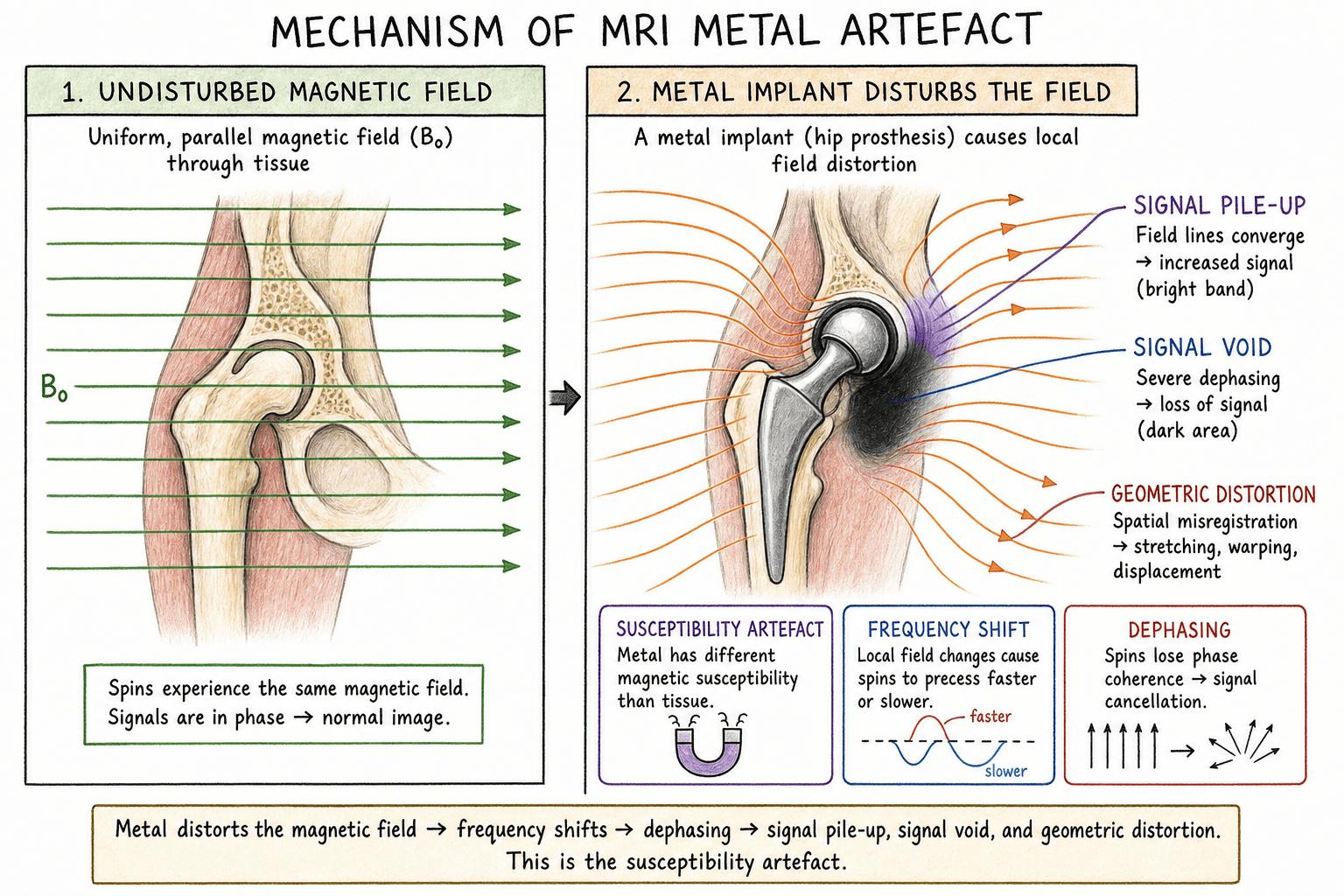
Susceptibility: Different magnetic properties cause local field distortion
Signal void: No signal from metal itself
Geometric distortion: Spatial mismapping of signal
Pile-up artefact: Signal displaced and concentrated
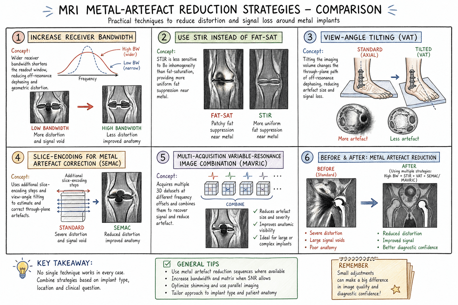
Key: MARS techniques address susceptibility and geometric distortion
- Metal artefact = signal void + geometric distortion
- Titanium causes less artefact than stainless steel or cobalt-chrome
- 1.5T preferred over 3T for metal artefact reduction
- MARS sequences: SEMAC, MAVRIC, VAT (View Angle Tilting)
- Increase bandwidth, use spin echo over gradient echo
- “Susceptibility artefact proportional to field strength
- “Stainless steel: 10x more artefact than titanium
- “Short tau inversion recovery (STIR) better than fat-sat near metal
- “Thinner slices and higher matrix reduce artefact
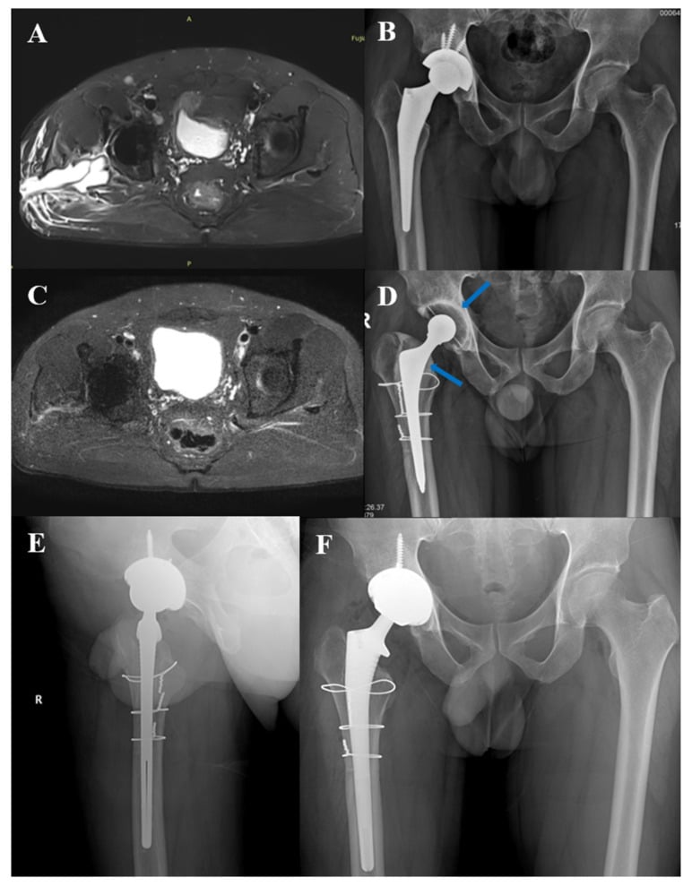
- “ALTR assessment around MoM hips requires MARS MRI
Understanding metal artefact reduction is increasingly important with the rising number of patients with orthopaedic implants. Know which materials cause most artefact (stainless steel worst), why 1.5T is preferred, and the basic MARS techniques. ALTR assessment around metal-on-metal hips is a common application.
Physics of Metal Artefact
- Cause
- No mobile protons in metal
- Appearance
- Black region at metal location
- Cause
- Local field inhomogeneity
- Appearance
- Blooming, signal distortion around metal
- Cause
- Frequency mismapping
- Appearance
- Spatial displacement of anatomy
- Cause
- Signal misregistration
- Appearance
- Bright bands adjacent to void
- Cause
- Off-resonance effects
- Appearance
- Incomplete fat suppression near metal
MARS = Material, Alignment, Resolution, SequenceFactors Affecting Artefact Severity
Hook:Artefact severity is proportional to field strength - use 1.5T over 3T when imaging around metal
Implant Material Properties
- Susceptibility
- Low
- Artefact Severity
- Minimal
- Common Uses
- Plates, screws, stems, spinal implants
- Susceptibility
- Moderate
- Artefact Severity
- Moderate-high
- Common Uses
- Femoral heads, tibial trays, bearing surfaces
- Susceptibility
- High
- Artefact Severity
- Severe
- Common Uses
- Older implants, some screws, wires
- Susceptibility
- Low
- Artefact Severity
- Minimal
- Common Uses
- Trabecular metal, acetabular augments
- Susceptibility
- Very low
- Artefact Severity
- Minimal
- Common Uses
- Ceramic-like bearing surfaces
- Susceptibility
- None
- Artefact Severity
- None
- Common Uses
- Spinal cages, radiolucent
Standard Protocol Optimisation
- Adjustment
- Use 1.5T over 3T
- Effect
- Artefact proportional to B0
- Adjustment
- Increase (wide bandwidth)
- Effect
- Reduces geometric distortion
- Adjustment
- Decrease (thin slices)
- Effect
- Reduces through-plane distortion
- Adjustment
- Increase (high resolution)
- Effect
- Improves spatial resolution
- Adjustment
- Spin echo over gradient echo
- Effect
- Less susceptibility-sensitive
- Adjustment
- STIR over chemical fat-sat
- Effect
- STIR works despite field inhomogeneity
- Adjustment
- Optimise (not too long)
- Effect
- Balance SNR and blurring
BLAST = Bandwidth up, Low field, Avoid gradient echo, Slices thin, STIRProtocol Levers to Cut Metal Artefact
Hook:These simple levers alone often suffice for small titanium implants; reserve full 3D SEMAC/MAVRIC for large cobalt-chrome arthroplasty.
Why Spin Echo, Not Gradient Echo, Near Metal
The protocol levers repeatedly say "spin echo over gradient echo," but the mechanism is exactly what an examiner probes. (General sequence physics is developed in the mri-imaging-principles topic; here is the metal-specific reason.) The dominant problem near an implant is the large static off-resonance (field inhomogeneity) from the metal's susceptibility, and whether the sequence can undo that dephasing decides the artefact.
- Gradient echo (GRE)
- Gradient reversal only - no refocusing pulse
- (Fast) spin echo (FSE / TSE)
- 180-degree refocusing pulse
- Gradient echo (GRE)
- Not refocused - T2*-weighted, dephasing persists
- (Fast) spin echo (FSE / TSE)
- Refocused - spins rephase at the echo, signal recovered
- Gradient echo (GRE)
- Severe blooming, large signal voids
- (Fast) spin echo (FSE / TSE)
- Much reduced
- Gradient echo (GRE)
- Avoid
- (Fast) spin echo (FSE / TSE)
- Workhorse, and the backbone of all 3D MARS sequences
Where the Distortion Goes: Frequency-Encode Direction, Bandwidth and the VAT/SEMAC Split
The physics tables name "geometric distortion: frequency mismapping" and "increase bandwidth reduces geometric distortion," and list VAT and SEMAC separately, but never assemble the directional logic that ties them together - the unifying concept the exam wants.
Metal off-resonance mismaps signal along the frequency-encode (readout) direction in-plane and along the slice-select direction through-plane, because both of those axes use frequency to localise signal; the phase-encode direction is largely spared. Three consequences follow:
- Receiver bandwidth. A higher receiver bandwidth spreads more hertz across each pixel, so a given off-resonance shift displaces signal by fewer pixels - less in-plane distortion and a smaller void. The price is signal-to-noise, which falls roughly with the square root of bandwidth; this is the trade-off behind "increase bandwidth when SNR allows."
- Steer the artefact. Because the in-plane displacement runs along the frequency-encode axis, swapping the frequency- and phase-encode directions moves the pile-up and void off the structure you care about.
- This is why VAT and SEMAC are different tools. View-angle tilting corrects the in-plane (readout-direction) distortion with an extra readout gradient; SEMAC adds slice-direction (z) phase encoding to correct the through-plane distortion; combining them (and MAVRIC's multi-frequency acquisition) covers both directions - the reason these are the definitive techniques for large cobalt-chrome arthroplasty.
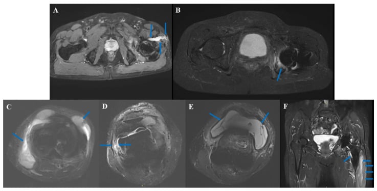
Differential Diagnosis of Periprosthetic Masses on MARS MRI
- Typical Patient
- MoM hip, raised Co/Cr ions
- MARS MRI Features
- Peri-articular cystic or solid mass, variable wall thickness, synovitis
- Discriminators
- Communicates with joint, abductor/tendon damage, ion levels elevated
- Typical Patient
- Pain, raised CRP/ESR, sinus
- MARS MRI Features
- Lamellated synovitis, fluid collections, reactive marrow oedema, sinus tract
- Discriminators
- Systemic inflammatory markers, positive aspirate; ions normal
- Typical Patient
- Older non-MoM bearing
- MARS MRI Features
- Expansile low-signal synovial masses, focal osteolysis around implant
- Discriminators
- Bearing is metal-on-poly/ceramic; ions normal
- Typical Patient
- Recent surgery or anticoagulation
- MARS MRI Features
- Well-defined collection, blood-degradation signal, no enhancing solid component
- Discriminators
- Temporal relation to surgery, resolves over time
- Typical Patient
- Lateral hip pain
- MARS MRI Features
- Fluid in greater trochanteric bursa, no intra-articular mass
- Discriminators
- Confined to bursa, no abductor destruction
- Typical Patient
- No bearing-related cause, growing mass
- MARS MRI Features
- Heterogeneous enhancing mass, may not respect joint planes
- Discriminators
- Independent of implant, biopsy required if atypical
Guidelines, Registries & Global Practice
- Region
- UK
- Position on Imaging
- Risk-stratified follow-up of MoM hips; cross-sectional imaging (MARS MRI or ultrasound) indicated for symptomatic patients or rising/raised metal ions
- Region
- US
- Position on Imaging
- Recommends clinical follow-up and considers cross-sectional imaging (MRI/US/CT) in symptomatic patients or with abnormal ion levels
- Region
- US
- Position on Imaging
- Supports a combined algorithm of symptoms, examination, radiographs, serum metal ions and MARS MRI for evaluating the painful MoM hip
- Region
- UK
- Position on Imaging
- Endorses surveillance pathways using metal ions plus MARS MRI or ultrasound for soft-tissue/pseudotumour assessment
- Region
- Europe
- Position on Imaging
- Aligns with stratified surveillance; cross-sectional imaging for symptomatic or high-risk implants
Advanced MARS Techniques
- Mechanism
- In-plane distortion correction
- Scan Time
- Moderate increase
- Availability
- Widely available
- Mechanism
- Through-plane encoding
- Scan Time
- Significant increase
- Availability
- GE, Siemens, Philips
- Mechanism
- Multi-frequency acquisition
- Scan Time
- Significant increase
- Availability
- GE
- Mechanism
- Combined SEMAC + MAVRIC
- Scan Time
- Long
- Availability
- GE
- Mechanism
- VAT + optimisation
- Scan Time
- Moderate increase
- Availability
- Siemens
Protocol Selection
- Field Strength
- 1.5T
- Key Sequences
- PD fat-sat, STIR, T1
- MARS Technique
- MARS (MAVRIC, SEMAC, WARP)
- Field Strength
- 1.5T
- Key Sequences
- PD, STIR
- MARS Technique
- VAT or MARS
- Field Strength
- 1.5T
- Key Sequences
- T1, T2, STIR sagittal/axial
- MARS Technique
- VAT, MARS if available
- Field Strength
- 1.5T
- Key Sequences
- STIR (oedema), T1
- MARS Technique
- Standard optimisation often sufficient
- Field Strength
- 1.5T
- Key Sequences
- PD fat-sat, STIR
- MARS Technique
- MARS if available
Controversies and Areas of Uncertainty
Clinical Imaging Applications
- MRI Appearance
- Cystic or solid mass adjacent to hip
- Significance
- May compress neurovascular structures
- MRI Appearance
- T2 bright, may have debris
- Significance
- Periarticular, trochanteric bursa
- MRI Appearance
- Oedema (T2 high) or atrophy (T1 fat)
- Significance
- Abductors commonly affected
- MRI Appearance
- Discontinuity, retraction
- Significance
- May affect surgical approach
- MRI Appearance
- Low signal debris, synovial thickening
- Significance
- Metal particle deposition
Clinical Decision Scenarios
Practise clinical reasoning and management decisions out loud
“A patient with a painful metal-on-metal hip replacement is referred for MRI. Blood cobalt level is 12 ppb (elevated). You are asked about optimal imaging.”
“A patient 2 years post lumbar fusion with persistent leg pain is referred for MRI. The spine surgeon wants to assess for recurrent disc herniation.”
“You are asked to explain why MRI around metal implants is challenging. An orthopaedic trainee asks what material causes the least artefact.”
Material Artefact Severity
- Titanium: Least artefact
- Tantalum/Oxinium: Low artefact
- Cobalt-chrome: Moderate-high
- Stainless steel: Severe (10x titanium)
Protocol Optimisation
- 1.5T over 3T (artefact proportional to B0)
- Spin echo over gradient echo
- Increase receiver bandwidth
- STIR not chemical fat-sat
- Thin slices, high matrix
MARS Techniques
- VAT: In-plane correction
- SEMAC: Through-plane encoding
- MAVRIC: Multi-frequency acquisition
- WARP: Siemens combined technique
Clinical Applications
- MoM hip ALTR surveillance
- Post-fusion spine assessment
- Periprosthetic soft tissue
- PJI soft tissue extent
Evidence Base
SEMAC — Original Slice-Encoding Technique
- SEMAC extends a view-angle-tilting (VAT) spin-echo sequence with additional z-phase encoding, resolving distorted excitation profiles that cause through-plane distortion.
- VAT suppresses in-plane distortion while z-phase encoding corrects through-plane distortion, so spins are repositioned to their true spatial locations.
- The method requires no additional hardware and was validated in phantom and in vivo spine and knee studies with feasible scan times.
MAVRIC vs Conventional FSE After Arthroplasty
- In 122 patients (74 hip, 27 shoulder, 21 knee arthroplasties), MAVRIC showed significantly better visualisation of synovium and periprosthetic bone than metal-artefact-reduction FSE at all three joints.
- Synovitis and periprosthetic osteolysis were detected only on MAVRIC images in a substantial proportion of subjects.
- Supraspinatus tendon tears in 44% of relevant subjects were seen only on MAVRIC and not on FSE.
MAVRIC-SL at 3T in Hip Arthroplasty
- In 21 hips, MAVRIC-SL reduced measured artefact area versus 2D FSE by 59.9% at the level of the hip and 31.3% at the femur (both significant).
- Joint capsule and obturator externus/iliopsoas attachment sites were better depicted, and abnormal findings were significantly better shown with MAVRIC-SL.
- MAVRIC-SL increased diagnostic confidence even at 3T, a field strength normally avoided around large metal implants.