Musculocutaneous Nerve Anatomy
Musculocutaneous palsy weakens the principal elbow flexors (biceps, brachialis) and supination (biceps), with sensory loss over the lateral forearm (lateral cutaneous nerve of forearm).
Brachioradialis is a strong elbow flexor supplied by the radial nerve, so some elbow flexion persists - patients are weak, not flexion-less. Testing flexion with the forearm pronated/neutral emphasises brachioradialis and can mask the deficit.
Overview
The musculocutaneous nerve is the smallest-named but conceptually tidy nerve of the arm: it does one compartment, three muscles, and one skin patch. Of the five terminal branches of the brachial plexus it is the one dedicated to elbow flexion and supination, which is why it dominates the nerve-transfer literature — restoring elbow flexion is the single highest priority after an upper brachial plexus injury, and the biceps branch of this nerve is the usual target. For exams it is worth holding three frames in mind at once: the anatomy (lateral cord → coracobrachialis → BBC → lateral forearm skin), the deficit (weak-but-not-absent flexion because brachioradialis compensates), and the surgery (a coracoid safe zone proximally, the LACN at the elbow, and the Oberlin transfer).
Origin & Course
Origin
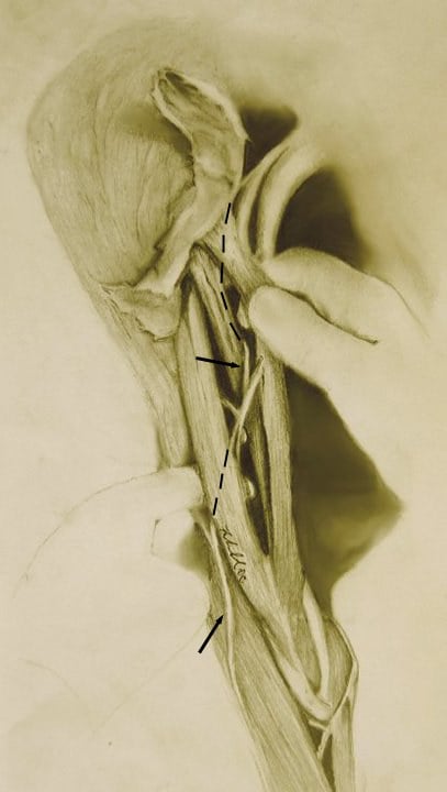
- The musculocutaneous nerve is the terminal continuation of the lateral cord of the brachial plexus, carrying fibres from the anterior rami of C5, C6 and (variably) C7.
- It arises in the axilla, lateral to the axillary artery.
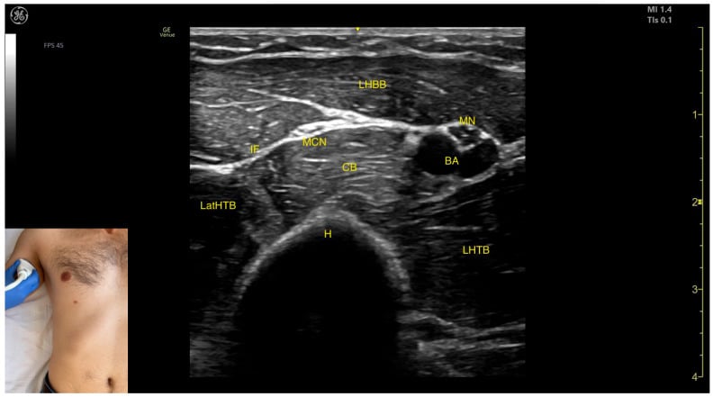
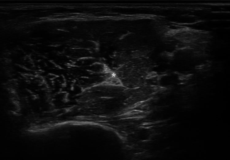
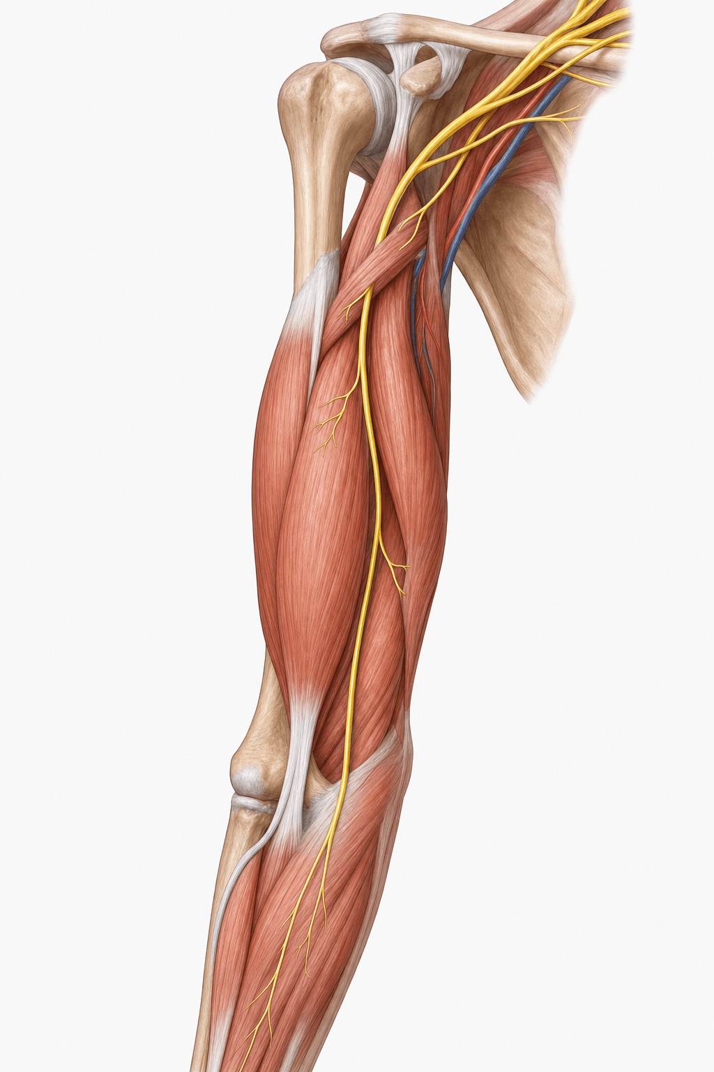
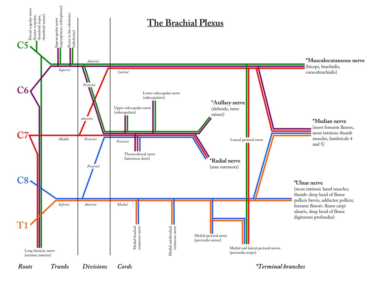
Innervation: Motor & Sensory Supply
The musculocutaneous nerve supplies the three muscles of the anterior (flexor) compartment of the arm: Biceps brachii, Brachialis, and Coracobrachialis.
- Coracobrachialis - flexes and adducts the arm at the shoulder.
- Biceps brachii - the principal supinator of the forearm and a strong elbow flexor (most powerful with the forearm supinated).
- Brachialis - the workhorse elbow flexor, active in all forearm positions (note: brachialis also receives a small radial-nerve contribution).
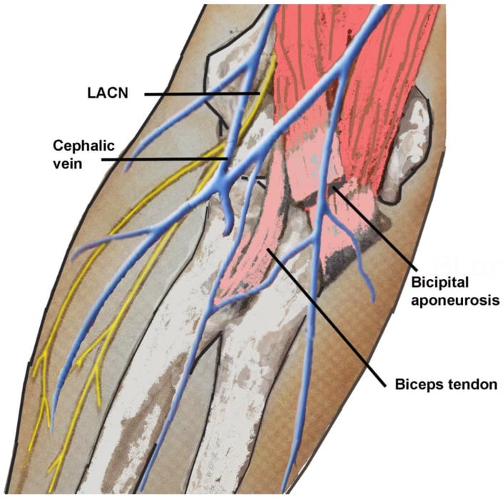
Sensory: the terminal lateral cutaneous nerve of the forearm supplies skin over the lateral (radial) aspect of the forearm.
Surgical Anatomy: Safe Zones, the LACN and Nerve Transfers
Three surgical themes turn this small nerve into a recurring exam topic.
The coracoid "safe zone" in anterior shoulder surgery
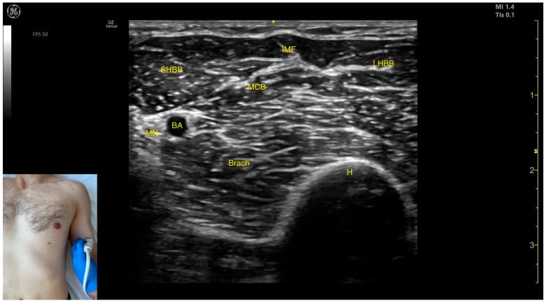
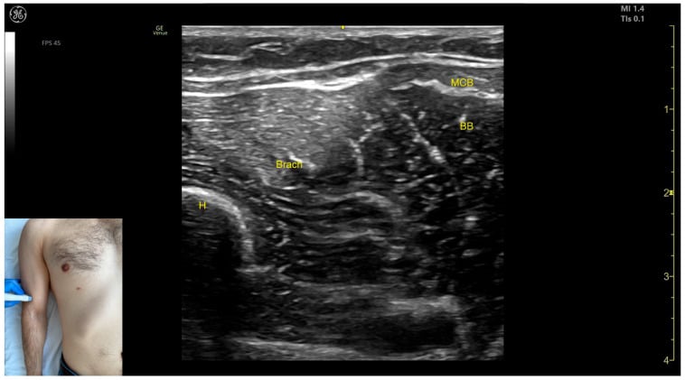
The musculocutaneous nerve enters the deep surface of coracobrachialis a variable distance below the coracoid — on average around 5 cm, but as little as 1.5–3 cm in some people. During the deltopectoral approach, Latarjet/coracoid transfer and anterior shoulder stabilisation, keeping dissection and retractors close to the coracoid and within the proximal few centimetres of conjoint tendon, and avoiding forceful medial retraction of coracobrachialis, protects the nerve. Because the entry point is variable, the "safe zone" is a guide, not a guarantee — the nerve can be tethered and injured by vigorous medial retraction even proximally.
The lateral cutaneous nerve of the forearm in distal biceps repair
At the elbow the terminal LACN emerges lateral to the biceps tendon, deep to the biceps aponeurosis — directly in the field of an anterior (single-incision) distal biceps tendon repair, where it is the most commonly injured nerve, producing lateral-forearm numbness or a painful neuroma. Careful retraction (and the option of a two-incision technique) reduces the risk. The LACN can also be entrapped where it pierces the deep fascia lateral to the biceps tendon, causing exercise-related lateral forearm pain in weightlifters.
Nerve transfers for elbow flexion
After an upper brachial plexus injury (C5–C6, Erb's pattern) with preserved hand function, restoring elbow flexion is the priority. The Oberlin transfer coapts a redundant ulnar nerve fascicle to the biceps branch of the musculocutaneous nerve; the double-fascicular (Mackinnon) transfer adds a median fascicle to the brachialis branch. Outcomes are good (most regain useful, often M4, elbow flexion), but success depends on accurate identification of the biceps and brachialis branches — and branching variation is common, so the branches are confirmed with a nerve stimulator intra-operatively.
Proximal coracoid · Mid arm · Distal biceps tendonWhere the musculocutaneous nerve is at surgical risk
Hook:Three danger points down the arm: the coracoid safe zone proximally, the muscle interval mid-arm, and the LACN at the distal biceps tendon.
A classic regional-anaesthesia trap that follows directly from the anatomy: in an axillary brachial plexus block the musculocutaneous nerve is frequently NOT anaesthetised, because by the level of the axilla it has usually already left the neurovascular sheath to pierce coracobrachialis — so local anaesthetic deposited around the axillary artery never reaches it. The result is preserved elbow flexion and supination and intact sensation over the lateral forearm (its LACN territory) despite an otherwise working block — a problem for forearm/elbow surgery and forearm tourniquet analgesia. The fix is to block it separately, depositing local anaesthetic into the body of coracobrachialis, or to use a more proximal supraclavicular/interscalene approach that catches it before it diverges. The exam point: if an axillary block leaves the lateral forearm sensate and elbow flexion strong, the musculocutaneous nerve has been missed — top it up in coracobrachialis.
BBCMusculocutaneous Nerve
Hook:The musculocutaneous nerve runs the 'BBC' of the anterior arm.
Clinical Correlations
Patterns & Causes
- Isolated injury is rare. When it occurs, consider anterior shoulder dislocation, anterior shoulder surgery (e.g. deltopectoral/coracoid procedures), distal biceps repair, deep arm lacerations, or strenuous/repetitive activity.
- More often the nerve is involved as part of an upper trunk / lateral cord brachial plexus injury.
- Deficit: weak elbow flexion and supination, with lateral forearm sensory loss; brachioradialis (radial nerve) preserves some flexion.
The biceps tendon reflex is the musculocutaneous nerve's reflex (C5/C6). Both its afferent and efferent limbs run in the musculocutaneous nerve, so a diminished or absent biceps jerk is a useful localizing sign of a musculocutaneous, lateral-cord or upper-trunk / C5-6 lesion (and of a C5/C6 root problem). Pair it with the brachioradialis (supinator) reflex (radial nerve, also C5/C6): a C5-6 root or upper-trunk lesion depresses both, whereas an isolated musculocutaneous lesion depresses the biceps jerk while the brachioradialis jerk is preserved — exactly mirroring the way brachioradialis preserves some elbow flexion. (Separately, an "inverted supinator reflex," where tapping brachioradialis produces finger flexion instead, points to a C5-6 cord/myelopathy level, not a peripheral musculocutaneous lesion.) The exam point: an absent biceps jerk with a normal brachioradialis jerk fits an isolated musculocutaneous nerve lesion.
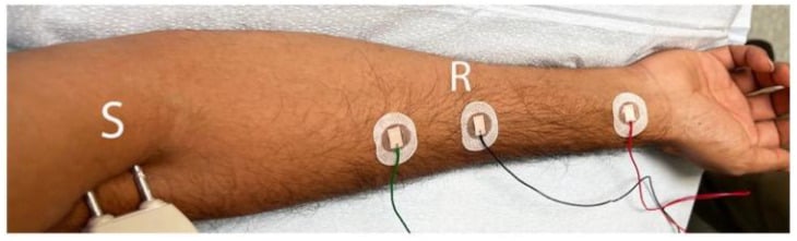
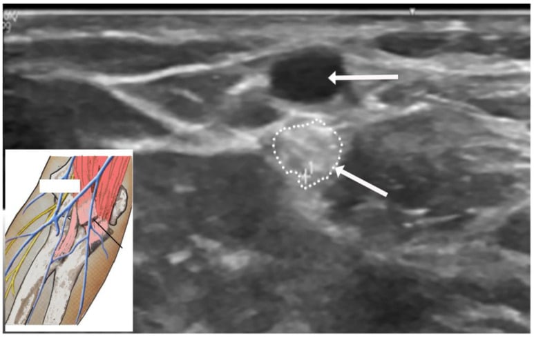
Guidelines, Registries & Global Practice
Global Practice Picture
Musculocutaneous nerve anatomy is foundational knowledge rather than a registry topic. Its clinical relevance is consistent worldwide: protect it during anterior shoulder and coracoid surgery, recognise its deficit pattern, and exploit its biceps branch as the recipient in elbow-flexion nerve transfers, where detailed branching knowledge and awareness of anatomic variation are essential.
Side-by-Side Synthesis
- Detail
- Lateral cord (C5, C6, C7)
- Detail
- Pierces coracobrachialis
- Detail
- Coracobrachialis, biceps brachii, brachialis (BBC)
- Detail
- Lateral cutaneous nerve of forearm (lateral forearm)
- Detail
- Non-piercing of coracobrachialis; median-nerve communications
- Detail
- At risk in anterior shoulder/coracoid + distal biceps surgery; Oberlin transfer recipient
Viva Scenarios
Practise clinical reasoning and management decisions out loud
“After an open anterior shoulder stabilisation, a patient has weak elbow flexion and numbness over the lateral forearm. What has happened and how do you assess it?”
“A 25-year-old has a C5-C6 (upper trunk) brachial plexus injury after a motorcycle crash, with no elbow flexion but normal hand function. Nine months on there is no recovery. How would you restore elbow flexion, and what anatomy must you respect?”
Anatomy
- Terminal branch of lateral cord (C5-C7)
- Pierces coracobrachialis
- Runs between biceps and brachialis
- Ends as lateral cutaneous nerve of forearm
Supply
- Motor: biceps, brachialis, coracobrachialis (BBC)
- Biceps = principal supinator + flexor
- Sensory: lateral forearm
- Brachioradialis (radial) compensates flexion
Clinical
- Deficit: weak flexion/supination + lateral forearm numbness (brachioradialis compensates)
- Coracoid safe zone: nerve enters coracobrachialis ~5 cm (min 1.5–3 cm) below the coracoid
- LACN = most commonly injured nerve in anterior distal biceps repair
- Erb's palsy (C5–6) involves it; Oberlin/double-fascicular transfer to biceps ± brachialis branch
- Variations common (non-piercing, median communication) — stimulate to confirm in transfers
Evidence Base
Both studies below were checked against their PubMed records and are cadaveric/clinical anatomy series, not outcome trials. The Bhardwaj clinical series is the one to quote: in 150 nerve-transfer patients the nerve pierced coracobrachialis in 92 percent and showed "classical" anatomy in 89 percent, with a single biceps branch in 77 percent — the practical message being that variation is common enough to demand intra-operative nerve-stimulator confirmation. The Erturk study is a FETAL series (note: not adult) included only to show how early these variations are established; adult non-piercing rates are lower (~8 percent). For a viva, the reproducible relations and the named surgical points (coracoid safe zone, LACN in distal biceps repair, Oberlin transfer) matter more than the exact percentages.
Anatomic Variations of the Musculocutaneous Nerve and Clinical Implications for Restoration of Elbow Flexion
- Prospective study of 150 patients undergoing nerve transfer for elbow flexion after brachial plexus injury
- The musculocutaneous nerve pierced coracobrachialis in 92% and showed 'classical' anatomy in 89.3%
- 76.6% had a single primary branch to biceps; 16.6% had a discrete branch to each biceps head; brachialis branch usually arose distal to the biceps branch
- Notable unreported variations were documented - vigilance is needed during transfer dissection
Anatomical Variations of the Musculocutaneous Nerve in the Human Fetus
- Fetal cadaveric dissection of 102 upper limbs from 51 human fetuses (17-40 weeks)
- In 13.7% the nerve did NOT pierce coracobrachialis
- The nerve gave 1 to 3 motor branches with variable terminal fringes; branch position scaled with arm length
- No significant side or sex differences