Brachial Plexus Anatomy
Overview
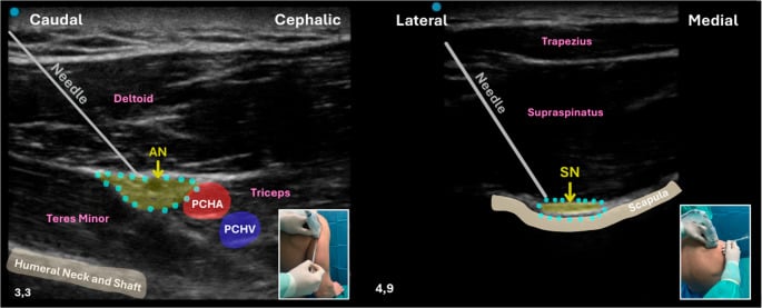
This is the map; the injuries are read off it. The adult traction injury and its reconstruction are on adult brachial plexus injury and the obstetric one on brachial plexus birth palsy - the same C5-6 upper trunk, a different mechanism and a different natural history. Compression rather than traction, at the same anatomical crossroads, is thoracic outlet syndrome. And the individual branches this map generates have their own pages where the lesion is isolated rather than plexal: the axillary nerve off the posterior cord and the suprascapular nerve off the upper trunk.
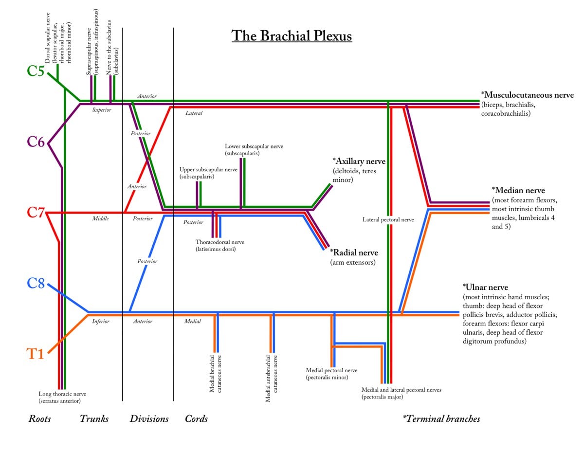
The brachial plexus is the single most-drawn structure in upper-limb vivas, so the goal is to make it automatic: a fixed five-level scaffold onto which you hang the few named branches that actually come off the roots and upper trunk, the posterior-cord ULTRA group, and the five terminal nerves. Once the map is secure, the clinical layer follows naturally — where a lesion sits (supra- versus infraclavicular), which pattern it produces (Erb's, Klumpke's, or whole-plexus), and the one question that changes the operation: is it preganglionic (avulsion) or postganglionic (rupture)? Build the map first, then read every plexus injury off it.
The Five Levels
Roots. The roots are the anterior (ventral) rami of C5-T1, emerging between scalenus anterior and scalenus medius. A prefixed plexus takes a contribution from C4; a postfixed plexus takes one from T2.
Trunks. The roots combine into three trunks, which lie in the posterior triangle of the neck - the supraclavicular fossa, and the reason root and trunk lesions are called supraclavicular. The upper trunk is C5 and C6, the middle trunk is C7 alone, and the lower trunk is C8 and T1.
Divisions. Each trunk divides into an anterior and a posterior division, six in all, and they lie behind the clavicle. No division has a named branch, so the clavicle marks a gap in the branch list.
Cords. The divisions regroup into three cords in the axilla, named by where each lies in relation to the axillary artery. The lateral cord is the anterior divisions of the upper and middle trunks; the posterior cord is the posterior divisions of all three trunks; the medial cord is the anterior division of the lower trunk alone, so its fibres are C8 and T1 only.
Branches. Five terminal nerves leave the cords: musculocutaneous, axillary, radial, median and ulnar.
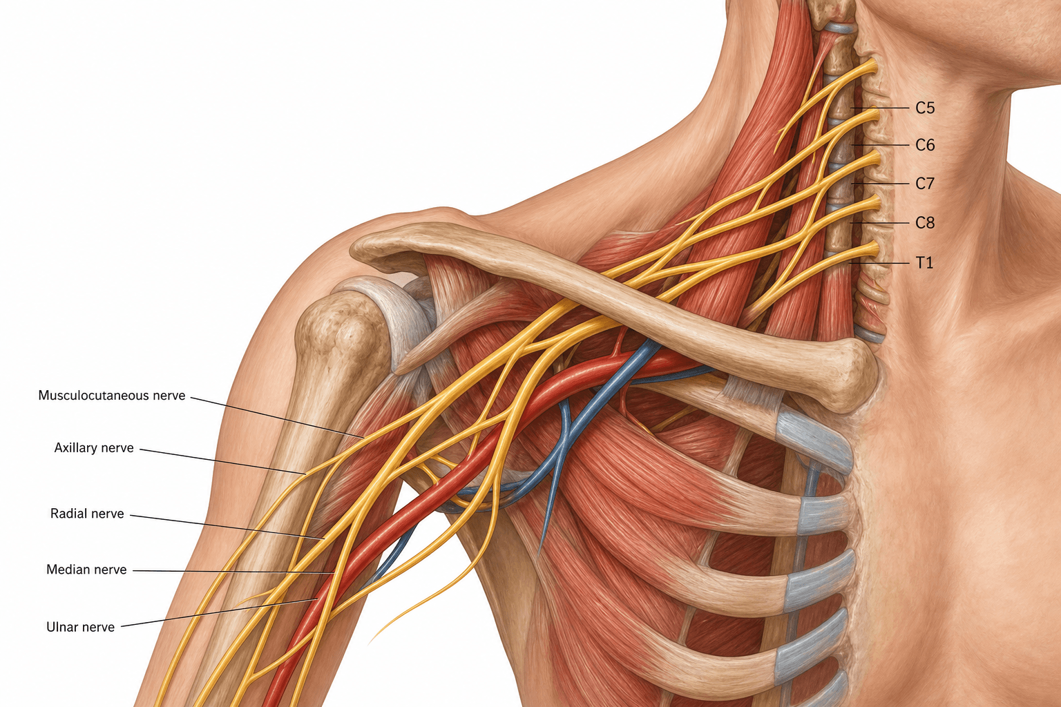
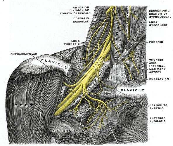
RTDCB — Randy Travis Drinks Cold BeerThe five levels, in order
Hook:Draw the five columns in this order before you draw a single nerve, and the branches have somewhere to hang.
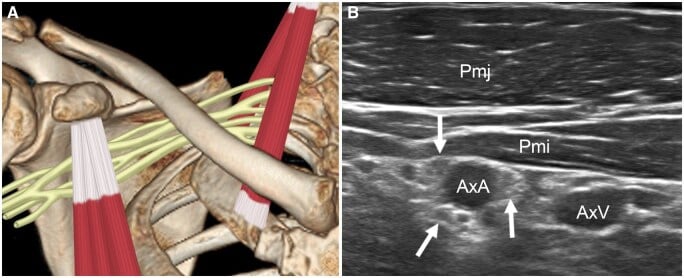
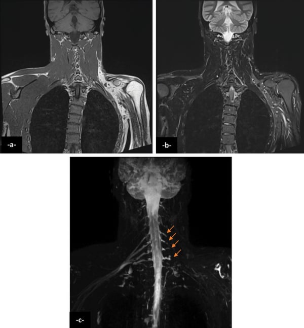
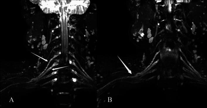
Seeing it on a scan. The three imaging levels below are the same three columns of the drawing: roots in the neck, trunks in the interscalene groove, cords around the axillary artery. Recognise each by its fixed landmarks rather than by trying to name a nerve on its own.
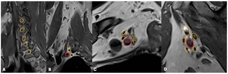
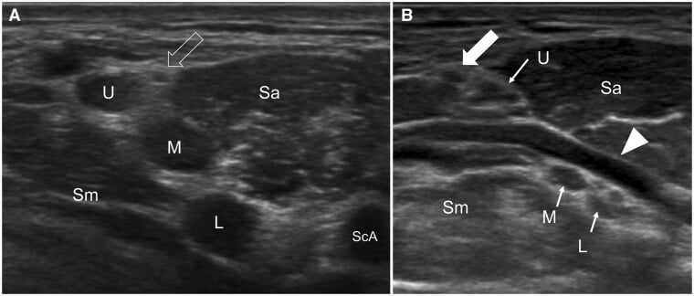
Nerve Branches at Each Level
A reliable viva answer names the branches by level, working top to bottom, and stops at each level before moving on.
- Roots: dorsal scapular nerve (C5 - rhomboids, levator scapulae); long thoracic nerve (C5-C7 - serratus anterior)
- Upper trunk: suprascapular nerve (supraspinatus, infraspinatus); nerve to subclavius
- Divisions: nothing named
- Lateral cord: lateral pectoral nerve; musculocutaneous nerve; lateral root of the median nerve
- Posterior cord: upper subscapular, lower subscapular, thoracodorsal, radial and axillary - the ULTRA group
- Medial cord: medial pectoral nerve; medial cutaneous nerve of the arm; medial cutaneous nerve of the forearm; ulnar nerve; medial root of the median nerve
The median nerve is the one branch built from two cords, a lateral root from the lateral cord and a medial root from the medial cord. Every other terminal nerve comes from a single cord, which is why the median is drawn last and from both sides.
ULTRAPosterior cord branches
Hook:All the posterior divisions feed the posterior cord, so its branches are 'ULTRA' — and they run the EXTENSORS plus the subscapularis/lat/teres group.
Clinical Correlations
Where the lesion sits. Supraclavicular injuries, at root or trunk level, are the most common, and they produce the two classic patterns below. Infraclavicular injuries, at cord or branch level, follow shoulder dislocation, humeral fracture or penetrating trauma, and tend to affect specific terminal nerves rather than a whole level.
Erb's palsy (C5-C6). An upper trunk injury from traction with the head pulled away from the shoulder - birth and motorcycle are the two mechanisms to quote - and the commonest obstetric and traction pattern. The abductors, external rotators and elbow flexors are lost, so the arm sits adducted and internally rotated with the elbow extended and the forearm pronated: the waiter's tip posture.
Klumpke's palsy (C8-T1). A lower trunk injury from traction with the arm pulled overhead. The intrinsic hand muscles go, giving an intrinsic-minus claw hand, and Horner's syndrome (ptosis, miosis, anhidrosis) is often associated, from T1 sympathetic involvement.
Burners and stingers. Transient upper-trunk traction injuries in contact sport - the Erb's level again, at the transient end of the same spectrum.
Localisation, Timing and Reconstruction
The anatomy is only useful if it localises the lesion and times the operation. Treatment is then individualised to the pattern - neurolysis, nerve grafting, nerve transfers (neurotisation), and tendon or free-muscle transfer or arthrodesis for late salvage - and it is the detailed plexus and terminal-branch anatomy that makes a transfer plannable, such as restoring elbow flexion through the musculocutaneous nerve.
Preganglionic (avulsion) versus postganglionic (rupture)
- Preganglionic (root avulsion)
- Proximal to the dorsal root ganglion (rootlets torn from cord)
- Postganglionic (rupture/lesion distal to DRG)
- Distal to the dorsal root ganglion
- Preganglionic (root avulsion)
- Often present (T1 sympathetic) — a red flag for lower-root avulsion
- Postganglionic (rupture/lesion distal to DRG)
- Absent
- Preganglionic (root avulsion)
- Suggests very proximal (root-level) injury of long thoracic/dorsal scapular
- Postganglionic (rupture/lesion distal to DRG)
- Spared
- Preganglionic (root avulsion)
- PRESERVED despite clinical anaesthesia (DRG and peripheral axon intact)
- Postganglionic (rupture/lesion distal to DRG)
- Absent (axon interrupted distal to DRG)
- Preganglionic (root avulsion)
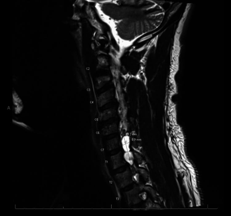
- Pseudomeningocele at the affected level
- Postganglionic (rupture/lesion distal to DRG)
- Often normal or shows a discrete rupture/neuroma
- Preganglionic (root avulsion)
- NOT directly repairable → nerve transfers (neurotisation)
- Postganglionic (rupture/lesion distal to DRG)
- Amenable to nerve grafting (or transfer)
The paradox to quote. In a preganglionic lesion the limb is clinically anaesthetic yet the SNAPs are preserved, because the sensory cell body in the dorsal root ganglion and its peripheral axon remain intact even though the central connection is torn.
Timing and nerve transfers
Timing. A sharp or penetrating laceration is explored early for primary repair. A closed traction injury that shows no clinical or electrical recovery by about 3 months is explored, and ideally operated on before 6-9 months, while the target muscle can still be reinnervated.
The transfers to know. Donor to target, each restoring one function:
- Spinal accessory to suprascapular - shoulder external rotation and abduction
- Oberlin transfer (an ulnar fascicle to the biceps branch of the musculocutaneous) and a median fascicle to the brachialis branch - elbow flexion
- Triceps branch of the radial to the axillary - deltoid
- Late salvage: tendon or free-functioning-muscle transfers, or arthrodesis
The lower trunk elsewhere. C8-T1 is also the target of thoracic outlet syndrome and of a Pancoast (superior sulcus) tumour. Both can present with intrinsic hand wasting, and the Pancoast tumour with Horner's syndrome as well - the same T1 sympathetic sign that flags a lower-root avulsion.
The intraplexal transfers above need a healthy donor root or nerve. In a pan-plexus injury with multiple root avulsions there may be none, so the examiner expects the extraplexal donors:
- Intercostal nerves (typically three, T3-T6) - to the musculocutaneous nerve for elbow flexion, or to a free muscle
- Phrenic nerve - a powerful donor, at the cost of diaphragmatic and respiratory function, so used cautiously and avoided in children and in respiratory compromise
- Contralateral C7 - for total avulsion, routed across (often through a vascularised ulnar nerve graft) to the median or musculocutaneous nerve
- Spinal accessory - the workhorse extraplexal donor, to the suprascapular nerve
When you cannot restore everything, the priority order is elbow flexion first, then shoulder stability with abduction and external rotation, then protective hand sensation and wrist and finger function. Hand function sits last because intrinsic recovery is rarely achieved in the adult, and is salvaged by transfers and fusions.
Three timing and testing points sit alongside the SNAP paradox and the pseudomeningocele.
Delay EMG and nerve conduction studies to about 3-4 weeks. Wallerian degeneration has to occur before denervation changes - fibrillations and positive sharp waves - appear, so a study done too early is falsely reassuring. SNAPs are also most reliable after this interval, and are then repeated serially to track reinnervation by the appearance of nascent motor units.
Intraoperative nerve action potential (NAP) recording across a neuroma-in-continuity decides the operation on the table: a present NAP means axons have crossed, and neurolysis alone is enough; an absent NAP means no useful regeneration, and the segment is resected and grafted.
The histamine triple-response (axon-flare) test is the bedside version of the SNAP paradox. Intradermal histamine normally produces a wheal and a flare, and the flare is an axon reflex that needs an intact peripheral sensory axon from the dorsal root ganglion. It is therefore preserved in a preganglionic avulsion despite clinical anaesthesia, and lost in a postganglionic lesion.
Guidelines, Registries & Global Practice
Global Practice Picture
Brachial plexus anatomy is universal core knowledge and the framework for diagnosing and reconstructing plexus injuries worldwide. The consistent principles are: classify the lesion by level (supra- vs infraclavicular, pre- vs postganglionic), recognise the Erb's and Klumpke's patterns, and use detailed terminal-branch anatomy to plan nerve transfers.
Side-by-Side Synthesis
- Key structures / branches
- Dorsal scapular (C5), long thoracic (C5-7)
- Key structures / branches
- Suprascapular + n. to subclavius (upper trunk)
- Key structures / branches
- Anterior + posterior; behind clavicle; no named branches
- Key structures / branches
- Named by axillary artery; posterior cord = ULTRA
- Key structures / branches
- Musculocutaneous, axillary, radial, median, ulnar
Viva Scenarios
Practise clinical reasoning and management decisions out loud
“The examiner asks you to draw the brachial plexus and describe the branch at each level.”
“A young motorcyclist has a complete flail, anaesthetic arm after a high-speed crash. How do you determine whether the injury is preganglionic or postganglionic, and how does that change management?”
Structure
- Roots C5-T1 → 3 trunks → 6 divisions → 3 cords → 5 branches
- Trunks: upper C5-6, middle C7, lower C8-T1
- Cords named by axillary artery
- Prefixed (C4) / postfixed (T2) variants
Branches
- Roots: dorsal scapular, long thoracic
- Upper trunk: suprascapular, n. to subclavius
- Posterior cord = ULTRA
- 5 terminal: MC, axillary, radial, median, ulnar
Clinical
- Erb's (C5-6): waiter's tip; Klumpke's (C8-T1): claw hand + Horner's
- Preganglionic avulsion: Horner's, winged scapula, pseudomeningocele, PRESERVED SNAPs → nerve transfers
- Postganglionic rupture: absent SNAPs → grafting
- Timing: explore sharp early; failed closed traction by ~3 mo (before ~6-9 mo)
- Transfers: SAN→suprascapular, Oberlin (ulnar→biceps), radial→axillary
Evidence Base
Both references (checked against PubMed) are reviews/anatomical series, not outcome trials — appropriate for a structural topic. Pejkova anchors the five-level scaffold, the five terminal branches and the injury-pattern classification that drive reconstruction. Bhardwaj's nerve-transfer series shows why the fine terminal-branch anatomy is not academic: variation is common and must be confirmed intra-operatively during neurotisation. For a viva the reproducible map, the branch-at-each-level list, the Erb/Klumpke patterns and the pre- versus postganglionic distinction matter more than any single citation.
Brachial Plexus Injuries - Review of the Anatomy and the Treatment Options
- Reviews the five anatomical sections of the brachial plexus: roots, trunks, divisions, cords and terminal branches
- The plexus ends in five terminal branches: musculocutaneous, median, axillary, radial and ulnar nerves
- Injury patterns classified as upper trunk, extended upper trunk, lower trunk, or whole-plexus ('swinging hand')
- Reconstruction (neurolysis, grafting, neurotisation, tendon/free-muscle transfer, arthrodesis) is chosen by injury type and deficit
Anatomic Variations of the Musculocutaneous Nerve and Clinical Implications for Restoration of Elbow Flexion
- Prospective study of 150 brachial plexus injury patients undergoing nerve transfer for elbow flexion
- Detailed the branching of the musculocutaneous nerve (a terminal branch of the lateral cord)
- Anatomic variation in plexus terminal branches is common and must be anticipated intra-operatively
- Knowledge of the branching pattern is key to safe, effective neurotisation