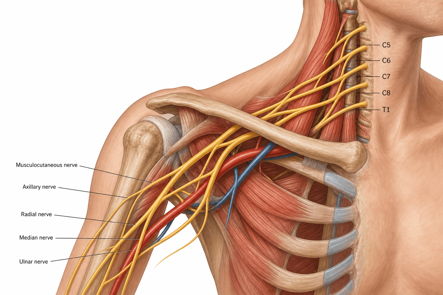
Roots → Trunks → Divisions → Cords → Branches
- Order: Roots, Trunks, Divisions, Cords, Branches ('Randy Travis Drinks Cold Beer').
- Roots are the anterior (ventral) rami of C5-T1; trunks are upper (C5-6), middle (C7) and lower (C8-T1).
- Each trunk splits into an anterior and posterior division behind the clavicle (6 divisions).
- The three cords (lateral, posterior, medial) are named by their position relative to the axillary artery.
- The five terminal branches are the musculocutaneous, axillary, radial, median and ulnar nerves.
- Upper plexus injury (C5-6) = Erb's palsy ('waiter's tip'); lower plexus injury (C8-T1) = Klumpke's palsy (claw hand, often with Horner's syndrome).
- “Posterior cord branches = 'ULTRA': Upper subscapular, Lower subscapular, Thoracodorsal, Radial, Axillary.
- “All posterior divisions form the posterior cord → the extensors (radial/axillary) are 'posterior'.
- “The only branches off the roots/trunks worth memorising: dorsal scapular & long thoracic (roots), suprascapular & nerve to subclavius (upper trunk).
Upper trunk injury (traction with the head pulled away from the shoulder - birth, motorcycle). Loss of abductors/external rotators and elbow flexors → the "waiter's tip" posture (adducted, internally rotated arm, extended elbow, pronated forearm).
Lower trunk injury (traction with the arm pulled overhead). Loss of the intrinsic hand muscles → a claw hand; often associated with Horner's syndrome (ptosis, miosis, anhidrosis) from T1 sympathetic involvement.
The Five Levels
Roots and Trunks
- Roots: the anterior (ventral) rami of C5-T1, emerging between scalenus anterior and medius. A prefixed plexus has a C4 contribution; a postfixed plexus has a T2 contribution.
- Trunks (in the posterior triangle / supraclavicular fossa):
- Upper trunk = C5 + C6
- Middle trunk = C7
- Lower trunk = C8 + T1
- Branches here: from the roots - dorsal scapular nerve (C5) and long thoracic nerve (C5-C7); from the upper trunk - the suprascapular nerve and the nerve to subclavius.
Branch-at-Each-Level Summary
A reliable viva answer names the branches by level, top to bottom.
- Roots: dorsal scapular nerve (C5 - rhomboids, levator scapulae); long thoracic nerve (C5-C7 - serratus anterior).
- Upper trunk: suprascapular nerve (supraspinatus, infraspinatus); nerve to subclavius.
- Lateral cord: lateral pectoral nerve; musculocutaneous nerve; lateral root of median.
- Posterior cord: upper subscapular, lower subscapular, thoracodorsal (latissimus dorsi), axillary (deltoid, teres minor), radial.
- Medial cord: medial pectoral nerve; medial cutaneous nerve of arm; medial cutaneous nerve of forearm; ulnar nerve; medial root of median.
Clinical Correlations
Supraclavicular vs Infraclavicular
- Supraclavicular injuries (roots/trunks) are the most common and include the classic upper (Erb's) and lower (Klumpke's) patterns.
- Infraclavicular injuries (cords/branches) follow shoulder dislocation, humeral fracture, or penetrating trauma and tend to affect specific terminal nerves.
- Preganglionic (root avulsion) vs postganglionic distinction guides reconstruction: avulsion is suggested by Horner's syndrome, winged scapula (long thoracic off the root), and pseudomeningocele on imaging, and is not amenable to direct repair (requires nerve transfer).
RTDCBThe Five Levels & Posterior Cord
Hook:Randy Travis Drinks Cold Beer; posterior cord = ULTRA.
Evidence Base
Brachial Plexus Anatomy & Treatment Overview
- Reviews the five anatomical sections of the brachial plexus: roots, trunks, divisions, cords and terminal branches
- The plexus ends in five terminal branches: musculocutaneous, median, axillary, radial and ulnar nerves
- Injury patterns classified as upper trunk, extended upper trunk, lower trunk, or whole-plexus ('swinging hand')
- Reconstruction (neurolysis, grafting, neurotisation, tendon/free-muscle transfer, arthrodesis) is chosen by injury type and deficit
Plexus Branching Detail for Nerve Transfer
- Prospective study of 150 brachial plexus injury patients undergoing nerve transfer for elbow flexion
- Detailed the branching of the musculocutaneous nerve (a terminal branch of the lateral cord)
- Anatomic variation in plexus terminal branches is common and must be anticipated intra-operatively
- Knowledge of the branching pattern is key to safe, effective neurotisation
Viva Scenarios
Practise clinical reasoning and management decisions out loud
“The examiner asks you to draw the brachial plexus and describe the branch at each level.”
Guidelines, Registries & Global Practice
Global Practice Picture
Brachial plexus anatomy is universal core knowledge and the framework for diagnosing and reconstructing plexus injuries worldwide. The consistent principles are: classify the lesion by level (supra- vs infraclavicular, pre- vs postganglionic), recognise the Erb's and Klumpke's patterns, and use detailed terminal-branch anatomy to plan nerve transfers.
Side-by-Side Synthesis
- Key structures / branches
- Dorsal scapular (C5), long thoracic (C5-7)
- Key structures / branches
- Suprascapular + n. to subclavius (upper trunk)
- Key structures / branches
- Anterior + posterior; behind clavicle; no named branches
- Key structures / branches
- Named by axillary artery; posterior cord = ULTRA
- Key structures / branches
- Musculocutaneous, axillary, radial, median, ulnar
Structure
- Roots C5-T1 → 3 trunks → 6 divisions → 3 cords → 5 branches
- Trunks: upper C5-6, middle C7, lower C8-T1
- Cords named by axillary artery
- Prefixed (C4) / postfixed (T2) variants
Branches
- Roots: dorsal scapular, long thoracic
- Upper trunk: suprascapular, n. to subclavius
- Posterior cord = ULTRA
- 5 terminal: MC, axillary, radial, median, ulnar
Clinical
- Erb's (C5-6): waiter's tip
- Klumpke's (C8-T1): claw hand + Horner's
- Avulsion signs: Horner's, winged scapula, pseudomeningocele
- Reconstruction: grafts, transfers, tendon transfer