High-energy traction injury of the young adult | Decide preganglionic versus postganglionic | Timing is everything | Build the arm with nerve transfers
- The injury: a high-energy traction injury, usually in a young man after a motorcycle crash - the force pulls the roots and trunks and can tear the root clean out of the spinal cord
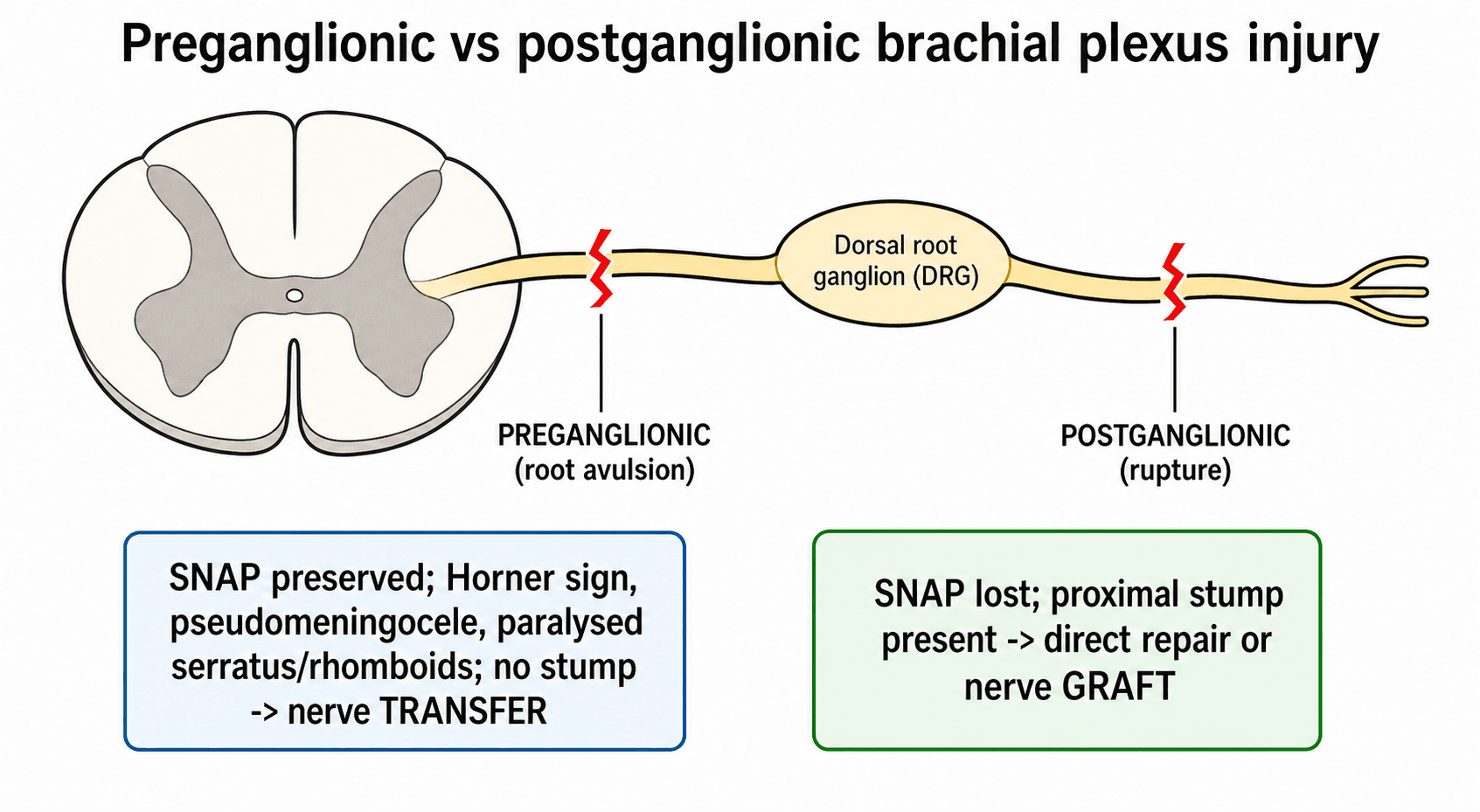
- The key split: a preganglionic (root avulsion) lesion is proximal to the dorsal root ganglion and cannot be repaired directly - it needs a nerve transfer; a postganglionic lesion is distal and may be grafted or may recover
- The clues to avulsion: a Horner's sign, winged scapula, paralysed rhomboids/serratus, preserved sensory nerve action potentials in an anaesthetic limb, and a pseudomeningocele on imaging - all point preganglionic
- Timing: reinnervate the muscle before it dies. Sharp/open injuries are explored early; closed traction injuries are usually reconstructed by 3 to 6 months, and results fall off sharply after about a year
- The priorities: restore elbow flexion first, then shoulder stability/abduction, then hand function - hand recovery is the hardest and least reliable goal
- “A Horner's sign (ptosis, miosis, anhidrosis) means a T1 root avulsion until proven otherwise - it is a marker of a severe, preganglionic, proximal injury
- “In a preganglionic injury the limb is anaesthetic but sensory nerve conduction is PRESERVED, because the cell body in the dorsal root ganglion is still connected to its peripheral axon - a classic exam discriminator
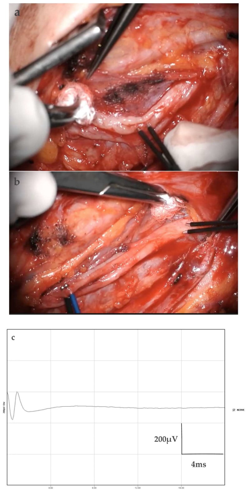
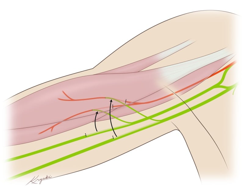
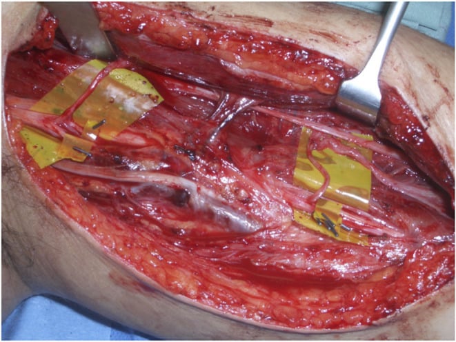
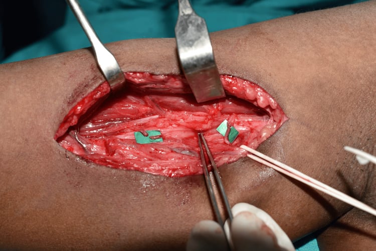
- “Elbow flexion is the first reconstructive priority, classically via an Oberlin (ulnar fascicle to biceps motor branch) transfer in upper plexus injury
- “Refer early to a specialist plexus centre - delayed referral is common and directly worsens the achievable outcome
Overview
The brachial plexus is the network of nerves that carries motor and sensory signals from the lower cervical and first thoracic spinal nerves out to the whole upper limb. An adult traumatic brachial plexus injury is usually a high-energy traction injury: the classic patient is a young man thrown from a motorcycle, whose head and shoulder are forced apart so violently that the nerves are stretched, ruptured, or pulled out of the spinal cord altogether.
What it costs. A partly or completely paralysed, numb, and often painful arm in a young, working person. Three questions run through everything that follows: where and how badly the plexus is injured, above all whether each root is preganglionic or postganglionic; whether there is still time to reinnervate the muscle; and in what order to rebuild the limb.
Not birth palsy. Brachial plexus birth palsy is a different obstetric traction problem in neonates, with its own classification (Narakas) and a high rate of spontaneous recovery. The adult injury is higher-energy, recovers far less spontaneously, and its surgical decisions are driven hard by timing and by the preganglionic/postganglionic distinction.
Pathophysiology and Applied Anatomy
The five sections. Roots, trunks, divisions, cords and branches, proximal to distal ("Read Those Damn Cadaver Books"). What matters clinically is which section is injured and what that predicts.
- Roots (C5, C6, C7, C8, T1): the spinal nerves emerging from the neck. The dorsal root ganglion (DRG) sits on the sensory root, and its position is the anatomical basis of the preganglionic/postganglionic split.
- Trunks: C5+C6 form the upper trunk, C7 alone the middle trunk, C8+T1 the lower trunk, all in the supraclavicular region.
- Divisions: each trunk splits into an anterior and a posterior division behind the clavicle.
- Cords (lateral, posterior, medial): named for their relation to the axillary artery, in the infraclavicular region.
- Branches: the terminal nerves - musculocutaneous, axillary, radial, median, ulnar.
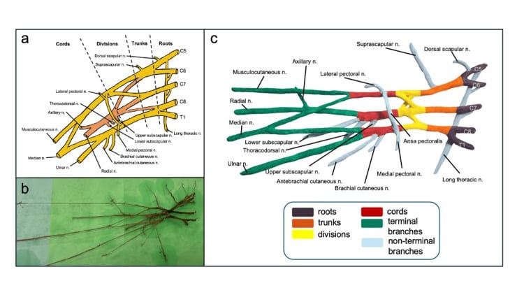
Branches that leave early are diagnostic flags. The dorsal scapular nerve (rhomboids) and the long thoracic nerve (serratus anterior) arise from the roots themselves, and the suprascapular nerve leaves the upper trunk. Paralysis of these muscles places the lesion very proximally, at root level, and points to a preganglionic injury.
The dorsal root ganglion. Sensory cell bodies live in the DRG. An injury proximal to it (preganglionic, a root avulsion) leaves the peripheral sensory axon attached to its living cell body, so the axon survives and the sensory nerve action potential (SNAP) is preserved even though no signal reaches the cord and the limb is anaesthetic. An injury distal to it (postganglionic, a rupture) disconnects the peripheral axon from its cell body, so the axon degenerates and the SNAP is lost. That one anatomical fact explains a whole cluster of findings, and it decides the operation: an avulsed root has no proximal stump to sew to and needs a nerve transfer, whereas a rupture leaves a proximal stump for direct repair or a nerve graft.
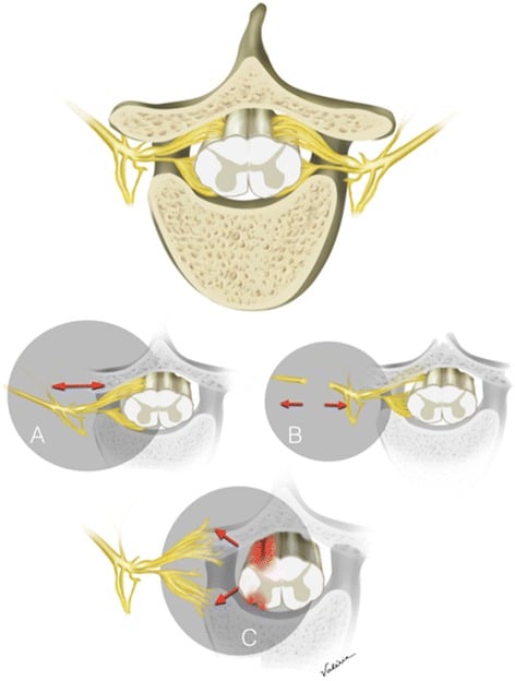
How the injury happens. Adult plexus injury is overwhelmingly a traction (stretch) injury. When the head and shoulder are forced apart, the classic motorcyclist thrown over the handlebars, the angle between neck and shoulder widens violently and the nerves are stretched beyond their limit.
Where the nerve fails depends on the force and the anatomy. The upper roots (C5-C6) are tethered and tend to rupture distal to the DRG (postganglionic), whereas the lower roots (C8-T1) are less supported and are more prone to being avulsed from the cord (preganglionic). This is why a Horner's sign, a T1 sign, signals a severe, proximal, lower-root injury.
The clock starts at injury. Once an axon is disconnected from its cell body it undergoes Wallerian degeneration, and the denervated muscle it supplied begins an irreversible clock toward fibrosis. Regenerating axons advance only slowly, on the order of about a millimetre a day, which is why reinnervation distance and timing dominate the surgical strategy: the closer and the sooner new axons can be delivered to a muscle, the better.
Other mechanisms. A sharp or open laceration divides the plexus cleanly, which favours early direct repair. An infraclavicular injury is often produced by a shoulder dislocation or a proximal humerus fracture dragging on the cords and branches.
Classification
Adult plexus injuries are classified in several overlapping ways, and they are used together: where along the plexus, how severe the nerve lesion, and which functional pattern.
The decisive split, because it determines whether direct repair is even possible.
- Preganglionic (root avulsion)
- Proximal to dorsal root ganglion - root torn from cord
- Postganglionic (distal to DRG)
- Distal to dorsal root ganglion - in trunk/cord/branch
- Preganglionic (root avulsion)
- No - there is no proximal stump to sew to
- Postganglionic (distal to DRG)
- Yes - if there is a healthy proximal stump
- Preganglionic (root avulsion)
- PRESERVED in an anaesthetic limb (cell body intact)
- Postganglionic (distal to DRG)
- Absent (peripheral sensory axon degenerates)
- Preganglionic (root avulsion)
- Horner's sign, paralysed rhomboids/serratus, pseudomeningocele, severe deafferentation pain
- Postganglionic (distal to DRG)
- Recovery may occur; signs localise more distally
- Preganglionic (root avulsion)
- Nerve TRANSFER (re-route a working donor)
- Postganglionic (distal to DRG)
- Neurolysis, direct repair, or nerve GRAFT
Clinical Assessment
The assessment is structured: history, then a systematic limb examination, then the special signs that localise the level.
History and resuscitation. These are high-energy poly-trauma patients, so assess and resuscitate the whole patient first. Then note the mechanism (motorcycle, fall, traction), any associated fractures and vascular injury, and the time since injury, which drives the surgical clock.
Look and feel. Inspect for posture (waiter's tip in upper injuries, a flail arm in pan-plexus), wasting, a winged scapula (serratus/long thoracic) and swelling or bruising. Map the sensory loss by dermatome.
Motor and reflex map. Grade every key muscle on the MRC scale by root and nerve so that the level can be plotted:
- Deltoid and supraspinatus - C5
- Biceps - C5-C6
- Wrist and finger extensors - C7
- Finger flexors - C8
- Intrinsics - T1
- Rhomboids and serratus - the proximal-branch flags
Vascular and special signs. Check the distal pulses and perfusion. Look hard for a Horner's sign: ptosis, miosis and facial anhidrosis on the injured side mean the T1 sympathetic outflow has been disrupted, a marker of a lower root avulsion and a severe, proximal injury that changes both the prognosis and the plan. Document the Tinel sign in the supraclavicular fossa; an advancing Tinel suggests a postganglionic lesion with regenerating axons.
A brachial plexus injury rarely travels alone. An associated subclavian or axillary artery injury can threaten the limb and must be identified and treated urgently; the dramatic neurological deficit can distract from a cold, pulseless hand. Exclude an associated cervical spine injury and other major trauma before focusing on the plexus.
Investigations
Investigations answer one question: for each root, is the lesion preganglionic or postganglionic, and is there anything to repair?
Nerve conduction studies and EMG are central, and are usually most informative from about 3 to 4 weeks, once Wallerian degeneration has declared itself. A preserved SNAP in an anaesthetic dermatome is the classic marker of a preganglionic lesion, for the reason set out in the anatomy: the DRG cell body is still attached to its peripheral axon.
EMG maps which muscles are denervated and, on serial testing, can show early reinnervation (nascent motor units) that argues for watchful waiting rather than surgery. Loss of paraspinal muscle (erector spinae) activity points to a very proximal, root-level lesion.
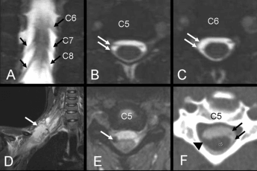
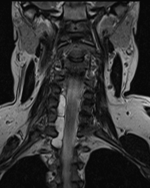
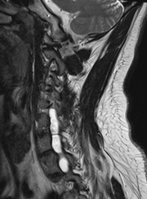
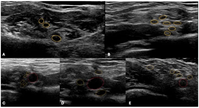
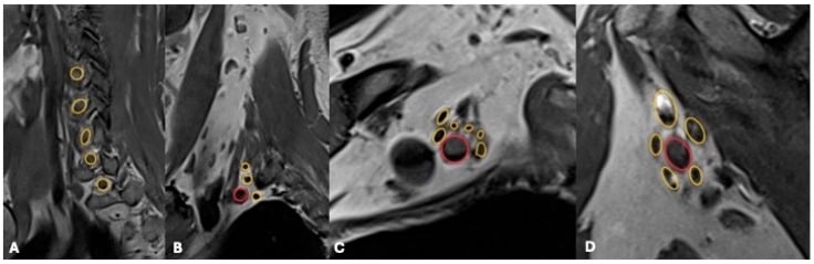
No single test is perfect, and imaging can miss avulsions (false negatives at a level that is in fact avulsed at exploration). The robust answer is to combine serial clinical examination, neurophysiology and imaging (MRI +/- CT myelography) to build a picture, and to accept that intra-operative exploration with nerve action potential recording is the final arbiter.
Management
Management is structured as who to operate on, when, and what to do, with the non-surgical care that runs alongside.
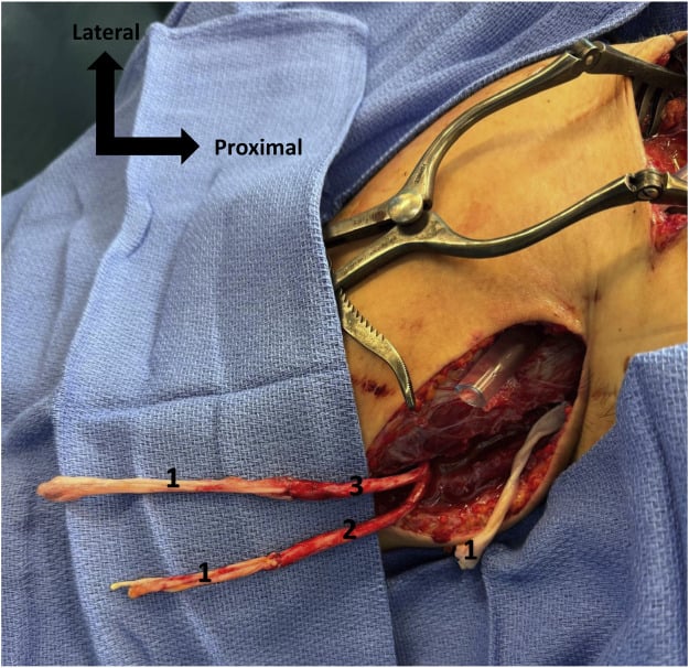
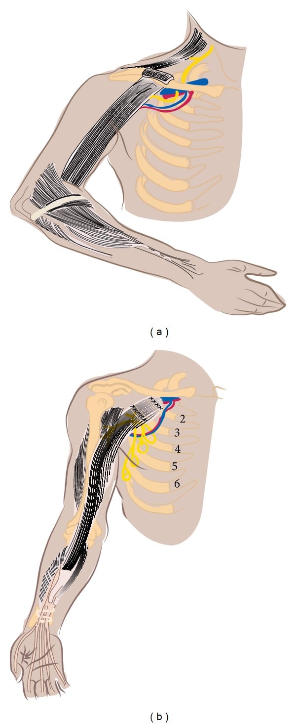
Timing dominates outcome. A denervated muscle has a limited time before it becomes fibrotic and can no longer be re-animated by reinnervation, so the whole strategy is to reconnect the nerve before that happens.
- Timing
- Immediate or early (primary repair)
- Reasoning
- Clean-cut ends can be repaired directly while they are identifiable
- Timing
- Urgent (with the vascular repair)
- Reasoning
- Limb perfusion takes priority; the plexus can be tagged/explored at the same time
- Timing
- Early (around 6-12 weeks)
- Reasoning
- No spontaneous recovery is possible - do not waste the muscle's time window
- Timing
- Explore/reconstruct by 3-6 months if no recovery
- Reasoning
- Allows neurapraxic/recovering lesions to declare themselves while staying inside the window
- Timing
- Reconstruct function, not nerves
- Reasoning
- Denervated muscle is lost - shift to tendon transfer, fusion, or free muscle transfer
Referral is a clinical decision. Delay is the commonest avoidable failure, and it is common. Across 84 adults with traumatic plexus injury assessed at two tertiary centres, 44% reached a plexus surgeon more than 3 months after injury, that is, only once the 3-6 month reconstruction window set above had already opened. Global palsy was the commonest pattern (39%), and a motorcycle mechanism and insurance or system factors predicted delay. The surgeon who first sees the patient rarely does the reconstruction, so the threshold for onward referral to a specialist plexus centre should be low and immediate rather than contingent on watching for recovery.
Deafferentation Pain and Its Management
Deafferentation (avulsion) pain is one of the most disabling features of a preganglionic injury and a major determinant of quality of life. It is a crushing or burning pain in the anaesthetic limb, generated by hyperactive deafferented neurons in the dorsal horn after the root is torn from the cord, and it is treated in tiers.
- Option
- Neuropathic agents - gabapentinoids (gabapentin, pregabalin), a tricyclic (amitriptyline) or an SNRI (duloxetine)
- Note
- Opioids are relatively ineffective for deafferentation pain and are avoided long term
- Option
- TENS, physiotherapy, and crucially a multidisciplinary pain service with psychological support
- Note
- A young patient's pain needs active, holistic management
- Option
- Successful reinnervation often reduces the pain
- Note
- A further reason to reconstruct early rather than wait
- Option
- Dorsal root entry zone (DREZ) lesioning - ablation of the hyperactive dorsal-horn neurons at the avulsed levels
- Note
- The most effective procedure specifically for root-avulsion pain; spinal cord stimulation, which works for many neuropathic pains, is generally less effective for avulsion pain because the dorsal columns themselves are disconnected
Complications
- Why it happens
- Severe avulsion, late surgery, distal (hand) targets
- Key point
- Hand reinnervation is the least reliable goal of all
- Why it happens
- Loss of sensory input after root avulsion
- Key point
- Severe, common, and a major hit to quality of life - treat actively
- Why it happens
- A flail limb left unmoved while awaiting reinnervation
- Key point
- Preventable with early, ongoing physiotherapy
- Why it happens
- Borrowing a working nerve/muscle
- Key point
- Choose expendable donors; deficit is usually minor but must be consented
- Why it happens
- Avulsion severity, late referral, target muscle loss
- Key point
- Salvage with tendon transfers, fusions, free muscle transfer, or prosthetics
- Why it happens
- Young, working patients with a disabled dominant limb
- Key point
- Often life-changing; needs holistic, multidisciplinary support
Prognostic Factors
Outcome turns on a handful of factors that are easy to list separately and hard to weigh together; the table pulls them into one framework.
- Favourable
- Upper plexus (C5-C6) - shoulder and elbow targets recover well
- Unfavourable
- Lower plexus (C8-T1) or pan-plexus - hand recovery is the least reliable goal of all
- Favourable
- Postganglionic with a healthy proximal stump (graftable)
- Unfavourable
- Multiple root avulsions - no proximal stump and few intraplexal donors
- Favourable
- Lower-energy or sharp/open (clean, early repair)
- Unfavourable
- High-energy traction (worse recovery, longer zone of injury)
- Favourable
- Reconstruction within the 3-6 month window
- Unfavourable
- Delay beyond about a year - denervated muscle is lost
- Favourable
- Proximal targets close to the donor (elbow, shoulder)
- Unfavourable
- Distal targets (hand intrinsics) - axons may not arrive before the muscle dies
- Favourable
- Several healthy expendable donors for transfer
- Unfavourable
- Few donors (extensive injury) forcing extraplexal donors or free muscle transfer
Why the hand comes last. Whatever the pattern, hand reinnervation is the hardest and least reliable goal, because the regenerating axons must travel the greatest distance and must arrive before the intrinsic muscles fibrose. That is why the reconstructive priorities run elbow flexion, then shoulder, then hand, and why honest counselling aims for a useful "helper arm" rather than a normal hand. High energy and delay are the two most modifiable predictors of a poor result, hence the emphasis on early referral.
Guidelines, Registries and Global Practice
- Specialist, multidisciplinary care is the consistent global message: traumatic adult plexus injuries do best when managed early in dedicated centres combining orthopaedic/hand, plastic, and peripheral-nerve neurosurgery, with physiotherapy and pain services. Delayed referral is documented as common and as a driver of poorer results.
- Timing principles are broadly agreed: explore sharp/open and vascular injuries early; reconstruct closed traction injuries inside roughly a 3 to 6 month window before denervated muscle is lost; shift to reconstructive (tendon transfer, fusion, free muscle transfer) options for late presentations.
- Nerve transfers have shifted global practice over recent decades: distal nerve transfers (such as the Oberlin transfer for elbow flexion and spinal-accessory-to-suprascapular transfer for the shoulder) are now widely used, shortening reinnervation distance and improving outcomes, while nerve grafting remains the answer for graftable postganglionic ruptures.
- Outcome variation reflects injury severity and access: upper-plexus injuries reliably regain elbow and shoulder function, whereas hand recovery after lower-plexus or pan-plexus injury remains poor everywhere - so realistic goal-setting (a useful helper arm) is a universal part of consent and care.
- Mechanism and demographics differ by region: motorcycle and road-traffic trauma dominate in many low- and middle-income settings, shaping a younger, predominantly male, higher-severity caseload - a global epidemiological pattern rather than any single country's experience.
MCQ Practice Points
High-yield, single-best-answer style points that examiners turn into questions.
Q: A young man has an anaesthetic arm after a motorcycle crash, yet nerve conduction shows preserved sensory action potentials. What does this mean? A: A preserved SNAP in an insensate limb indicates a preganglionic (root avulsion) lesion - the dorsal root ganglion cell body keeps the peripheral sensory axon alive even though no signal reaches the cord. It points toward nerve transfer, not graft.
Q: Which clinical sign best indicates a T1 root avulsion? A: A Horner's sign (ptosis, miosis, anhidrosis) - it reflects disrupted T1 sympathetic outflow and flags a severe, proximal, lower-root preganglionic injury.
Q: Is a pseudomeningocele proof of root avulsion? A: No. It is a strong but imperfect sign - it is absent in a substantial proportion of genuine avulsions, so imaging is never a substitute for operative exploration with nerve action potential testing.
Q: What is the first reconstructive priority and the workhorse procedure for it? A: Elbow flexion, classically restored by the Oberlin transfer (ulnar nerve fascicle to the biceps motor branch) in an upper plexus injury with an intact C8-T1.
Q: Why does timing dominate the plan in a closed traction injury? A: Denervated muscle has a time limit before irreversible fibrosis - so closed traction injuries are generally reconstructed by 3 to 6 months, and beyond roughly a year the strategy shifts to tendon transfers, joint fusions, or free functioning muscle transfer.
Exam Viva Scenarios
Practise clinical reasoning and management decisions out loud
“A 24-year-old man is brought in after a high-speed motorcycle crash. His right arm hangs limp and he cannot move it. How do you assess and work him up?”
“The examiner asks you to explain the difference between a preganglionic and a postganglionic brachial plexus injury and why it changes your operation.”
The Big Picture
- High-energy traction injury, typically a young man on a motorcycle
- Decide preganglionic (avulsion) vs postganglionic FIRST
- Anatomy: Roots, Trunks, Divisions, Cords, Branches
- Patterns: upper (C5-6, best) / lower (C8-T1, worst) / pan-plexus
Signs of Avulsion (Preganglionic)
- Horner's sign (ptosis, miosis, anhidrosis) = T1 avulsion
- Preserved SNAP in an anaesthetic limb
- Paralysed rhomboids and serratus; winged scapula
- Pseudomeningocele on imaging; severe deafferentation pain
Investigations
- NCS/EMG from ~3-4 weeks (SNAP, denervation, reinnervation)
- MRI plexus + CT myelography for root avulsion
- Plain films/CT for fractures; chest film for phrenic palsy
- Exploration with NAP testing is the final arbiter
Management
- Sharp/open and vascular injuries = early/urgent surgery
- Closed traction: reconstruct by ~3-6 months (window!)
- Ladder: Neurolysis - Graft - Transfer - Free muscle (FFMT)
- Priorities: elbow flexion (Oberlin), then shoulder, then hand
Evidence Base
The following studies support the key teaching points above.
Axial T2-DRIVE MR Myelography Is Highly Accurate for Preganglionic Injury
- Prospective study of 24 patients planned for brachial plexus reconstruction, with operative exploration as the reference standard
- Axial T2-DRIVE MR myelography accurately identified the specific avulsed roots in each patient (sensitivity 100%, specificity 97.1%, accuracy 98.1%)
- Absent or pauci-rootlet appearance was the most reliable sign of avulsion
- Pseudomeningoceles were present in only ~68% of avulsions, so the authors argue they should be a supporting (ancillary) sign rather than the primary one
Delayed Referral Is Common and Worsens the Achievable Outcome
- 84 adults with traumatic brachial plexus injury evaluated at two tertiary referral centres in a large US metropolitan area
- Nearly half (44%) presented to a plexus surgeon in a delayed time frame (more than 3 months from injury)
- Global palsy was the most common pattern (39%), followed by upper-trunk and infraclavicular patterns
- Motorcycle-accident mechanism and certain system/insurance factors were associated with delayed referral
Reconstruction Can Restore Useful Elbow Flexion Even in Older Patients
- 58 patients older than 50 underwent nerve grafting, nerve transfer, or free functioning muscle transfer for elbow flexion after traumatic brachial plexus injury
- Mean elbow flexion grade improved from 0.26 to 2.63; 60% achieved functional flexion of M3 or better, versus none preoperatively
- More patients reached M3 or better with nerve transfers (69%) than with free functioning muscle transfer (43%)
- High-energy injuries did worse, but age itself did not correlate with the flexion grade achieved
Global Plexus Injury: Nerve Transfers Restore Elbow and Shoulder, Hand Stays Hard
- Concise review of surgical approaches for traumatic global (pan-plexus) brachial plexus injury, which predominantly affects adults
- Primary reconstruction relies mainly on single or multiple nerve transfers, with or without nerve grafts, to power targeted muscles
- Outcomes are best for restoring elbow flexion and shoulder abduction; wrist and hand function remain the hardest to recover
- Free functioning muscle transfer is the main option for delayed presentations or after failed nerve transfer