The Engine of Key Pinch
- Two heads: transverse from the palmar shaft of the third metacarpal, oblique from the capitate and bases of the second and third metacarpals.
- Both heads insert on the ulnar sesamoid and ulnar base of the thumb proximal phalanx, with an expansion into the extensor hood.
- Innervation is the deep (motor) branch of the ulnar nerve, C8-T1 — adductor pollicis is its terminal muscle.
- The radial artery enters the palm between the two heads to complete the deep palmar arch.
- The adductor aponeurosis is the structure that interposes in a Stener lesion after complete thumb ulnar collateral ligament rupture.
- “Froment sign: thumb IP flexion during key pinch — flexor pollicis longus (anterior interosseous) substituting for a paralysed adductor.
- “Jeanne sign: compensatory thumb MCP hyperextension during the same pinch — the other half of the same story.
- “Adductor pollicis is spared in a pure median nerve palsy; it is the muscle that proves the ulnar nerve is the problem.
- “The oblique head shares the deep motor branch with the deep head of flexor pollicis brevis — hence the confusing 'dual innervation' of the thenar eminence.
Overview
Adductor pollicis is a flat, triangular, two-headed muscle occupying the deep plane of the palm between the thumb ray and the middle finger. With the first dorsal interosseous it generates the compressive force of key (lateral) pinch, the grip used to turn a key, hold a card or fasten a button.
Surgically it matters for four reasons that recur endlessly in the viva:
- It is the terminal muscle of the deep branch of the ulnar nerve, and the muscle whose failure defines ulnar motor palsy at the wrist or elbow.
- Its aponeurosis is the tissue that converts a repairable thumb ulnar collateral ligament (UCL) tear into an irreparable one, the Stener lesion.
- Its contracture produces the adducted first web that dominates the spastic and post-traumatic hand.
- The deep palmar arch and the deep ulnar motor branch both run in intimate contact with it, so every release of the muscle is a neurovascular exercise.
Attachments, Innervation and Relations
Transverse head (larger, distal). Arises from the palmar (anterior) surface of the distal two-thirds of the shaft of the third metacarpal, along a ridge that runs from just distal to the base to the metacarpal neck. Its fibres run transversely and radially, converging on the thumb.
Oblique head (proximal, deeper). Arises from the capitate, the palmar carpal ligaments and the bases of the second and third metacarpals, frequently with a slip from the sheath of the flexor carpi radialis tendon and the trapezoid, and with some fibres from the deep transverse metacarpal ligament complex. A small deep slip passes to the radial sesamoid and blends with flexor pollicis brevis; this is the source of much of the confusion about which "head" of FPB is ulnar-innervated.
Insertion. The two heads converge into a single tendon containing the ulnar sesamoid of the thumb metacarpophalangeal (MCP) joint, and insert on the ulnar side of the base of the proximal phalanx and on the sesamoid itself. A broad fibrous sheet, the adductor aponeurosis, sweeps dorsally over the ulnar aspect of the MCP joint to blend with the extensor pollicis longus (EPL) hood, which is why adductor pollicis contributes to thumb IP extension and why its aponeurosis lies exactly superficial to the UCL.
The gap between the heads. Distally the two heads are separated by a triangular defect through which the radial artery passes from the first dorsal interosseous space into the palm to complete the deep palmar arch, and through which the princeps pollicis artery arises.
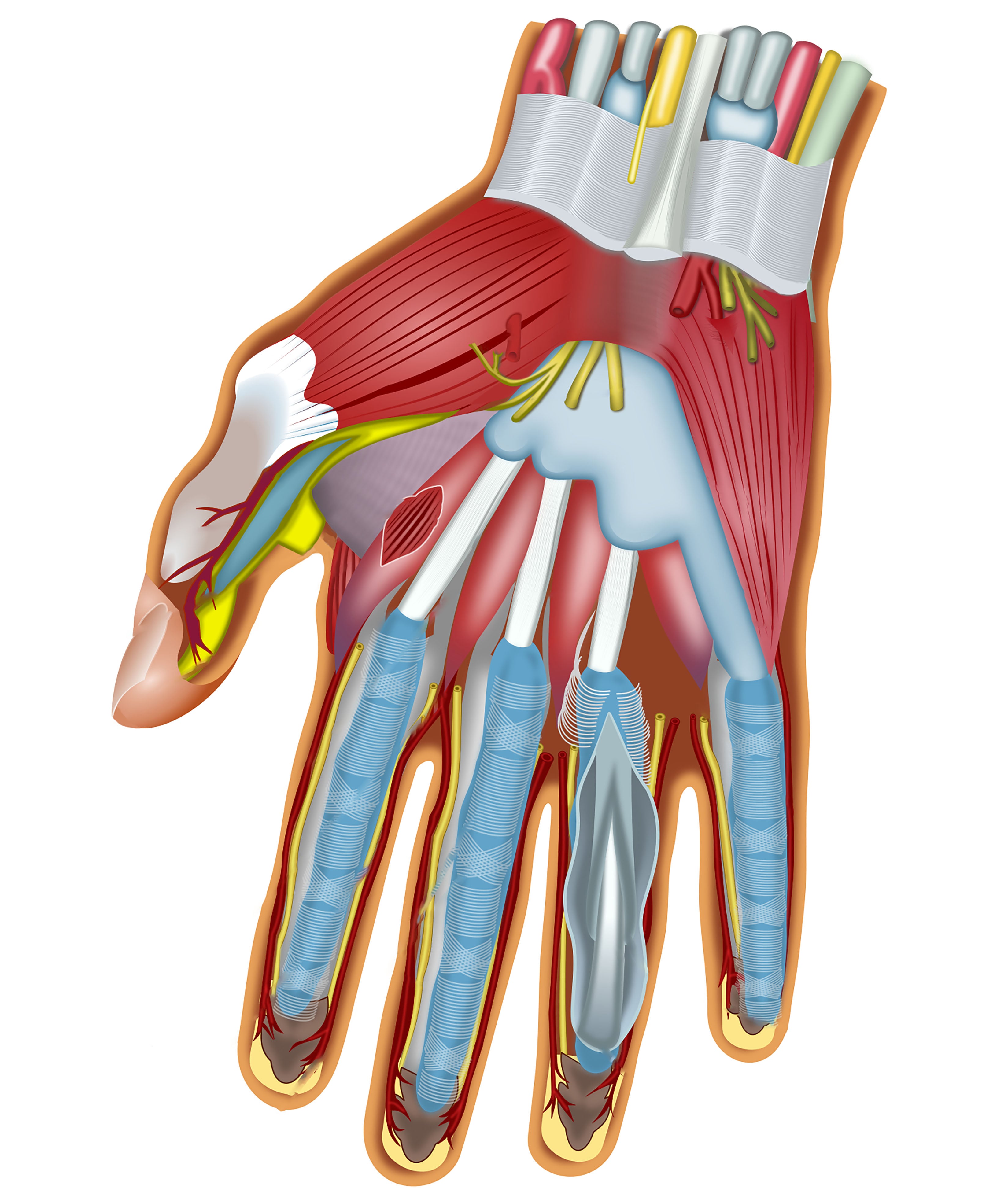
The deep ulnar motor branch and the deep palmar arch share the dorsal surface of the muscle, and both are casualties of the same mistakes.
The nerve is divided in deep palmar penetrating trauma and in over-enthusiastic release of the oblique head from the capitate. The result is clawing of the ring and little fingers plus loss of key pinch, with normal sensation: a purely motor deficit.
The arch bleeds when the interval between the heads is opened; the bleeding is deep, brisk and difficult to control from a palmar approach. Release the transverse head subperiosteally off the third metacarpal shaft, never bluntly through the interval.
Action and Biomechanics
- Action
- Adduction in the plane of the palm
- Mechanism
- Large moment arm about the ulnar-directed axis
- Clinical correlate
- Key pinch compression
- Action
- Flexion component
- Mechanism
- Line of pull passes palmar to the flexion-extension axis
- Clinical correlate
- Contributes to thumb-in-palm posture
- Action
- Flexion and ulnar deviation
- Mechanism
- Insertion on ulnar sesamoid and ulnar base of P1
- Clinical correlate
- Chronic overload produces MCP ulnar deviation and radial collateral strain
- Action
- Extension
- Mechanism
- Aponeurotic expansion into the EPL hood
- Clinical correlate
- Thumb IP extension is preserved in isolated EPL loss via intrinsics
- Action
- Indirect stabilisation
- Mechanism
- Transverse head arises from the third metacarpal, anchoring the central pillar
- Clinical correlate
- Pinch collapses if third metacarpal is unstable
Key pinch. A compression couple between the thumb pulp and the radial side of the index proximal phalanx, supplied by two muscles. Adductor pollicis pulls the thumb metacarpal ulnarwards, in the plane of the palm, with a large moment arm about the trapeziometacarpal (TMC) joint; the first dorsal interosseous simultaneously abducts the index radially into the thumb as the opposing post, and without it the index simply collapses ulnarwards and pinch is lost even with a normal thumb. Both are supplied by the deep branch of the ulnar nerve, which is why ulnar palsy costs approximately 70-80% of key pinch strength while grip strength, mostly extrinsic flexors, falls far less.
Force. Adductor pollicis and the first dorsal interosseous stand apart from the other intrinsics: their physiological cross-sectional areas are comparable to those of extrinsic muscles and much greater than the rest of the intrinsic group. The moment arm for palmar adduction at the TMC joint is large and relatively constant through range, so this is a strength muscle rather than a positioning muscle.
Length-tension and the adducted thumb. The muscle is short and its excursion small, of the order of 2-3 cm, so it contractures rapidly when the thumb is held adducted. This is why the thumb is splinted in palmar abduction after nerve injury, burns or spasticity: not because the joint stiffens first, but because the adductor shortens. Even when the muscle is paralysed the first web narrows over months, as the unopposed but still-present passive tension and habitual posture shorten the soft tissues and the paralysed muscle fibroses in a short position.
Synergists and antagonists. The first dorsal interosseous acts as the index-side post, flexor pollicis brevis adds the MCP flexion component, and the first palmar interosseous contributes where present. The antagonists are abductor pollicis longus and abductor pollicis brevis for palmar and radial abduction, extensor pollicis brevis, and opponens pollicis, which places the thumb rather than compressing it.
When it fails. Key pinch collapses, and flexor pollicis longus (FPL, anterior interosseous nerve, median) and extensor pollicis longus are recruited to substitute. FPL is a poor substitute because its vector flexes the interphalangeal (IP) joint rather than compressing the pulp, so the thumb IP joint visibly flexes: Froment sign (Jules Froment, 1915), the "signe du journal". To lengthen the FPL lever and pull harder, the patient simultaneously hyperextends the thumb MCP joint using extensor pollicis longus and brevis and by allowing the incompetent volar plate to give: Jeanne sign. Chronic Jeanne sign leads to fixed MCP hyperextension with a lax volar plate, and eventually to a Z-thumb posture.
Surface Anatomy and Examination
Palpation. The muscle is deep; only its distal transverse fibres are reliably palpable. Place the pulp of your index finger in the patient's first web space from the palmar side and your thumb dorsally, and ask the patient to squeeze a card: the firm, contracting mass you feel palmar and deep is adductor pollicis, and the mass you feel dorsally is the first dorsal interosseous. Wasting is seen as hollowing of the first web space from the dorsal aspect, although this appearance is contributed to substantially by atrophy of the first dorsal interosseous, which is more superficial and dorsal and the earliest muscle to waste.
Isolation testing. Forearm pronated, hand flat on the table, thumb adducted against the radial border of the index; resist attempted thumb abduction while palpating the first web. Flexor pollicis brevis (partly median), the first dorsal interosseous and simple wedging of the thumb metacarpal against a stiff index can all fake adduction, which is why the dynamic Froment test, which forces a functional demand, outperforms static muscle testing.
Froment sign. Ask the patient to grip a sheet of paper between the thumb pulp and the radial border of the index proximal phalanx, and withdraw it steadily with equal force on both sides. Watch the thumb interphalangeal joint, not the paper. A normal hand holds the paper with the IP joint straight or gently extended and the MCP flexed, pure adductor pollicis compression; the test is positive when the IP joint flexes as flexor pollicis longus substitutes for the paralysed adductor.
Jeanne sign. Perform the same manoeuvre and watch the thumb metacarpophalangeal joint instead. Compensatory MCP hyperextension during attempted key pinch is positive and indicates adductor pollicis and deep flexor pollicis brevis weakness. A fixed, non-correctable hyperextension means chronic volar plate attenuation and predicts failure of tendon transfer alone.
An examiner asking "what else would you see?" wants Jeanne when you have offered Froment, and vice versa.
Quantification. Use a pinch meter rather than manual grading, compared with the contralateral hand and with age- and sex-matched norms (Mathiowetz), because decisions about tendon transfer for pinch are driven by recorded values rather than a subjective account. Serial key pinch measurements are the single most informative outcome measure in ulnar nerve recovery and after adductorplasty. Record the first web angle as well, thumb metacarpal to index metacarpal in maximum palmar abduction: normal is approximately 45-60 degrees, and anything less than 30 degrees is a functionally significant contracture.
False positives. Pain in the thumb MCP joint from arthritis or UCL injury produces guarding that mimics Froment sign, so examine for tenderness first. A fixed IP flexion contracture from Dupuytren disease or prior trauma looks like a positive Froment sign at rest; the test is only valid if the resting posture is normal and the deformity appears with effort.
Complications
- Mechanism
- Blunt dissection dorsal to the oblique head or deep palmar penetrating trauma
- Prevention
- Stay subperiosteal at the metacarpal bases; know the nerve lies on the dorsal surface
- Salvage
- Direct repair or graft; late — adductorplasty and index abduction transfer
- Mechanism
- Dissection between the adductor heads
- Prevention
- Approach the transverse head from its bony origin, not through the interval
- Salvage
- Direct repair or ligation if the superficial arch is intact — confirm with an Allen test pre-operatively
- Mechanism
- Skin flap elevation too deep dorsally
- Prevention
- Subdermal flap elevation under vision; identify and protect two or three constant branches
- Salvage
- Neuroma excision and transposition into muscle or bone
- Mechanism
- Inadequate skin lengthening or failure to hold the position
- Prevention
- Plan skin first; transarticular wire for 3-4 weeks and night splinting for 3 months
- Salvage
- Repeat release with a larger flap; consider free tissue in the burned web
- Mechanism
- Release of both heads plus the first dorsal interosseous origin
- Prevention
- Release stepwise and stop when the web opens
- Salvage
- Adductorplasty once the web is supple
- Mechanism
- Transfer into an unstable MCP joint or a tight web
- Prevention
- Correct contracture and stabilise the MCP first
- Salvage
- MCP arthrodesis plus re-tensioning of the transfer
- Mechanism
- Failure to repair the adductor aponeurosis after Stener repair
- Prevention
- Leave a 2-3 mm cuff dorsally and repair it
- Salvage
- Delayed ligament reconstruction with a free tendon graft or MCP fusion
Clinical Relevance
High versus low lesion. In a high lesion, at the elbow or cubital tunnel, adductor pollicis, all interossei, the ulnar two lumbricals, the hypothenar muscles and flexor digitorum profundus (FDP) to ring and little are affected; clawing is less marked because the FDP is also weak, the ulnar paradox. In a low lesion, at the wrist or Guyon canal, FDP is spared, so the profundus pulls the distal interphalangeal joints into flexion against paralysed intrinsics and clawing is more marked. Froment sign is present in both.
Recovery order. Because adductor pollicis is the terminal muscle of the deep branch, it is the last to reinnervate. A persistent Froment sign at 12-18 months after a proximal repair does not mean failure of the repair, but it is a poor prognostic sign for pinch.
Electrodiagnosis. The first dorsal interosseous and abductor digiti minimi are the conventional recording sites, but an adductor pollicis compound muscle action potential is useful when the deep branch is selectively involved. A normal abductor digiti minimi response with abnormal first dorsal interosseous and adductor pollicis responses localises the lesion to the deep motor branch distal to the hypothenar takeoff: Guyon canal zone 2 or a distal palmar lesion.
Surgical Relevance
First web space release. The release is sequential, and it stops as soon as the web opens.
- 1Step 1 — Skin
Assess the skin envelope. A tight web needs a four-flap Z-plasty or a dorsal transposition flap; a single Z-plasty gains roughly 75% length, a four-flap Z-plasty gains substantially more and is preferred for the web.
Do the skin plan before releasing muscle — the release will demand skin
- 2Step 2 — Fascia
Release the first dorsal interosseous fascia and the deep fascia of the first web from a dorsal approach.
Often gains 10-20 degrees alone
- 3Step 3 — Transverse head
Release the transverse head of adductor pollicis subperiosteally from the palmar surface of the third metacarpal shaft, working distal to proximal and staying strictly on bone.
The largest single gain in abduction
- 4Step 4 — Oblique head
If still tight, release the oblique head from the second and third metacarpal bases and the capitate. Stop as soon as the web opens — the deep motor branch and deep arch lie immediately dorsal.
Reserve for severe contracture
- 5Step 5 — First dorsal interosseous
Release the origin of the first dorsal interosseous from the first metacarpal only if abduction remains limited.
Costs index abduction strength — a real functional loss
- 6Step 6 — Joint
Trapeziometacarpal capsulotomy if the joint itself is contracted; consider arthrodesis in the low-demand or severely spastic hand.
Last resort
- 7Step 7 — Hold
Hold in maximum palmar abduction with a thumb spica or a temporary transarticular Kirschner wire for 3-4 weeks, then a night splint for at least 3 months.
Recurrence is the rule if the position is not held
- Deep branch of the ulnar nerve: crosses the palm on the dorsal surface of the adductor, roughly 1-2 cm distal to the distal edge of the transverse carpal ligament and approximately 2 cm radial to the hook of hamate as it turns radially. When releasing the oblique head, stay on bone at the metacarpal bases and never sweep a blunt instrument dorsally.
- Deep palmar arch: lies approximately 1 cm proximal to the superficial arch, at the level of the metacarpal bases; the radial artery reaches it by passing between the adductor heads roughly at the level of the proximal third of the second intermetacarpal space.
- Princeps pollicis artery: deep to the oblique head along the ulnar border of the first metacarpal, and the commonest bleeder in a first web release performed from the palmar side.
- Dorsal sensory branches of the radial nerve: cross the dorsal first web subcutaneously, two or three branches constant. Injury produces a painful neuroma in the web that ruins an otherwise successful release, so elevate skin flaps in the subdermal plane under direct vision.
- Ulnar digital nerve to the thumb: runs on the palmar-ulnar aspect of the thumb, crossing superficial to the adductor insertion. It is the nerve at risk in a UCL repair approached too palmarwards.
Adductorplasty restores key pinch by tendon transfer. The options:
- Donor
- Extensor carpi radialis brevis with an interposition tendon graft
- Route
- Through the interspace between the second and third metacarpals, deep to adductor pollicis, sutured to the adductor pollicis tendon; proximal end tunnelled subcutaneously across the dorsum of the wrist
- Notes
- The workhorse; strong, expendable donor, good vector
- Donor
- Brachioradialis
- Route
- Subcutaneously across the palm to the thumb
- Notes
- Large excursion after full release of its fascial attachments; useful in combined palsy
- Donor
- FDS of ring finger
- Route
- Through the third intermetacarpal space
- Notes
- Convenient, but risks swan-neck at the donor PIP and quadriga if harvested distally
- Donor
- EIP
- Route
- Around the ulnar border of the hand or through the intermetacarpal space
- Notes
- Modest force; often better used for opposition
- Donor
- Extensor carpi radialis longus or accessory abductor pollicis longus slip
- Route
- To the first dorsal interosseous insertion
- Notes
- Restores the index post rather than the thumb vector
The insertion point. In Smith's original description the graft is sutured to the tendon of adductor pollicis itself, which carries the pull to the ulnar sesamoid and the ulnar base of the thumb proximal phalanx. Securing the transfer to the adductor tendon or its footprint reproduces the native vector; a transfer taken only to the metacarpal is weaker and less effective. In Smith's series pinch in the affected hand averaged 25% of the normal side before operation and approximately doubled afterwards, a useful benchmark for what reconstruction can and cannot achieve.
The prerequisites. A supple first web and a stable thumb MCP joint. A transfer into a hand with a fixed adduction contracture or a hyperextending MCP will fail, since a tendon transfer alone will not restore pinch across a collapsing joint; release and stabilise first, transfer second, often as staged procedures. Where Jeanne sign is fixed the MCP is stabilised by volar plate capsulodesis, sesamoid arthrodesis, or MCP arthrodesis in 10-15 degrees of flexion, which is reliable and is the usual choice in the low-demand or spastic hand.
Third metacarpal shaft plating. The transverse head origin is elevated from the palmar surface. This is a safe subperiosteal plane if kept strictly on bone, but the muscle is a genuine obstacle to palmar plating and the dorsal approach is preferred.
Pollicisation and toe transfer. The adductor insertion is preserved and reattached to reconstruct the ulnar side of the new thumb; it is the single most important motor for pinch in a reconstructed thumb.
Guidelines, Registries & Global Practice
Anatomical variation across populations
- The Riche-Cannieu anastomosis between the recurrent median motor branch and the deep ulnar branch is reported at high frequency in cadaveric series worldwide, with quoted prevalence for any communication commonly between 50% and 77%. Series differ largely in how strictly a "communication" is defined; the clinically meaningful complete crossover is rare in all populations.
- Accessory slips of the oblique head to the radial sesamoid and to flexor pollicis brevis are common and explain the inconsistent descriptions of flexor pollicis brevis innervation across textbooks.
- The presence of a first palmar interosseous (a true fourth palmar interosseous acting on the thumb) is variably described; where absent, its adduction role is subsumed by the oblique head of adductor pollicis.
Practice differences in thumb UCL injury
- Consensus across AO, BOA/BSSH and North American teaching is that a complete rupture with a Stener lesion warrants operative repair, and that an incomplete tear is managed in a thumb spica for four to six weeks.
- Where practice diverges is the routine use of imaging. Ultrasound-first pathways are widespread in European and Australasian units where musculoskeletal ultrasound expertise is embedded; MRI-first pathways predominate where ultrasound expertise is less consistent. Both approaches are defensible provided the clinical stress test is performed after radiographs.
- Suture anchor versus bone tunnel fixation shows no consistent superiority; anchor use is driven by availability and cost, and bone tunnels remain the standard in resource-limited settings with equivalent reported results.
Ulnar nerve reconstruction practice
- There is growing international adoption of supercharged end-to-side anterior interosseous to ulnar motor nerve transfer for high ulnar lesions, on the rationale that reinnervation of adductor pollicis and the interossei by native regenerating axons across a long distance is unreliable. Evidence is largely cohort level; the operation is now widely offered in specialist upper-limb units.
- Where nerve reconstruction is unavailable or has failed, tendon transfer for key pinch and index abduction remains the definitive, globally applicable solution and is not dependent on advanced technology.
High- versus limited-resource practice
- Well-resourced: early imaging for UCL injury, suture anchors, nerve conduction studies and MRI for pure motor ulnar deficits, staged nerve transfer.
- Limited-resource: clinical stress testing after radiographs, bone tunnel ligament repair, and a strong reliance on the Froment and Jeanne signs and on serial pinch measurement for both diagnosis and follow-up. Tendon transfer, requiring no implants, is the mainstay of ulnar palsy reconstruction.
Related pages: Thumb UCL Injuries and Gamekeeper's Thumb are where the adductor aponeurosis stops being anatomy and becomes the reason an operation is needed - the Stener lesion is defined by this muscle's fascia. Ulnar Nerve Anatomy and Ulnar Nerve Palsy cover the deep motor branch for which adductor pollicis is the terminal muscle, and therefore the Froment and Jeanne signs; Cubital Tunnel Syndrome and Ulnar Tunnel Syndrome (Guyon's Canal) are the two compression sites, and the level determines whether sensation is spared. Median Nerve Anatomy matters here for the opposite reason - the Riche-Cannieu communication between the two nerves is why a Froment sign can be absent despite a divided ulnar nerve. Intrinsic Minus and Intrinsic Plus Hand sets the wider pattern of intrinsic loss of which first web space wasting is the most visible sign, and Thumb CMC Arthritis is the commonest competing cause of a painful weak pinch in an older hand.
MCQ Practice Points
Q: Where does the transverse head of adductor pollicis arise? A: The palmar surface of the distal two-thirds of the third metacarpal shaft. The oblique head arises from the capitate and the bases of the second and third metacarpals.
Q: Which muscle is the terminal muscle of the deep branch of the ulnar nerve? A: Adductor pollicis — usually entering the oblique head. It is therefore the last intrinsic to reinnervate after a proximal ulnar nerve repair.
Q: Which artery passes between the two heads of adductor pollicis? A: The radial artery, on its way to complete the deep palmar arch. The princeps pollicis arises at this point.
Q: A patient hyperextends the thumb MCP joint during key pinch. Which sign is this? A: Jeanne sign. Interphalangeal flexion in the same manoeuvre is Froment sign. Both reflect adductor pollicis palsy.
Q: What structure interposes in a Stener lesion? A: The adductor aponeurosis — its proximal edge slides distal to the retracted ulnar collateral ligament stump, preventing healing.
Q: Adductor pollicis inserts in relation to which sesamoid? A: The ulnar sesamoid of the thumb metacarpophalangeal joint. Flexor pollicis brevis inserts via the radial sesamoid.
Q: Does adductor pollicis have its own compartment? A: Yes. In the ten-compartment model of the hand, adductor pollicis has a dedicated compartment, released through a dorsal first web incision.
Q: A patient cannot oppose the thumb but has a normal Froment test. Which nerve? A: Median. Adductor pollicis (ulnar) is intact; opponens pollicis (recurrent median motor branch) is not.
Q: Where did Smith secure the graft in his adductorplasty? A: To the tendon of adductor pollicis, which carries the pull to the ulnar sesamoid and the ulnar base of the thumb proximal phalanx. A transfer taken only to the metacarpal is weaker.
Q: Which structure separates the thenar from the midpalmar space? A: The thenar septum, which runs from the palmar fascia to the third metacarpal along the origin of the transverse head of adductor pollicis.
Exam Viva Scenarios
Practise clinical reasoning and management decisions out loud
“A 52-year-old carpenter cannot turn a key. On examination the thumb interphalangeal joint flexes when he grips a card. Sensation over the little finger is intact. Take me through your assessment.”
“A 30-year-old fell while skiing with a pole in her hand. The thumb metacarpophalangeal joint is swollen and tender ulnarly, and you feel a lump proximal to the joint. Radiographs are normal. What now?”
“A 19-year-old with spastic hemiplegic cerebral palsy has a thumb held tightly in the palm, preventing hygiene and grasp. The metacarpophalangeal joint hyperextends when you pull the thumb out. How do you plan treatment?”
Anatomy
- Transverse head: palmar third metacarpal shaft
- Oblique head: capitate, bases of 2nd and 3rd MC
- Insert: ulnar sesamoid and ulnar base of thumb P1
- Aponeurosis blends with the EPL hood
Neurovascular
- Deep branch of ulnar nerve, C8-T1 — terminal muscle
- Nerve lies on the dorsal surface with the deep arch
- Radial artery passes between the two heads
- Princeps pollicis arises deep to the oblique head
Clinical Signs
- Froment: thumb IP flexion on key pinch
- Jeanne: thumb MCP hyperextension on key pinch
- Normal in isolated median palsy
- Loses 70-80% of key pinch when paralysed
Surgery
- Stener lesion: aponeurosis blocks UCL healing
- Web release: skin, fascia, transverse head, oblique head
- Smith adductorplasty: ECRB plus graft to the adductor pollicis tendon
- Stabilise a hyperextending MCP before any transfer
Evidence Base
The Stener Lesion: Displacement of the Ruptured Thumb Ulnar Collateral Ligament
- Anatomical and operative study of complete ulnar collateral ligament ruptures of the thumb metacarpophalangeal joint
- In the majority of complete ruptures the distal ligament stump displaces proximal and superficial to the adductor aponeurosis
- The interposed aponeurosis prevents contact between the ligament and its bony insertion, so the tear cannot heal in a splint
- Provided the anatomical rationale for operative repair rather than immobilisation in complete tears
- Stener also published an indexed clinical and anatomical study of the skeletal injuries accompanying thumb ulnar collateral ligament rupture (Acta Chir Scand 1963, PMID 13983826)
Cannieu-Riche Anastomosis of the Ulnar to Median Nerve in the Hand
- Cadaveric report of a communication between the proximal deep branch of the ulnar nerve supplying ADDUCTOR POLLICIS and a ramus of the recurrent median branch to the superficial head of flexor pollicis brevis
- The clinical consequence is preservation of some or all thenar function from the ULNAR nerve when the median nerve is divided - and the mirror image in ulnar injury
- The paper reviews reported incidence, the resulting double innervation, and proposes a classification of the types of this connection
- The authors' explicit warning is that ignorance of the anomaly produces obscure clinical, surgical and electroneuromyographic findings
- Prevalence of some thenar communicating branch is high across the cadaveric literature, but a functionally complete crossover producing an all-ulnar or all-median hand is uncommon
Grip and Pinch Strength: Normative Data for Adults
- 310 male and 328 female adults aged 20 to 94 tested with standardised positioning and instructions
- Grip measured by dynamometer and tip, key and palmar pinch by pinch gauge, stratified into 12 age groups for each sex
- Highest grip strength occurred in the 25 to 39 age groups; tip, key and palmar pinch were relatively stable from 20 to 59 years with a gradual decline from 60 to 79
- Grip strength correlated highly with age, whereas pinch strength showed only low to moderate correlation with age
- Average hand strength differed only minimally between right-handed and left-handed subjects
Architectural Design of the Human Intrinsic Hand Muscles
- Twenty muscles studied (18 intrinsics and 2 thumb extrinsics, 180 muscles in total): length, mass, pennation angle, fibre length and sarcomere length measured, with physiological cross-sectional area calculated
- The first dorsal interosseous and adductor pollicis had physiological cross-sectional areas comparable to extrinsic muscles and much greater than the other intrinsic muscles
- The interossei had relatively high physiological cross-sectional areas with low fibre length to muscle length ratios, indicating adaptation for high force and low excursion
- The lumbricals had an extremely high fibre length to muscle length ratio, implying a design toward high excursion
- Intrinsic muscle lengths were relatively similar to one another, interpreted as a space constraint within the hand
Extensor Carpi Radialis Brevis Tendon Transfer for Thumb Adduction: A Study of Power Pinch
- 18 patients with weak pinch treated by extensor carpi radialis brevis adductorplasty with an interposition tendon graft
- The graft is passed distally through the interspace between the second and third metacarpals, deep to adductor pollicis, and sutured to the adductor pollicis tendon; proximally it is tunnelled subcutaneously at the dorsum of the wrist to the detached extensor carpi radialis brevis
- Tendon length is set so that the radial side of the thumb lies in the plane of the palm with the wrist straight; the graft is made slightly longer in combined median and ulnar palsy
- Mean tip-to-side pinch was 25% of the normal hand before operation and approximately doubled afterwards
- 8 of the 18 had a simultaneous transfer for index finger abduction; no patient had difficulty with phase conversion and none lost wrist function
Diagnostic Accuracy of Ultrasound and MRI in Detecting Stener Lesions: Systematic Review and Meta-analysis
- Systematic review and bivariate random-effects meta-analysis; reference standard was surgical exploration or clinical joint stability
- Nine ultrasound studies (315 thumbs) and six MRI studies (107 thumbs) met inclusion
- Pooled ULTRASOUND sensitivity 95%, specificity 94%
- Pooled MRI sensitivity 93%, specificity 98%
- The authors' conclusion is that ultrasound is an appropriate FIRST-LINE modality - it is dynamic, immediately available and performs comparably to MRI
- Plain radiographs remain mandatory first to identify a displaced bony avulsion, which changes the operation