Shoulder Pain | Infraspinatus Wasting | Notch-Specific Compression
- Suprascapular nerve arises from upper trunk (C5-C6), travels through suprascapular notch under transverse scapular ligament
- Spinoglenoid notch entrapment spares supraspinatus, isolates infraspinatus atrophy and weakness
- Paralabral ganglion cysts from superior labral tears are the leading compressive aetiology at spinoglenoid notch
- Infraspinatus wasting is the hallmark clinical sign; external rotation weakness at 0 and 90 degrees
- EMG supports the diagnosis but frank denervation appears in only about a third of confirmed cases - abnormal motor unit action potentials are the commoner finding, so a study without fibrillations does not exclude it; MRI identifies ganglion and labral tear; decompression indicated for progressive deficit or failed conservative care
- “Suprascapular notch lesion = supraspinatus + infraspinatus wasting
- “Spinoglenoid notch lesion = infraspinatus only wasting (supraspinatus spared)
- “Always image the labrum when isolated infraspinatus atrophy present
- “Positive lag sign and hornblower sign indicate advanced infraspinatus dysfunction
Overview and Epidemiology
Suprascapular nerve entrapment is an under-recognised cause of shoulder pain and weakness, and it is frequently misdiagnosed as rotator cuff pathology or cervical radiculopathy. Early recognition is what prevents irreversible muscle atrophy, and surgical decompression yields good outcomes when it is performed before advanced fatty infiltration.
Who. Overhead athletes, weightlifters, swimmers and manual labourers. The disability is significant in athletes and manual workers alike.
Two sites. The nerve can be caught at the suprascapular notch or, more distally, at the spinoglenoid notch. The two sites produce distinct clinical pictures, and paralabral ganglion cysts are the most common compressive lesion at the spinoglenoid notch.
Anatomy and Pathophysiology
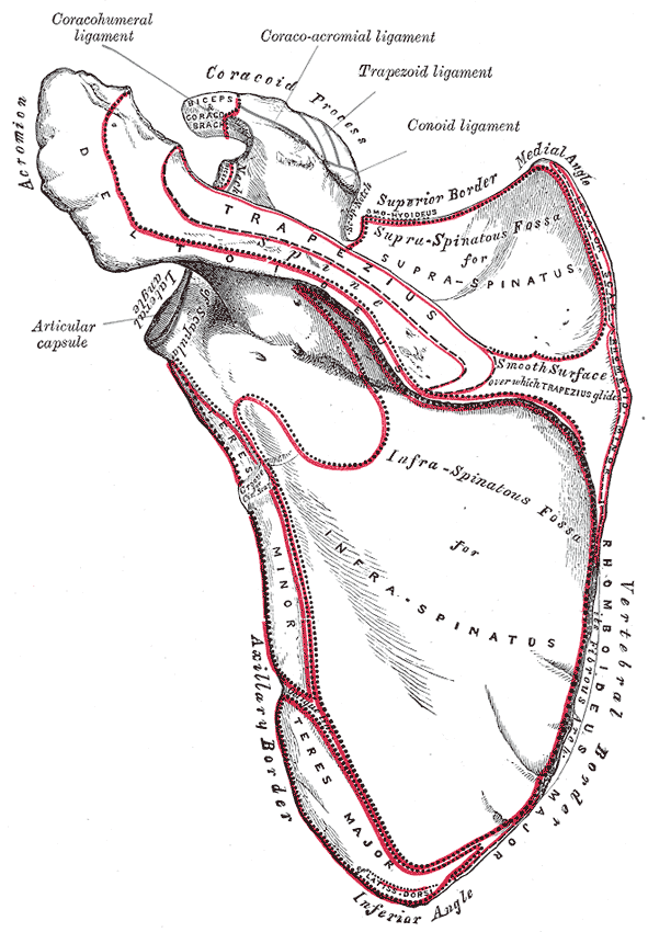
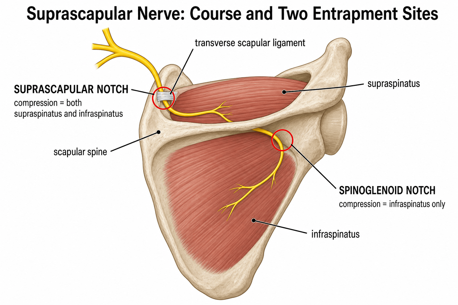
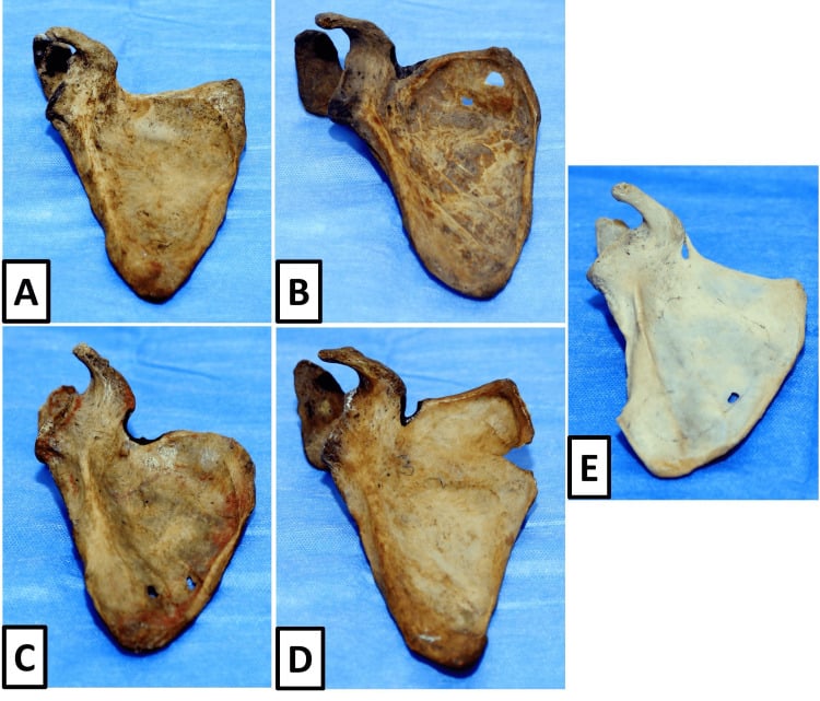
The course. The suprascapular nerve (C5-C6, occasionally C4) arises from the upper trunk of the brachial plexus. It travels posteriorly through the suprascapular notch at the superior border of the scapula, beneath the transverse scapular ligament, and gives motor branches to supraspinatus. It then continues around the scapular spine through the spinoglenoid notch at the base of the spine, under the spinoglenoid ligament, to innervate infraspinatus.
Why the level matters. Compression at the suprascapular notch denervates both supraspinatus and infraspinatus. Compression at the spinoglenoid notch spares supraspinatus and isolates infraspinatus, so a preserved supraspinatus is the key sign pointing to distal compression. The nerve is relatively fixed at both notches, which makes it vulnerable to traction and mass effect.
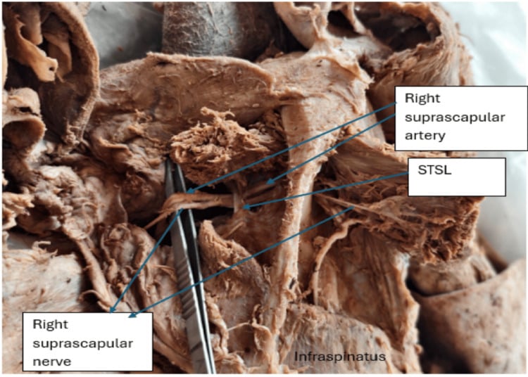
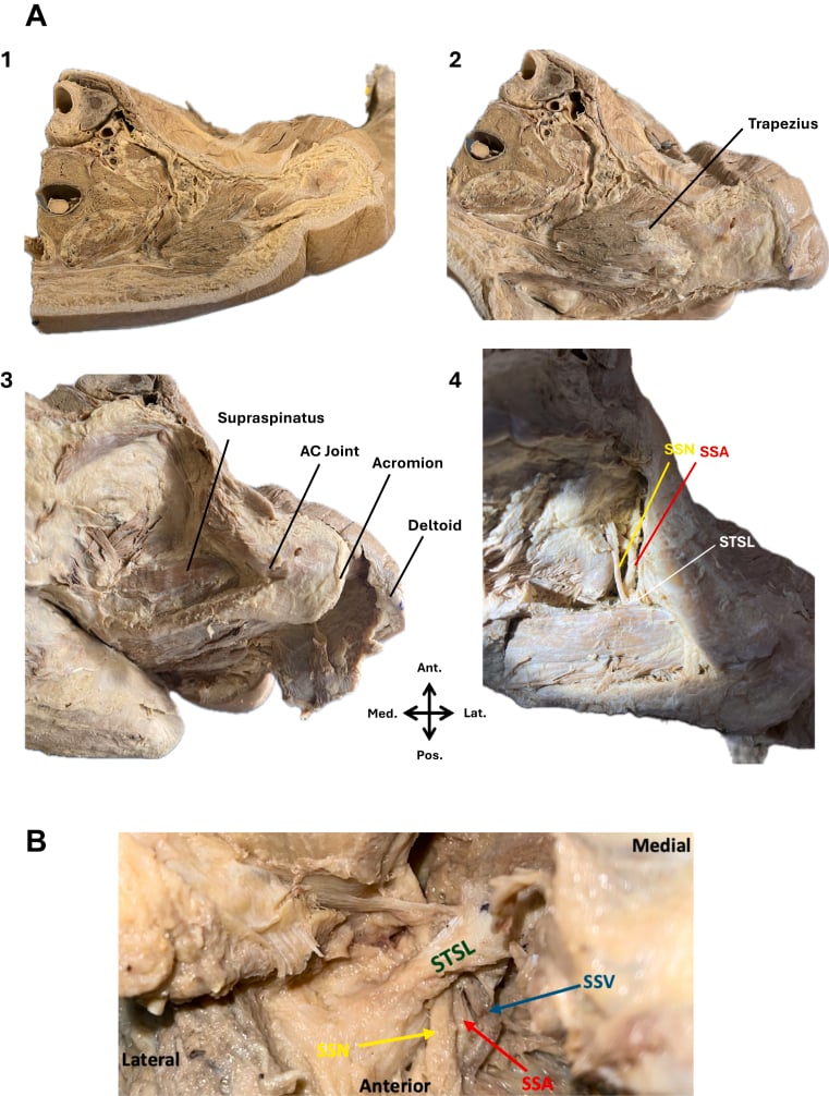
Not purely motor. The nerve is predominantly motor, but it carries articular (sensory) branches to the acromioclavicular joint, the subacromial bursa and the posterosuperior glenohumeral joint capsule. Together these supply roughly 70 percent of the sensory innervation of the shoulder joint. That is the anatomical basis of the suprascapular nerve block, infiltrated at the suprascapular notch or the floor of the supraspinatus fossa, and used for analgesia in:
- Adhesive capsulitis
- Rheumatoid and osteoarthritic shoulder pain
- Post-operative pain
- Palliation of malignant shoulder pain
The paralabral ganglion. A superior or posterosuperior labral tear allows synovial fluid to track into the spinoglenoid region. The fluid accumulates into an expanding cyst that compresses the nerve against the notch, and infraspinatus denervation and wasting follow. Recurrence is high if the labral tear is not addressed, which is why cyst decompression alone is not enough and the operation includes labral repair.
Traction from a massive cuff tear. A distinct, examinable mechanism beyond the two compressive notches: a massive, medially retracted supraspinatus/infraspinatus tear tethers and tractions the suprascapular nerve. As the musculotendinous junction retracts medially, the nerve is drawn medially and its angle at the suprascapular notch increases, producing a traction neuropathy that compounds the wasting of the torn cuff.
This is the likely reason EMG abnormalities are commoner with massive cuff tears (as in the Boykin series), and it is the rationale for nerve assessment in that setting. Restoring the musculotendinous unit by repairing the retracted tear can relieve the traction, and is generally preferred to an isolated nerve release. Distinguish this traction mechanism from true notch entrapment by a discrete compressive lesion.
What happens to the muscle. Chronic compression leads to muscle atrophy followed by fatty infiltration (Goutallier stages). Irreversible changes begin after 6-12 months of untreated compression.
Classification and Types
Distinguishing the notch level is the single most important step in planning surgery.
- Suprascapular Notch
- Supraspinatus + infraspinatus
- Spinoglenoid Notch
- Infraspinatus only
- Suprascapular Notch
- Both fossae
- Spinoglenoid Notch
- Infraspinatus fossa only
- Suprascapular Notch
- Ligament entrapment, traction, tumour
- Spinoglenoid Notch
- Paralabral ganglion cyst, labral tear
- Suprascapular Notch
- Deep, poorly localised posterosuperior shoulder ache from the articular branches
- Spinoglenoid Notch
- Typically less pain than a notch lesion, because fewer articular branches are affected
- Suprascapular Notch
- Ligament release +/- notchplasty
- Spinoglenoid Notch
- Cyst decompression + labral repair
By aetiology. Repetitive traction in overhead athletes, weightlifters and swimmers is one mechanism. The nerve can also be compressed by a thickened or ossified transverse scapular ligament, a ganglion, fracture callus, or a space-occupying lesion such as a tumour or varix. Entrapment can also be iatrogenic, after distal clavicle excision or shoulder surgery. Ganglion cysts account for the majority of surgically treated spinoglenoid notch cases.
- Mechanism
- Thickened transverse scapular ligament
- Associated Findings
- No mass on MRI
- Treatment Notes
- Open or arthroscopic ligament release
- Mechanism
- Labral tear with cyst formation
- Associated Findings
- MRI shows cyst and labral defect
- Treatment Notes
- Arthroscopic labral repair + cyst drainage
- Mechanism
- Scapular fracture or callus
- Associated Findings
- History of trauma, callus on CT
- Treatment Notes
- Decompression of callus mass
- Mechanism
- Tumour, varix, or aneurysm
- Associated Findings
- MRI or CT shows mass
- Treatment Notes
- Oncological or vascular consultation
Clinical Assessment
History. The pain is deep, aching and posterior, related to activity and worse at night. Weakness shows as difficulty with external rotation, overhead lifting and throwing. Onset is insidious in ganglion cases and acute after trauma or repetitive stress. Ask about previous treatment: failed physiotherapy, injections or rotator cuff repairs.
Inspection. Compare the two sides. Infraspinatus fossa wasting is the hallmark, and supraspinatus wastes as well if the lesion is proximal.
Pain without numbness. Expect no cutaneous numbness at either site, because the suprascapular nerve supplies no skin. Its articular branches produce a deep, poorly localised posterosuperior ache instead, so the discriminating pattern is pain without numbness. Demonstrable dermatomal or patch numbness points elsewhere: C5 radiculopathy, or the axillary nerve for the regimental badge area.
Examination tests. External rotation is weak at the side and in 90 degrees of abduction.
- Infraspinatus strength. Seated, arm at the side, elbow flexed 90 degrees, resist external rotation. Weakness against the contralateral side indicates infraspinatus dysfunction.
- External rotation lag sign. Passively externally rotate to maximum and release. A lag greater than 10 degrees indicates significant infraspinatus weakness.
- Hornblower sign. The patient brings the hand to the mouth with the elbow elevated. It is positive if the arm drops into internal rotation (infraspinatus failure).
- Jobe test. Positive if supraspinatus is involved.
- Notch palpation. Tenderness 2 cm medial to the posterolateral acromion (suprascapular notch) or at the base of the scapular spine (spinoglenoid notch).
- Wasting Pattern
- Infraspinatus +/- supraspinatus
- Discriminating Finding
- Notch tenderness, positive lag sign
- Key Test / Imaging
- EMG abnormal (motor unit changes more often than frank denervation); MRI ganglion or ligament
- Wasting Pattern
- Supraspinatus +/- infraspinatus
- Discriminating Finding
- Positive impingement, weakness on Jobe
- Key Test / Imaging
- Ultrasound or MRI shows tendon tear
- Wasting Pattern
- Deltoid, biceps, brachioradialis
- Discriminating Finding
- Neck pain, dermatomal sensory loss
- Key Test / Imaging
- Cervical MRI, EMG myotomal pattern
- Wasting Pattern
- Patchy, often multiple nerves
- Discriminating Finding
- Acute severe pain followed by weakness
- Key Test / Imaging
- EMG shows axonal neuropathy, no mass
Isolated infraspinatus atrophy with a normal supraspinatus is almost pathognomonic for spinoglenoid notch compression by a paralabral ganglion. Always obtain an MRI with intra-articular contrast or high-resolution sequences to visualise the labral tear and cyst. Missing the labral pathology leads to cyst recurrence after isolated decompression.
Investigations
Radiographs. AP, axillary and scapular Y views of the shoulder. They are usually normal and serve to rule out other bony pathology; look for ossification at the notch, fracture callus and bony tumours.
EMG. Indicated in every suspected case before surgery; it confirms the diagnosis and localises the level. The findings are fibrillations, positive sharp waves and reduced recruitment in infraspinatus, and in supraspinatus as well if the lesion is proximal. Fibrillations and positive sharp waves appear 3-4 weeks after onset, so study at least 3-4 weeks after symptoms begin. A normal early EMG does not exclude entrapment; repeat it if suspicion is high.
Denervation is the minority finding. In the Boykin referral series, frank denervation was present in only 33% of electrodiagnostically confirmed cases, while the commonest abnormality was far subtler: abnormal motor unit action potentials, in 88%. A study reported without fibrillations therefore does not exclude the diagnosis, and waiting for denervation before imaging will miss most of it. Do not make MRI conditional on finding denervation on EMG.
Suprascapular neuropathy in a shoulder referral practice
- Of 92 patients undergoing electrodiagnostic testing for suspected SSN, 40 (43%) were confirmed; SSN was diagnosed in 4.3% of all new patients
- Patients with a massive rotator cuff tear were significantly more likely to have an abnormal study (p=0.006)
- Abnormal motor unit action potentials were the commonest finding (88%); frank denervation was seen in only 33%
- The diagnostic parameters the authors say surgeons should know: mean latency in studies reported as diagnostic was 2.90 plus or minus 0.08 ms for supraspinatus and 3.78 plus or minus 0.14 ms for infraspinatus
- The 43% figure is the yield among patients already suspected clinically or radiographically - 40 of 92 (42%) of those tested - and is not a population prevalence
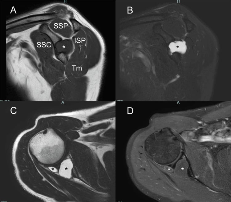
MRI. The key investigation, indicated in all cases with a positive EMG or high clinical suspicion. Axial and coronal oblique T2 fat-saturated sequences, with an MR arthrogram if a labral tear is suspected. It shows the size and location of a ganglion cyst, the labral tear, muscle atrophy and fatty infiltration, and ligament thickening. Always comment on labral integrity and the Goutallier grade of fatty infiltration.
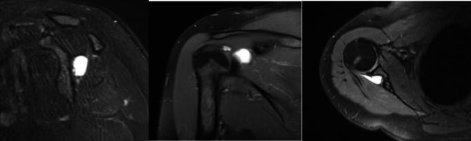
Intraosseous cyst. A less common compressive mechanism is a multiloculated intraosseous cyst extending to the spinoglenoid notch. T1, fluid-sensitive and contrast-enhanced sequences distinguish its cystic content, its enhancing margins and its relation to the suprascapular nerve.
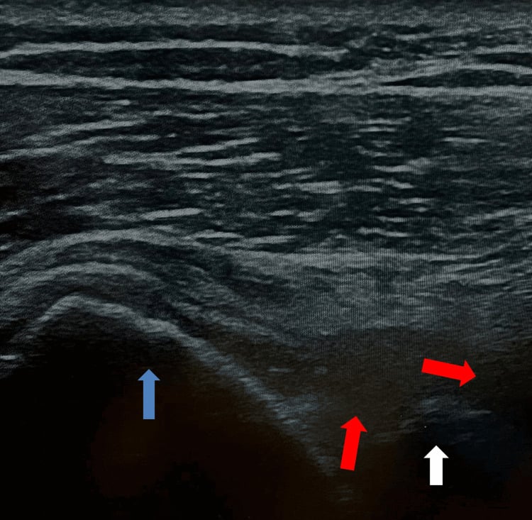
Ultrasound. Ultrasound can demonstrate a cystic lesion at the spinoglenoid notch and guide aspiration or diagnostic injection. MRI better maps the labral communication and muscle denervation.
CT. Reserved for selected cases: bony notch narrowing, fracture callus or tumour. It gives better bone detail for planning a notchplasty.
The combination of isolated infraspinatus atrophy plus a spinoglenoid notch ganglion on MRI is diagnostic in its own right.
Management
Conservative first for most. Non-operative care is first line for most cases and is appropriate for idiopathic or traction-related entrapment without a space-occupying lesion. The aim is to reduce nerve irritation, maintain muscle function and allow spontaneous recovery in traction or idiopathic cases.
Weeks 0-6. Avoid repetitive overhead loading, heavy lifting and contact sports; light daily activities and scapular stabilisation exercises are allowed. Physiotherapy focuses on periscapular strengthening and posture correction.
Weeks 6-12. Repeat the clinical examination for pain, strength and progression of atrophy, and reassess every 6 weeks with objective strength testing. A short course of NSAIDs or analgesia treats a flare. Repeat the EMG, and if there is no improvement at 3 months, consider surgery.
The decision at 3 months. Continue non-operative care when symptoms are mild and stable, compliance is good and there is no mass lesion. Surgery is indicated for:
- Failed conservative care (more than 3 months)
- Progressive weakness or worsening atrophy
- Persistent pain affecting function
- A compressive lesion confirmed on MRI, such as a ganglion
- Acute traumatic entrapment with documented denervation
The ganglion does not wait. A documented ganglion cyst causing progressive infraspinatus atrophy is best managed surgically, because delay risks irreversible fatty infiltration.
Muscle quality sets the ceiling. Assess fatty infiltration before decompression. Advanced fatty change (greater than Goutallier grade 2) predicts poorer functional recovery after decompression, and advanced fatty infiltration (Goutallier greater than 3) is listed as a contraindication to suprascapular notch release in the approach table below.
- Diagnosis
- Suprascapular notch compression, no ganglion
- Treatment
- Transverse scapular ligament release +/- notchplasty
- Key Pearl
- Address ligament first; check for space-occupying lesion
- Diagnosis
- Spinoglenoid notch + paralabral ganglion on MRI
- Treatment
- Arthroscopic labral repair + cyst decompression
- Key Pearl
- Treat the labral tear to prevent recurrence
- Diagnosis
- Confirmed entrapment on EMG/MRI
- Treatment
- Surgical decompression at affected notch
- Key Pearl
- Delay increases irreversible atrophy risk
Surgical Technique
Choosing the approach. The notch and the pathology decide between an arthroscopic and an open approach.
- Preferred Approach
- Arthroscopic or open ligament release
- Key Steps
- Divide transverse scapular ligament, notchplasty if narrow
- Contraindications
- Advanced fatty infiltration (Goutallier greater than 3)
- Preferred Approach
- Arthroscopic labral repair + cyst decompression
- Key Steps
- Posterior portal, labral repair, cyst drainage, spinoglenoid ligament release
- Contraindications
- Large extra-articular cyst requiring open approach
Suprascapular notch release. Identify the superior transverse scapular ligament, protect the suprascapular artery, and divide the ligament in a controlled way under direct vision, and add a notchplasty if the notch is narrow. Adequate release is confirmed without traction or thermal injury to the nerve.
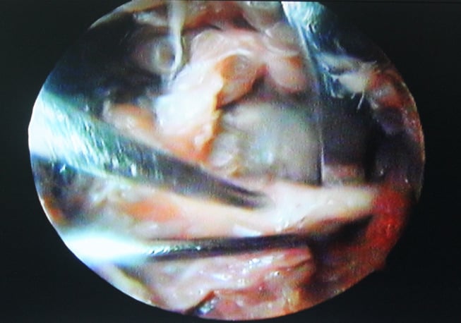
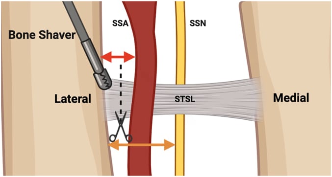
Spinoglenoid ganglion, arthroscopic. Beach chair or lateral decubitus with the arm in traction. A posterior viewing portal, an anterior working portal, and an accessory posterolateral portal for access to the cyst.
- Address the labral tear with suture anchors
- Decompress the cyst by incising the capsule and draining its contents
- Release the spinoglenoid ligament if it is thickened
- Confirm the nerve is free of compression
The axillary nerve (inferior), the suprascapular artery (medial) and the articular cartilage.
After surgery. Sling for 4-6 weeks with early passive range of motion, then progressive strengthening after the labrum has healed.
Arthroscopic suprascapular nerve release: indications and technique
- Described indications and surgical technique for arthroscopic suprascapular nerve release at both notches
- Emphasized addressing compressive pathology such as ganglion cysts and ligament release
- Good outcomes with arthroscopic approach in selected patients
Complications
Complications arise both from the untreated entrapment and from surgical decompression.
Of the untreated entrapment.
- Irreversible atrophy of infraspinatus, with or without supraspinatus, with fatty infiltration (Goutallier)
- Permanent external-rotation weakness and loss of overhead and throwing power
- Secondary impingement and scapular dyskinesis, because loss of infraspinatus alters scapular mechanics and force couples
- Chronic posterior shoulder pain and reduced sporting or occupational capacity
- Cyst enlargement with intra-osseous extension, or recurrent effusion, if a labral tear is left untreated
Of surgical decompression.
- Iatrogenic nerve injury to the suprascapular nerve itself, or to the nearby axillary nerve (inferior) during spinoglenoid work
- Suprascapular artery injury causing bleeding; the artery runs over the ligament at the notch
- Incomplete decompression, with the ligament not fully divided or the notch still stenotic, and persistent symptoms
- Persistent weakness despite a technically successful release, when advanced fatty infiltration was present before surgery
- Stiffness or failure of labral healing, and the standard arthroscopic risks of infection and chondral injury
Recurrence of the ganglion cyst is the commonest failure, almost always from an unrepaired labral tear. Recurrent infraspinatus symptoms after spinoglenoid cyst decompression are, until proven otherwise, due to an unaddressed labral tear allowing the cyst to reform via a one-way valve. This is why labral repair - not cyst drainage alone - is the operation. Counsel and image accordingly before any revision.
Guidelines, Registries & Global Practice
- Overhead athletes (tennis, volleyball, swimming) have the highest incidence worldwide
- Ganglion cysts are the leading cause of surgically treated spinoglenoid entrapment in all regions
- Idiopathic ligamentous compression predominates in manual labourers and older patients
- Missed diagnosis is common; many patients undergo unsuccessful rotator cuff surgery before correct identification
- High-resource centres: Routine pre-operative EMG plus high-resolution MRI; arthroscopic decompression standard
- Limited-resource settings: Clinical diagnosis and basic MRI; open ligament release remains effective
- Universal principle: Outcome depends on timely decompression before irreversible fatty infiltration
- Surgery: Concentrated in shoulder-specialist units; revision rates higher when labral pathology is missed
- Diagnosis emphasis
- EMG confirmation plus MRI for mass lesion
- Conservative care
- Activity modification and physiotherapy 3 months
- Surgical indications
- Progressive deficit or compressive mass
- Diagnosis emphasis
- Clinical + EMG; MRI for ganglion and labrum
- Conservative care
- Physiotherapy-led; early referral if no improvement
- Surgical indications
- Failed conservative or documented cyst
- Diagnosis emphasis
- High index of suspicion in overhead athletes
- Conservative care
- Individualised; shorter conservative trial in athletes
- Surgical indications
- Early surgery for ganglion cysts in young patients
- Diagnosis emphasis
- EMG and MR arthrogram for labral assessment
- Conservative care
- Sport-specific rehabilitation protocols
- Surgical indications
- Decompression before Goutallier grade 3
There is no dedicated international registry for suprascapular nerve entrapment. Evidence is derived from case series and small prospective cohorts. The consistent message across guidelines is that EMG confirms the diagnosis, MRI identifies treatable compressive pathology (especially ganglion cysts), and surgical decompression is effective when performed before advanced muscle degeneration. Always document pre-operative muscle quality and counsel patients on realistic strength recovery.
Controversies & Areas of Uncertainty
Arthroscopic techniques allow simultaneous labral repair and cyst decompression with less morbidity, but open release remains reliable for isolated suprascapular notch ligament division. No randomised trials exist; choice depends on surgeon experience and pathology.
Most authors recommend 3 months of non-operative treatment before surgery, yet athletes with documented ganglion cysts and progressive atrophy may benefit from earlier intervention. The precise threshold for "failed conservative care" is not standardised.
Some surgeons routinely perform bony notchplasty after ligament release; others reserve it for visibly stenotic notches. No comparative data define the added benefit or risk of routine bony enlargement.
Recurrent symptoms after cyst decompression are usually due to untreated labral tears. Revision rates and functional results after re-operation are poorly documented in the literature.
Exam Viva Scenarios
Practise clinical reasoning and management decisions out loud
“A 32-year-old right-hand-dominant tennis player presents with 4 months of posterior shoulder pain and progressive weakness in external rotation. Examination shows visible wasting of the infraspinatus fossa with preserved supraspinatus. External rotation strength is 4-/5 at the side and in 90 degrees abduction. MRI reveals a 2.5 cm spinoglenoid notch ganglion cyst with a superior labral tear. EMG confirms denervation limited to infraspinatus. What is your diagnosis and recommended treatment?”
“A 45-year-old manual labourer presents with 9 months of deep aching shoulder pain and progressive weakness. He has wasting of both supraspinatus and infraspinatus fossae. External rotation and abduction strength are reduced. No labral tear or ganglion is seen on MRI. EMG shows denervation of both supra- and infraspinatus with normal cervical paraspinals. The transverse scapular ligament appears thickened. What is your diagnosis and surgical plan?”
Key Anatomy
- Suprascapular nerve (C5-C6) passes under transverse scapular ligament at suprascapular notch
- Spinoglenoid notch lies at base of scapular spine; compression here spares supraspinatus
- Paralabral ganglion from labral tear is the most common compressive lesion at spinoglenoid notch
- Nerve is fixed at both notches, making it vulnerable to traction and mass effect
Clinical Diagnosis
- Infraspinatus wasting +/- supraspinatus wasting localises the notch level
- External rotation weakness at side and 90 degrees abduction; positive lag and hornblower signs
- Deep posterior shoulder pain, worse with overhead activity and at night
- Always examine for labral tear signs when isolated infraspinatus atrophy present
Investigations
- EMG localises the level (perform greater than 3 weeks after onset), but frank denervation appears in only about a third of confirmed cases - abnormal motor unit action potentials are the commoner finding
- MRI identifies ganglion cyst, labral tear, and Goutallier fatty infiltration grade
- MR arthrogram improves labral tear detection when spinoglenoid ganglion suspected
- Normal early EMG does not exclude entrapment; repeat if high clinical suspicion
Treatment Algorithm
- Conservative first for idiopathic/traction cases: activity modification, physiotherapy 3 months
- Surgery indicated for progressive deficit, compressive mass, or failed conservative care
- Suprascapular notch: transverse scapular ligament release +/- notchplasty
- Spinoglenoid notch + ganglion: arthroscopic labral repair + cyst decompression + ligament release
Surgical Pearls
- Always address labral tear when decompressing a ganglion cyst to prevent recurrence
- Assess Goutallier grade pre-operatively; greater than grade 2 predicts poorer strength recovery
- Post-op sling 4-6 weeks, early passive ROM, scapular stabilisation, then strengthening
- Document pre-operative EMG and muscle quality; counsel on realistic functional gains
Complications & Failure
- Recurrent cyst if labral tear not repaired (most common failure mode)
- Persistent pain or weakness if advanced fatty infiltration present pre-operatively
- Nerve injury during arthroscopic decompression (rare with careful portal placement)
- Stiffness or scapular dyskinesis if rehabilitation is inadequate
Evidence Base and Key Trials
Suprascapular nerve entrapment at the spinoglenoid notch
- PUBMED INDEXES THIS 1982 PAPER WITH NO ABSTRACT, so the points below reflect the entity as attributed to Aiello in the subsequent literature rather than quotations from the authors; title, authors, journal, year and DOI were resolved against PubMed
- The indexed MeSH terms do support the substance: Ligaments/pathology, HYPERTROPHY, Nerve Compression Syndromes/pathology, Scapula/pathology, Sports Medicine, in a middle-aged male - consistent with spinoglenoid compression by a hypertrophied ligament in an athlete
- It is credited with the first clear description of isolated infraspinatus atrophy from spinoglenoid notch compression, and with establishing the notch-specific clinical and EMG pattern
Suprascapular nerve entrapment: a meta-analysis
- Literature review (1959-2001) of 88 cases meeting inclusion criteria; entrapment is rare and occurs mainly in patients under 40 years
- A ganglion compressing the nerve was significantly commoner in males, and typically caused ISOLATED infraspinatus atrophy
- Combined supra- and infraspinatus atrophy was more typical of LIGAMENTOUS compression, and a history of trauma pointed to the ligament as the cause