The Cuff Tendon That Defines Rotator Cuff Surgery
- Origin: medial two-thirds of the supraspinous fossa plus the supraspinous fascia; the muscle is pennate with a dominant anterior tendon.
- Insertion: superior facet of the greater tuberosity, footprint abutting the articular margin with a bare area of roughly 1 mm.
- Innervation: suprascapular nerve (C5, C6, variably C4) via the suprascapular notch, under the superior transverse scapular ligament.
- The critical zone lies about 1 cm medial to the insertion and is hypovascular on the articular side.
- The rotator cable stress-shields the crescent, which is why some large tears still elevate.
- “Army under the bridge, Navy over: the suprascapular nerve passes UNDER the superior transverse scapular ligament, the artery passes OVER it.
- “Mochizuki showed the supraspinatus footprint is far smaller than classically taught; the infraspinatus occupies much of the superior facet.
- “Supraspinatus does not merely initiate abduction; it is active throughout the arc and contributes roughly a third of abduction torque.
- “Acromiohumeral interval less than 7 mm indicates a chronic, likely massive, cuff-deficient shoulder.
Overview
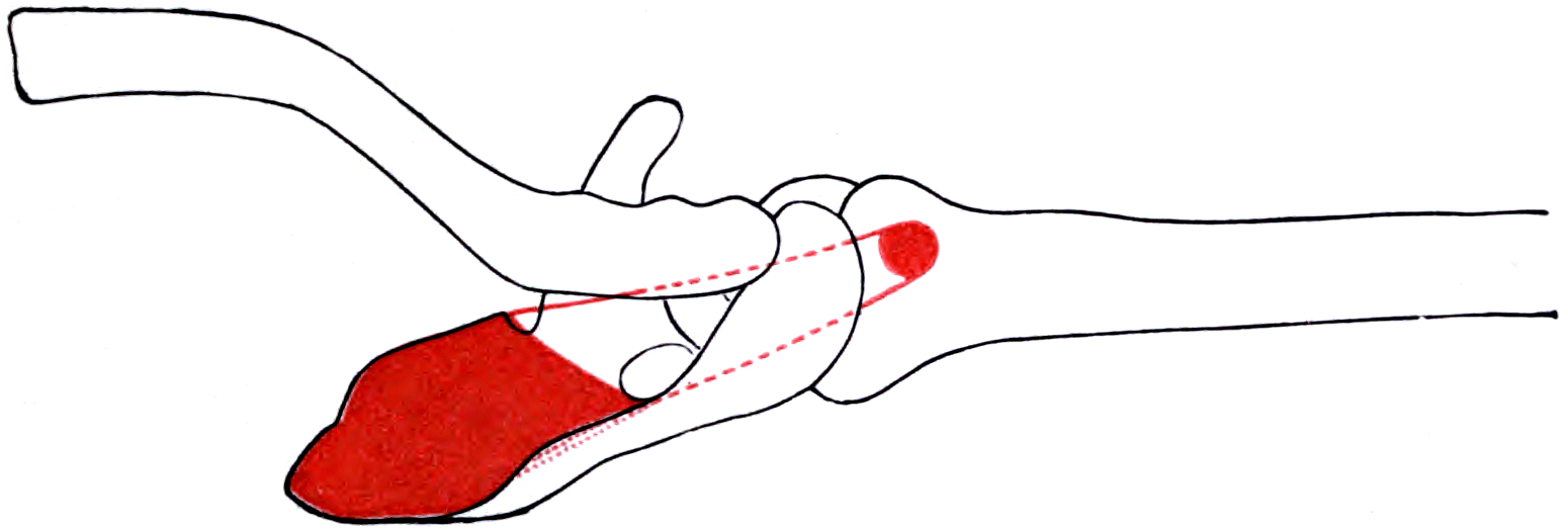
The supraspinatus fills the supraspinous fossa above the scapular spine, passes beneath the coracoacromial arch, and inserts on the superior facet of the greater tuberosity. It is the smallest of the four cuff muscles by cross-sectional area yet accounts for the overwhelming majority of rotator cuff surgical volume, because it sits in the narrowest passage in the shoulder and carries the highest tensile load per unit area.
Two ideas dominate every viva on this tendon. The first is that the supraspinatus is a compressor, not simply an elevator: its line of pull is almost horizontal in the scapular plane, so its dominant vector presses the humeral head into the glenoid concavity and creates the fulcrum against which the deltoid works. The second is that the tendon is part of a continuous cuff-capsule hood, not an isolated strap — its fibres interdigitate with the infraspinatus posteriorly and with the coracohumeral and superior glenohumeral ligament complex anteriorly at the rotator interval.
Examiners will push on two mechanical models. Know both and know when each applies.
- Concavity compression (Lippitt and Matsen). The cuff compresses the head into a glenoid whose effective depth is doubled by the labrum. Stability is proportional to compressive force and to concavity depth. The supraspinatus is the superior arm of that compressive ring; when it fails, the deltoid's superiorly directed vector is no longer resisted and the head migrates upward.
- The suspension bridge (Burkhart). The rotator cable is a thick, cord-like condensation running from just posterior to the biceps groove, arcing medial to the cuff insertion, to the inferior border of the infraspinatus. It behaves like the cable of a suspension bridge: load is transmitted from the muscle bellies to the two bony anchors, stress-shielding the thin crescent of insertional tissue lateral to it.
- Why this matters clinically. A tear confined to the crescent leaves the cable intact, so force transmission continues around the defect and the patient still elevates. A tear that detaches an anchor of the cable, or that propagates through the cable, destroys the transmission path and produces pseudoparalysis out of proportion to tear size.
Army under the bridge, Navy overSuprascapular Notch Relations
Hook:Explains why a ganglion at the suprascapular notch denervates both muscles and one at the spinoglenoid notch denervates only infraspinatus.
Attachments, Innervation and Relations
Origin
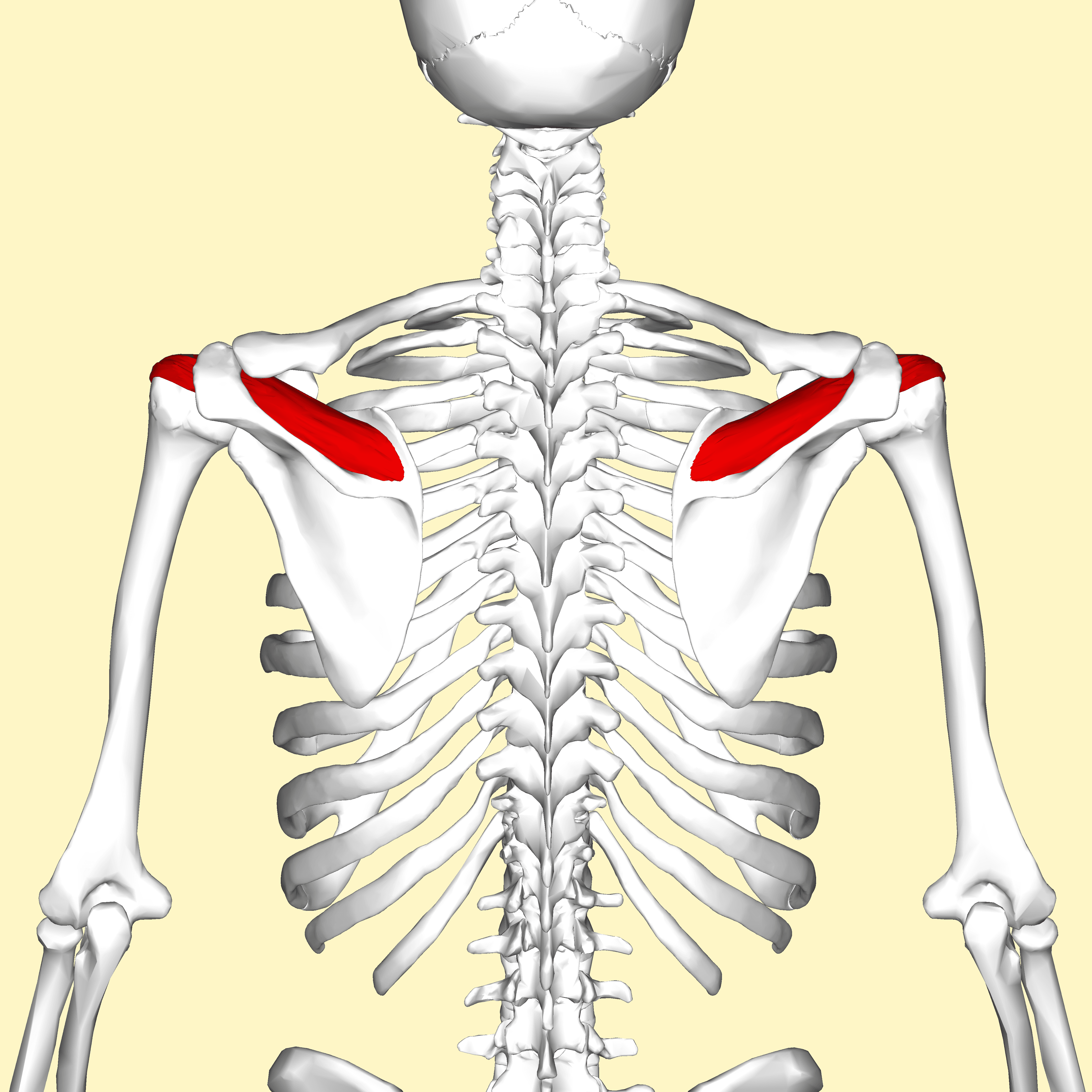
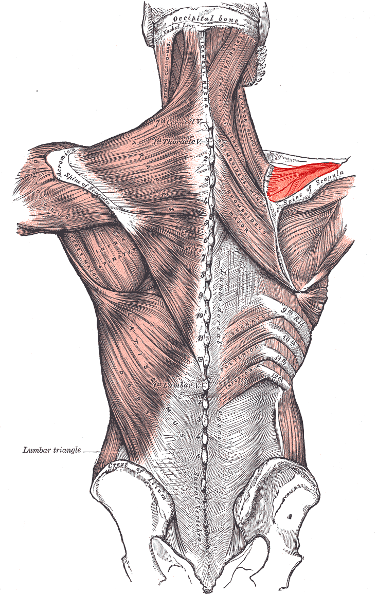
- Medial two-thirds of the supraspinous fossa on the dorsal scapula, above the scapular spine.
- Additional fibres from the deep surface of the supraspinous fascia, which roofs the fossa and converts it into an osteofascial compartment.
- The muscle is bipennate with a dominant, thick anterior tendon that is roughly cylindrical and carries most of the load, and a thinner, flatter posterior muscular portion. Load is not distributed evenly: the anterior tendon has the highest stiffness and ultimate strength, which is why tears typically begin at the anterior insertion just behind the biceps.
Insertion — the footprint
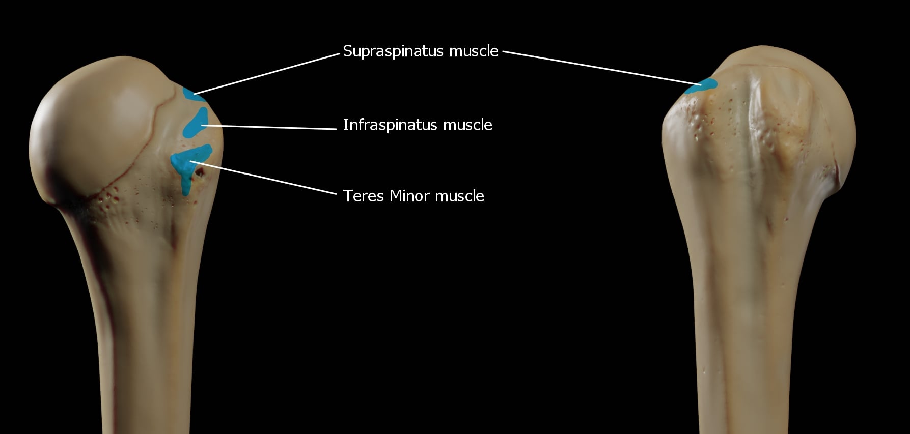
- Superior facet of the greater tuberosity, with the medial edge of the footprint essentially abutting the articular cartilage margin; cadaveric work places the bare area between cartilage and footprint at under 1 mm anteriorly.
- Classical dimensions: the supraspinatus footprint measures approximately 23 mm anterior-to-posterior and 12 to 16 mm medial-to-lateral, on a superior facet whose total surface is a rough trapezoid.
- Refined dimensions (Mochizuki and colleagues, JBJS Am 2008): careful separation of supraspinatus from infraspinatus fibres shows the true supraspinatus insertion is a small triangle of roughly 12.6 mm anterior-to-posterior and 6.9 mm medial-to-lateral at the anterior part of the superior facet, with the infraspinatus occupying the remainder of the superior facet as well as the middle facet. This is the single most examinable modern anatomy finding on this tendon.
- Surgical translation: the footprint you must restore is broader than a single row of anchors placed at the articular margin will cover; the medial-to-lateral dimension is the reason double-row and transosseous-equivalent constructs were developed.
- Some fibres continue over the bicipital groove and blend with the transverse humeral ligament and the lateral wall of the biceps pulley.
The leash that limits mobilisation.
- In 84 per cent of shoulders there are no more than two motor branches to supraspinatus, arising under the transverse scapular ligament or just distal to it.
- The supraspinatus motor branches lie a mean of 3 cm from the origin of the long head of biceps; the infraspinatus branches a mean of 2 cm from the posterior glenoid rim.
- A standard anterosuperior approach allows only about 1 cm of safe lateral advancement; a Debeyre-type advancement up to 3 cm, limited by branch tension.
- A repaired but denervated cuff is a worse outcome than an unrepaired one.
The limit of the deltoid split.
- In a mini-open or lateral transdeltoid repair, the anterior branch crosses the deep surface of the deltoid roughly 5 to 7 cm distal to the lateral acromion.
- Place a stay suture at the distal apex of the split before retracting.
- Splits carried distally, or aggressive retraction, denervate the anterior and middle deltoid.
Action and Biomechanics
Actions by plane
- Scapular-plane elevation (abduction): the supraspinatus is active through the entire arc, not just the first 30 degrees. It contributes roughly one third of abduction torque.
- Concavity compression: its resultant vector is largely medially directed (compressive) with a smaller superior component, so it seats the head rather than lifting it.
- External rotation: a minor contribution from the posterior fibres.
- Capsular tensioning: as part of the cuff-capsule hood, it tensions the superior capsule and, with the coracohumeral ligament, resists inferior subluxation of the adducted arm.
Moment arm and length-tension
- The supraspinatus moment arm for abduction is largest in the lower range and diminishes as the arm elevates, while the deltoid's moment arm grows. The two are therefore complementary: the cuff dominates initiation and stabilisation, the deltoid dominates power at higher angles.
- With the arm at the side, the deltoid's line of pull is nearly parallel to the humeral shaft, so almost all its force is shear (superiorly directed). Without supraspinatus compression, that shear translates the head superiorly rather than rotating it — the mechanical basis of proximal migration.
Synergists and antagonists
- Synergists: deltoid (elevation), infraspinatus and teres minor and subscapularis (compression), long head of biceps (a secondary head depressor, often hypertrophied in chronic cuff deficiency).
- Antagonists: latissimus dorsi, pectoralis major, teres major (adductors and depressors).
This is the classic examiner probe. Four mechanisms, all worth stating.
- The transverse force couple is intact. Subscapularis anteriorly is balanced by infraspinatus and teres minor posteriorly. Their equal and opposite pull still centres the head, so the deltoid retains a stable fulcrum.
- The rotator cable is intact. If the tear lies within the crescent, load is still transmitted along the cable to its anterior and posterior bony anchors. Burkhart's term for this is a functional or balanced tear.
- Deltoid compensation. The deltoid can elevate a shoulder that has a stable fulcrum, and hypertrophies to do so.
- The long head of biceps acts as a secondary depressor in the cuff-deficient shoulder.
Conversely, pseudoparalysis appears when the tear extends anteriorly into the rotator interval and subscapularis, or posteriorly beyond the infraspinatus, unbalancing the couple and detaching a cable anchor. Tear size alone does not predict function — tear configuration does.
What happens when it fails
- Loss of concavity compression allows superior translation, narrowing the acromiohumeral interval and producing secondary abrasion of the tendon and of the undersurface of the acromion.
- Chronic loss leads to fatty infiltration and myotendinous retraction, both of which shorten the muscle and reduce the excursion available for repair.
- End stage is rotator cuff tear arthropathy: acetabularisation of the acromion, femoralisation of the humeral head, and loss of the glenohumeral fulcrum.
Surface Anatomy and Examination
Palpation and positioning
- The supraspinous fossa is palpated above the scapular spine; muscle bulk here is compared side to side, since wasting is the earliest sign of a chronic tear or of suprascapular neuropathy.
- The tendon insertion is brought out from under the acromion by placing the arm in extension, adduction and internal rotation (the hand behind the back position), which delivers the superior facet anterolaterally where it can be palpated just anterior to the anterolateral acromial corner.
- The suprascapular notch is tender to deep palpation just medial to the base of the coracoid, above the scapular spine.
Named tests
Isolation test for supraspinatus.
- Arm at 90 degrees in the scapular plane (30 degrees anterior to the coronal plane), full internal rotation, thumb down.
- Resist downward pressure.
- Positive: weakness or pain.
- Caveat: the empty-can position is provocative and pain, not weakness, often limits the patient. The full-can position (external rotation, thumb up) generates comparable supraspinatus activity with less pain and less deltoid substitution.
Test of a large tear, not a small one.
- Passively elevate to 90 degrees or above, then ask the patient to lower the arm slowly.
- Positive: the arm drops or descends in a jerky, uncontrolled manner.
- Interpretation: highly specific for a large full-thickness tear, poorly sensitive for small ones.
Localises to supraspinatus plus upper infraspinatus.
- Elbow 90 degrees, arm at the side, passively place near-maximal external rotation, then release.
- Positive: the arm springs back into internal rotation.
- A lag of 5 to 10 degrees suggests supraspinatus involvement; larger lags implicate the infraspinatus.
Bursal, not tendon-specific.
- Neer: passive forced elevation with the scapula stabilised.
- Hawkins-Kennedy: 90 degrees forward flexion then forced internal rotation.
- Both are sensitive and non-specific; a positive test localises pain to the subacromial space but says nothing about tendon integrity.
Tabulated examination summary
- How to perform
- 90 degrees scapular plane, thumb up, resist depression
- Positive finding
- Weakness against resistance
- What it means
- Supraspinatus deficiency with less pain confounding
- False positives
- Deltoid substitution if scapula not stabilised
- How to perform
- 90 degrees scapular plane, thumb down, resist depression
- Positive finding
- Pain or weakness
- What it means
- Supraspinatus load, but provocative
- False positives
- Bursitis, calcific deposit, acromioclavicular arthropathy
- How to perform
- Lower the elevated arm slowly
- Positive finding
- Uncontrolled drop
- What it means
- Large full-thickness tear
- False positives
- Pain inhibition in acute calcific tendinopathy
- How to perform
- Passive near-maximal ER at the side, release
- Positive finding
- Arm springs into internal rotation
- What it means
- Supraspinatus plus upper infraspinatus tear
- False positives
- Stiffness limits the passive position and masks the lag
- How to perform
- Reassess power after subacromial local anaesthetic
- Positive finding
- Weakness persists
- What it means
- Structural tear rather than pain inhibition
- False positives
- Inadvertent intra-articular or intratendinous injection
Grading and pitfalls
- Grade power with the scapula stabilised; scapular hitching is the commonest source of a falsely normal test.
- False positives: pain inhibition in calcific tendinopathy, adhesive capsulitis and acromioclavicular arthropathy all produce apparent weakness.
- False negatives: in a cable-intact crescent tear, strength testing may be entirely normal.
Complications
Failure of healing
- Re-tear is the dominant complication. Rates rise steeply with tear size, age over 65, Goutallier grade 3 or 4, diabetes and smoking. Massive tears repaired in older patients have re-tear rates reported well above 50 per cent in structural imaging series.
- Structural failure and clinical failure are dissociated: many patients with an imaging re-tear report satisfactory pain relief and function, particularly if the repair heals partially and restores the cable.
- Prevention: tension-free anatomic repair, adequate release without nerve traction, footprint preparation to bleeding bone, glycaemic control, smoking cessation, and protected rehabilitation.
Suprascapular neuropathy
- Mechanism: traction from over-vigorous medial mobilisation, or from lateralising a chronically retracted muscle back to the footprint (repairing a Patte 3 tear can itself tension the nerve).
- Presentation: failure to regain strength despite an intact repair on imaging; wasting of both fossae if the lesion is at the suprascapular notch.
- Avoidance: respect the measured limits — the infraspinatus branches sit only 2 cm from the posterior glenoid rim, and safe lateral advancement is of the order of 1 to 3 cm depending on the technique. Accept a partial repair rather than force excursion.
Deltoid dehiscence
- The specific complication of the open anterosuperior approach. Repair the deltoid origin to bone through drill holes in the acromion, not to fascia alone, and protect it for six weeks.
- Established dehiscence is difficult to reconstruct and produces permanent weakness; it is one of the strongest arguments for arthroscopic and mini-open techniques.
Stiffness
- Post-operative stiffness is common and usually resolves; the risk is higher in patients with diabetes, in small tears repaired in a pre-operatively stiff shoulder, and after prolonged immobilisation.
- The competing risk is re-tear from early aggressive motion. Current practice weights protection of the repair more heavily in large tears and early motion more heavily in small ones.
Iatrogenic cartilage and implant complications
- Medial-row anchors placed too vertically penetrate the articular surface.
- Anchor pull-out in an osteoporotic tuberosity; consider a transosseous or hybrid construct.
- Knot impingement against the acromion if bulky knots sit on the bursal surface.
Missed associated pathology
- An unaddressed long head of biceps lesion or subscapularis tear is a frequent cause of persistent pain after a technically satisfactory supraspinatus repair.
- Always inspect the biceps pulley and the subscapularis footprint with the arm internally rotated before completing the case.
Clinical Relevance
The spectrum of pathology
- Typical Patient
- Any age, overhead worker
- Discriminating Feature
- Painful arc 60 to 120 degrees, no true weakness
- Imaging Hallmark
- Bursal fluid, no tendon defect
- Typical Patient
- Female 30 to 50
- Discriminating Feature
- Acute, disabling, night pain out of proportion
- Imaging Hallmark
- Dense deposit at the critical zone on radiograph
- Typical Patient
- Throwers, younger patients
- Discriminating Feature
- Pain at late cocking, retained strength
- Imaging Hallmark
- Undersurface fibre disruption, contrast tracking into tendon
- Typical Patient
- Manual worker, older
- Discriminating Feature
- Pain reproduced by Hawkins, weakness less prominent
- Imaging Hallmark
- Superficial fibre loss with bursal reaction
- Typical Patient
- 50 to 70
- Discriminating Feature
- Weakness variable, cable intact so elevation preserved
- Imaging Hallmark
- Full-thickness defect lateral to the cable
- Typical Patient
- Over 65
- Discriminating Feature
- Pseudoparalysis if cable anchors lost
- Imaging Hallmark
- Retraction to the glenoid, Goutallier 3 to 4, tangent sign positive
- Typical Patient
- Over 70
- Discriminating Feature
- Anterosuperior escape, painful pseudoparalysis
- Imaging Hallmark
- Acromiohumeral interval less than 7 mm, acetabularisation
- Typical Patient
- Volleyball, overhead athletes
- Discriminating Feature
- Painless wasting, no numbness anywhere
- Imaging Hallmark
- Denervation oedema then fatty change on MRI
Classification of partial-thickness tears
Retraction and muscle quality
- Grades
- Stage 1 near the insertion; Stage 2 to the level of the humeral head; Stage 3 to the glenoid
- What It Measures
- Coronal retraction of the tendon stump
- Surgical Consequence
- Stage 3 usually needs releases and may be irreparable
- Grades
- 0 no fat, 1 fatty streaks, 2 fat less than muscle, 3 fat equals muscle, 4 fat exceeds muscle
- What It Measures
- Fatty infiltration of the muscle belly
- Surgical Consequence
- Stage 3 to 4 is irreversible and predicts re-tear and poor function
- Grades
- Positive or negative
- What It Measures
- Whether the supraspinatus belly crosses a line along the superior borders of the coracoid and scapular spine on the sagittal Y view
- Surgical Consequence
- Positive tangent sign indicates significant atrophy and a poor healing environment
- Grades
- Stage 1 greater than 0.6; Stage 2 0.4 to 0.6; Stage 3 less than 0.4
- What It Measures
- Proportion of the supraspinous fossa filled by muscle on the sagittal Y view
- Surgical Consequence
- Falling ratio parallels irreparability
- Grades
- Type I flat, Type II curved, Type III hooked
- What It Measures
- Shape of the undersurface of the acromion on the outlet view
- Surgical Consequence
- Type III associates with full-thickness tears; causation remains debated
Imaging
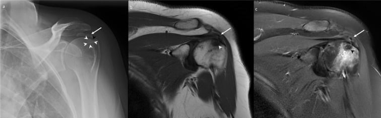
- Radiographs: true anteroposterior (Grashey), scapular Y outlet, axillary. Look for the acromiohumeral interval (normal 7 to 14 mm; less than 7 mm implies significant cuff loss), greater tuberosity sclerosis and cyst formation, a calcific deposit at the critical zone, and acetabularisation in arthropathy.
- Ultrasound: operator-dependent but accurate for full-thickness tears in trained hands, and uniquely capable of dynamic assessment of subacromial gliding.
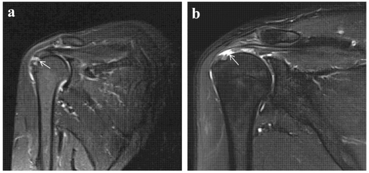
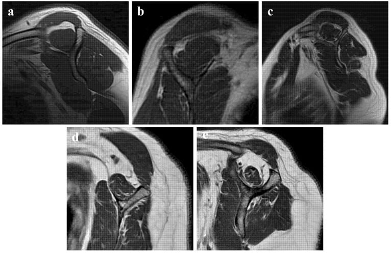
- MRI: fat-suppressed T2 or proton-density coronal oblique for the tendon and footprint; sagittal oblique at the level of the scapular spine, medial to the coracoid, for Goutallier grading and the tangent sign; axial for the biceps and subscapularis. Look also for a paralabral cyst at the spinoglenoid notch if there is posterior wasting.
- MR arthrography improves detection of articular-sided partial tears through contrast tracking into the tendon substance.
Calcific tendinopathy sits in the critical zone, roughly 1 cm medial to the insertion, and is a cell-mediated, self-limiting process passing through formative, resting and resorptive phases. The resorptive phase is the most painful and is exactly when the deposit looks fluffy and ill-defined on radiographs — the phase in which ultrasound-guided needling or lavage is most effective. A dense, well-defined deposit in a comfortable patient is in the formative phase and should be left alone.
Surgical Relevance
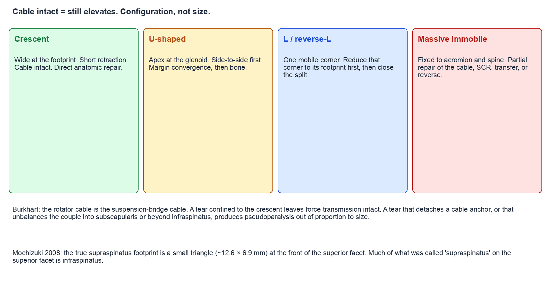
Tear patterns and the repair configuration each demands
- Geometry
- Wide at the footprint, short medial-to-lateral retraction
- Mobility
- Excellent lateral mobility
- Repair Strategy
- Direct anatomic repair to the footprint; single or double row
- Geometry
- Apex retracted medially, often to or past the glenoid
- Mobility
- Mobile side to side but not laterally
- Repair Strategy
- Margin convergence first (side-to-side sutures from medial to lateral), then anchor the converged margin to bone
- Geometry
- One leaf detached from the footprint, the other split longitudinally
- Mobility
- Asymmetric; one corner is the mobile leading edge
- Repair Strategy
- Identify the corner, reduce it to its anatomic footprint corner first, then close the longitudinal limb
- Geometry
- Fixed retraction with adhesions to the acromion and scapular spine
- Mobility
- Poor in all directions
- Repair Strategy
- Interval slides, partial repair to restore the cable, superior capsular reconstruction, tendon transfer, or reverse arthroplasty
In a U-shaped tear, pulling the apex laterally to bone creates enormous tension at the free margin and a high failure rate. Margin convergence places side-to-side sutures from medial to lateral between the anterior and posterior leaves. As the leaves converge, the apex of the U migrates laterally toward the footprint under almost no tension, and the residual defect becomes a small crescent that can be anchored anatomically. Burkhart's biomechanical point is that this dramatically reduces strain at the free margin.
Interval slides
- Anterior interval slide: release through the rotator interval, between supraspinatus and the coracohumeral ligament complex down to the base of the coracoid, freeing the anterior leaf.
- Posterior interval slide: release between supraspinatus and infraspinatus, developed medially toward the scapular spine.
- Constraint: both slides are limited by the suprascapular nerve, whose infraspinatus branches lie a mean of only 2 cm from the posterior glenoid rim. Warner's cadaveric work showed a standard anterosuperior approach permits about 1 cm of safe lateral advancement and a formal muscle advancement up to 3 cm before the motor branches are placed on stretch, sometimes less. Interval slides purchase excursion at a real neurological cost and are a last resort.
Fixation constructs
- Description
- One row of anchors at the lateral edge of the articular cartilage or mid-footprint
- Advantage
- Lower cost, fewer implants, less tension, simplest
- Limitation
- Restores a narrower footprint contact area
- Description
- Medial row at the articular margin plus lateral row at the tuberosity
- Advantage
- Greater footprint coverage and higher initial fixation strength
- Limitation
- More implants, higher cost, medial-row failure mode
- Description
- Medial mattress sutures bridged over the tendon to lateral knotless anchors
- Advantage
- Best pressurised footprint contact and self-reinforcing
- Limitation
- Risk of medial cuff failure at the mattress row if over-tensioned, and strangulation of an already hypovascular tendon
- Description
- Sutures through tuberosity tunnels, open or with an arthroscopic device
- Advantage
- No implants, low cost, marrow access
- Limitation
- Cut-out in an osteoporotic tuberosity
Place anchors at a deadman angle of 45 degrees or less to the line of pull. Too vertical an anchor pulls out; too lateral a medial-row anchor leaves an exposed medial footprint. In osteoporotic bone the tuberosity cortex is thin and the anchor should be seated fully below the cortical rim. Marrow venting of the footprint after preparation exposes the repair to a bone-derived healing response without destabilising the tuberosity — light decortication only, since aggressive burring removes the cortical bone the anchor needs.
Approaches that use or divide the tendon
- Arthroscopic: posterior viewing portal in the soft spot 2 cm inferior and 1 cm medial to the posterolateral acromial corner; lateral working portal 2 to 3 cm lateral to the acromial edge in line with the posterior clavicle; anterior portal through the rotator interval. Lateral portals placed more than about 5 cm distal to the acromion approach the axillary nerve.
- Mini-open lateral transdeltoid: deltoid split from the anterolateral acromial corner extending no more than 5 cm distally, with a stay suture at the apex. Suits crescent tears with a mobile lateral edge.
- Open anterosuperior (Neer): deltoid detached from the anterior acromion with a cuff of tissue or a small bone flake for secure repair. Provides unrivalled access to a massive tear but risks deltoid dehiscence, which is functionally catastrophic and difficult to salvage.
The tendon as a donor and as a reconstructed structure
- The supraspinatus is not used as a transfer donor — it is the structure being reconstructed.
- Superior capsular reconstruction (fascia lata autograft or dermal allograft from the superior glenoid to the greater tuberosity) restores the superior restraint when the tendon is irreparable, re-establishing a fulcrum without a muscle.
- Tendon transfers substitute for it: lower trapezius transfer with an interposition graft, or latissimus dorsi transfer, for irreparable posterosuperior tears in a younger patient with an intact subscapularis and a functioning deltoid.
- Subacromial balloon spacers and reverse arthroplasty occupy the salvage end.
- Younger than about 60
- Intact subscapularis and functioning deltoid
- No glenohumeral arthritis
- Preserved passive elevation
- Pain with weakness rather than true pseudoparalysis
- Older than about 70
- Established cuff tear arthropathy with acetabularisation
- True pseudoparalysis with anterosuperior escape
- Goutallier 4 with a positive tangent sign
- Failed previous repair or reconstruction
Guidelines, Registries & Global Practice
Anatomical variation across populations
- The acromial shape described by Bigliani (flat, curved, hooked) varies in reported prevalence between cadaveric populations and imaging cohorts, and the direction of causation between hooked morphology and cuff tearing remains contested. Os acromiale, an unfused acromial epiphysis, is present in a small percentage of shoulders with reported population differences, and is a recognised cause of persistent pain after decompression.
- The suprascapular notch is morphologically variable, ranging from a shallow depression to a complete bony foramen when the superior transverse scapular ligament ossifies. A complete foramen occurs in a small minority of scapulae and is associated with suprascapular neuropathy.
- Prevalence of asymptomatic full-thickness cuff tears rises steeply with age and is substantial in populations over 60 in every region studied — an essential caveat when attributing symptoms to an imaging finding.
Side-by-side guidance
- Position on Supraspinatus and Cuff Disease
- Evidence-based guidance supports non-operative management as an initial option for many degenerative full-thickness tears, and repair for acute traumatic tears in active patients; routine isolated acromioplasty is not supported.
- Position on Supraspinatus and Cuff Disease
- Recommend a structured non-operative programme first for atraumatic tears, with early specialist referral for acute traumatic full-thickness tears; subacromial decompression alone is discouraged for isolated impingement pain.
- Position on Supraspinatus and Cuff Disease
- Emphasises anatomic footprint restoration, tension-free repair, and protection of the suprascapular nerve during medial mobilisation.
- Position on Supraspinatus and Cuff Disease
- Support arthroscopic repair where expertise exists, and reserve tendon transfer, superior capsular reconstruction and reverse arthroplasty for irreparable disease stratified by age and subscapularis integrity.
Registry and outcome signals
- National joint registries (AOANJRR, NJR, AJRR) record reverse total shoulder arthroplasty as the fastest-growing shoulder arthroplasty category, with the cuff-deficient shoulder as a principal indication and good medium-term survivorship.
- Registries do not capture cuff repair as a joint replacement procedure, so structural re-tear data come from institutional imaging cohorts rather than registries; the consistent signal across those cohorts is that age, tear size and muscle quality dominate the outcome, and that the choice between single-row and double-row fixation influences structural healing more than patient-reported outcome.
- Randomised trials of arthroscopic subacromial decompression against placebo surgery for subacromial pain without a full-thickness tear have shifted international practice sharply away from isolated decompression.
High-resource versus limited-resource practice
- Well-resourced settings: routine MRI, arthroscopic repair with transosseous-equivalent constructs, superior capsular reconstruction and reverse arthroplasty for salvage.
- Limited-resource settings: ultrasound is an accurate and inexpensive substitute for MRI in trained hands; open or mini-open transosseous repair through bone tunnels achieves comparable results without implant cost; physiotherapy-led care is appropriate first-line management for atraumatic tears, supported by the same evidence used in high-income settings.
MCQ Practice Points
Q: What is the relationship of the suprascapular nerve and artery to the superior transverse scapular ligament? A: The nerve passes under the ligament, the artery passes over it. At the spinoglenoid notch, both pass under the inferior transverse (spinoglenoid) ligament.
Q: What are the accepted dimensions of the supraspinatus footprint? A: Classically about 23 mm anterior-to-posterior and 12 to 16 mm medial-to-lateral. Mochizuki's refined dissection assigns only a small anterior triangle of roughly 12.6 mm by 6.9 mm to supraspinatus, with infraspinatus occupying the rest of the superior facet.
Q: How much bare bone lies between the articular cartilage margin and the medial edge of the supraspinatus footprint? A: Under 1 mm anteriorly — the footprint effectively abuts the cartilage. This is why the medial row of anchors sits at the articular margin.
Q: Where is Codman's critical zone and what makes it critical? A: Approximately 1 cm medial to the insertion, at the watershed between ascending osseous vessels and descending muscular vessels. Rathbun and Macnab showed the hypovascularity is position dependent — the wringing-out phenomenon in adduction.
Q: Which structure is stress-shielded by the rotator cable? A: The rotator crescent, the thin insertional sheet lateral to the cable. Its avascularity and its stress-shielded state together explain why degenerative tears begin there.
Q: What did Warner's cadaveric study establish about the limits of cuff mobilisation? A: The infraspinatus motor branches lie a mean of 2 cm from the posterior glenoid rim and the supraspinatus branches a mean of 3 cm from the origin of the long head of biceps. A standard anterosuperior approach permits only about 1 cm of safe lateral advancement; a Debeyre-type muscle advancement up to 3 cm, limited by tension in the motor branches — and sometimes less.
Q: What is the normal acromiohumeral interval and what does narrowing signify? A: Normal is 7 to 14 mm. Less than 7 mm indicates significant chronic cuff deficiency with superior migration and is a poor prognostic sign for repair.
Q: What depth defines an Ellman grade III partial tear? A: Greater than 6 mm, corresponding to more than 50 per cent of a normal insertional tendon thickness of about 10 to 12 mm.
Q: How is the tangent sign assessed and what does a positive sign mean? A: On the sagittal oblique MRI at the level of the scapular spine, draw a line along the superior borders of the coracoid and the scapular spine. If the supraspinatus belly does not cross that line, the sign is positive, indicating significant atrophy and a poor healing environment.
Q: Why is the full-can test often preferred to the empty-can test? A: Both generate comparable supraspinatus activity, but the empty-can (internally rotated) position is provocative, so pain rather than true weakness limits the patient and the deltoid substitutes. The full-can position gives a cleaner strength assessment.
Exam Viva Scenarios
Practise clinical reasoning and management decisions out loud
“A 68-year-old retired plumber has an MRI showing a full-thickness supraspinatus tear retracted to the level of the humeral head, with Goutallier grade 2 change. He elevates to 160 degrees actively and his main complaint is night pain. Explain the discrepancy between the imaging and his function.”
“You are repairing a Patte stage 3 supraspinatus tear arthroscopically. After bursal and articular releases the tendon still will not reach the footprint. Your assistant suggests continuing the posterior interval slide medially until it does. What is your response?”
“A 24-year-old competitive volleyball player has obvious wasting of the infraspinous fossa, near-normal supraspinous bulk, weak external rotation and normal sensation over the whole limb. Where is the lesion and how does this differ from the cuff pathology you have been describing?”
Attachments
- Origin: medial two-thirds supraspinous fossa plus fascia
- Insertion: superior facet, greater tuberosity
- Footprint: about 23 mm AP by 12 to 16 mm ML (classical)
- Mochizuki: true supraspinatus triangle about 12.6 by 6.9 mm
- Bare area to cartilage: under 1 mm
Nerve and Vessels
- Suprascapular nerve C5, C6 (variable C4), upper trunk
- Nerve under, artery over the superior transverse ligament
- Two motor branches within 1 cm of the notch
- Notch 3 cm medial to superior glenoid rim
- Spinoglenoid notch 2 cm medial to posterior glenoid rim
- No cutaneous sensory territory
Biomechanics
- Concavity compression, not simple elevation
- Active through the whole abduction arc
- Rotator cable stress-shields the crescent
- Cable-intact tear can retain full elevation
- Loss leads to superior migration and arthropathy
Grading
- Ellman I under 3 mm, II 3 to 6 mm, III over 6 mm
- Patte 1 near insertion, 2 to head, 3 to glenoid
- Goutallier 0 to 4; 3 to 4 irreversible
- Tangent sign positive equals significant atrophy
- Acromiohumeral interval under 7 mm equals cuff deficiency
Surgical Rules
- Warner: about 1 cm safe advancement standard approach, up to 3 cm with muscle advancement
- Deltoid split no more than 5 cm from lateral acromion
- Deadman anchor angle 45 degrees or less
- Margin convergence before lateral anchoring in U-shaped tears
- Always inspect the biceps pulley and subscapularis
Evidence Base
Humeral Insertion of the Supraspinatus and Infraspinatus: New Anatomical Findings Regarding the Footprint of the Rotator Cuff
- 113 shoulders from 64 cadavers: insertion areas studied in 97, muscular portions removed in 16 to isolate the tendinous portion, and precise footprint measurements taken in a further 26
- The supraspinatus inserted into the superiormost area of the LESSER tuberosity in 21 per cent of specimens - it does not always stop at the greater tuberosity
- The supraspinatus insertion is a small triangular area of roughly 12.6 mm anterior-posterior and 6.9 mm medial-lateral
- The infraspinatus footprint is a trapezoid occupying much of the superior facet in addition to the middle facet
- The supraspinatus footprint is therefore considerably smaller than classically described
The Rotator Crescent and Rotator Cable: An Anatomic Description of the Shoulder's Suspension Bridge
- Described a thick cable-like condensation arcing medial to the cuff insertion from the biceps to the inferior infraspinatus
- The thinner crescent lateral to the cable is stress-shielded by it
- Load is transmitted from muscle to the anterior and posterior bony anchors of the cable
- Explains preserved function in tears confined to the crescent
The Microvascular Pattern of the Rotator Cuff
- Note: this 1970 paper has no abstract indexed on PubMed, so the summary below reflects the finding for which it is universally cited rather than a quotable abstract
- Perfusion studies of cadaveric shoulders injected in different arm positions
- Described a zone of hypovascularity near the supraspinatus insertion that filled or emptied depending on arm position
- The wringing-out phenomenon: with the arm adducted the tendon is draped over the humeral head and its vessels are compressed
The Microvascular Pattern of the Supraspinatus Tendon
- Injection studies comparing articular and bursal surfaces of the supraspinatus
- The articular side of the tendon is markedly less vascular than the bursal side
- The hypovascularity is most pronounced near the insertion
Anatomy and Relationships of the Suprascapular Nerve: Anatomical Constraints to Mobilisation of the Supraspinatus and Infraspinatus Muscles
- 31 shoulders in 18 cadavera dissected to define the limits of cuff mobilisation for chronic massive retracted tears
- Two SEPARATE 84 per cent findings, often merged in the retelling: in 26 of 31 shoulders there were no more than two motor branches to supraspinatus (the first always the larger), and in 26 of 31 the first motor branch originated underneath the transverse scapular ligament or just distal to it
- In one shoulder (3 per cent) the first motor branch passed OVER the ligament - the variant that matters if the ligament is being divided
- The infraspinatus had three or four motor branches of the same size in 15 of 31 (48 per cent), and the supraspinatus branches were fewer, usually smaller and significantly SHORTER than those to infraspinatus
- Mean distance from the origin of the long head of biceps to the supraspinatus motor branches 3 cm; from the posterior glenoid rim to the infraspinatus motor branches 2 cm
- A standard anterosuperior approach permitted only 1 cm of safe lateral advancement; a Debeyre-type advancement up to 3 cm, limited by tension in the motor branches, and sometimes less
Effectiveness of Physical Therapy in Treating Atraumatic Full-Thickness Rotator Cuff Tears (MOON Shoulder Group)
- Multicentre prospective cohort of patients with atraumatic full-thickness cuff tears treated with a structured physiotherapy programme
- Approximately three quarters had not elected surgery at two years
- Patients who improved did so within the first twelve weeks
- Failure to improve by twelve weeks predicted eventual surgery
Fatty Muscle Degeneration in Cuff Ruptures: Pre- and Postoperative Evaluation by CT Scan
- Pre-operative CT grading fatty degeneration in five stages in 63 patients scheduled for cuff repair, compared with post-operative evaluation at a mean of 17.7 months in 57
- Infraspinatus fatty degeneration occurred with large anterosuperior tears even when the infraspinatus tendon itself was not torn, and worsened with time
- The subscapularis rarely degenerated, and when it did so only moderately, even when its tendon was NOT torn
- Infraspinatus degeneration NEVER regressed after repair and increased in three patients; it correlated with impaired active external rotation before and after surgery
- Recurrence occurred in 25 per cent of supraspinatus repairs but was never observed for infraspinatus, and infraspinatus degeneration had a HIGHLY NEGATIVE influence on the outcome of supraspinatus repair - the authors conclude it is preferable to operate on wide tears before irreversible muscular damage occurs
- After effective repair, moderate supraspinatus degeneration regressed in six of 14 patients — so the process is not uniformly irreversible, although advanced degeneration behaves as though it is