Rotator Cuff Arthropathy | Grammont Principles | Deltoid-Powered | 10 Year Survivorship over 90 percent
- Grammont Principle: Medialize center of rotation to glenoid surface. Distalize humerus to tension deltoid.
- Cuff Tear Arthropathy: Superior migration of humeral head. Femoralization. Acetabularization.
- Deltoid Function: RTSA converts deltoid from a short muscle with poor mechanical advantage to efficient arm elevator.
- Scapular Notching: Most common complication (around 68 percent in Grammont medialized series). Inferior baseplate tilt and lateralization help prevent.
- Acromial Fractures: Risk 2-7 percent. Increased deltoid tension. May be stress or traumatic.
- “RTSA increases deltoid moment arm by 30-40 percent through medialization of center of rotation
- “Scapular notching reduced by inferior tilt of baseplate and larger glenosphere
- “Intact deltoid is ABSOLUTE requirement - check axillary nerve function preoperatively
- “Active external rotation preserved if teres minor intact (often limited postoperatively)
Medialization and distalization are fundamental. Center of rotation moved to glenoid surface eliminates eccentric loading and maximizes deltoid efficiency. Lengthening the arm tensions deltoid for improved elevation. Vivas will ask you to explain these biomechanical principles.
Most common complication. Caused by mechanical impingement of humeral component on inferior glenoid rim. Prevented by: inferior baseplate tilt (10-15 degrees), larger glenosphere (38-42mm), inferior glenosphere overhang, lateralized designs.
RTSA is deltoid-powered. Intact deltoid and axillary nerve are ABSOLUTE requirements. Preoperatively assess: deltoid bulk, axillary nerve function, prior surgical approaches (deltopectoral vs lateral). Previous deltoid detachment is relative contraindication.
Most patients have limited active external rotation postoperatively because posterior cuff (infraspinatus, teres minor) is deficient. If teres minor intact, ER preserved. Latissimus dorsi transfer can improve ER in select cases.
- Anatomic TSA
- Intact or repairable
- Reverse TSA
- Deficient or irreparable
- Key Pearl
- RTSA does not require rotator cuff
- Anatomic TSA
- Rotator cuff
- Reverse TSA
- Deltoid
- Key Pearl
- Deltoid MUST be intact for RTSA
- Anatomic TSA
- Native (lateral to glenoid)
- Reverse TSA
- Medialized to glenoid surface
- Key Pearl
- Grammont principle
- Anatomic TSA
- Contraindicated
- Reverse TSA
- Primary indication
- Key Pearl
- Superior migration means cuff deficient
- Anatomic TSA
- Preserved
- Reverse TSA
- Often limited
- Key Pearl
- Posterior cuff deficiency limits ER
- Anatomic TSA
- Major concern (rocking horse)
- Reverse TSA
- Less eccentric loading
- Key Pearl
- Medialization reduces glenoid torque
DELTOIDDELTOID - Essential Preoperative Checks
Hook:Before RTSA, check the DELTOID - the entire procedure depends on it!
Overview and Epidemiology
Reverse total shoulder arthroplasty (RTSA) has revolutionized the management of complex shoulder conditions, particularly rotator cuff arthropathy. By reversing the ball-and-socket anatomy, placing a convex glenosphere on the glenoid and a concave socket on the humerus, RTSA converts the deltoid muscle into the primary arm elevator.
Epidemiology:
- Exponential growth in utilization over the past decade (over 300 percent increase)
- Now represents over 60 percent of all shoulder arthroplasties in some series
- Mean patient age 65-75 years, though indications expanding to younger patients
- Female predominance (60-65 percent) reflecting rotator cuff arthropathy demographics
RTSA indications have expanded beyond rotator cuff arthropathy to include: acute proximal humerus fractures in the elderly, failed hemiarthroplasty, revision of failed anatomic TSA, tumor reconstruction, and inflammatory arthritis with cuff deficiency. However, the core indication remains cuff tear arthropathy with pseudoparalysis.
Historical Development:
- Professor Paul Grammont (Lyon, France) developed the modern design in 1985
- Key innovation was the Grammont principle: medialization of center of rotation
- Earlier designs (Neer Mark I, II) failed due to glenoid loosening from eccentric loading
- Modern semi-constrained designs achieve over 90 percent 10-year survivorship
Anatomy and Pathophysiology
Rotator Cuff Tear Arthropathy (CTA):
Cuff tear arthropathy represents the end-stage of massive rotator cuff tear with secondary degenerative changes. It is the most common indication for RTSA.
Pathophysiological Cascade:
- Massive rotator cuff tear: Usually supraspinatus and infraspinatus
- Loss of force couple: Deltoid pulls humeral head superiorly unopposed
- Superior migration: Humeral head articulates with undersurface of acromion
- Mechanical changes: Increased joint reaction forces, synovial fluid changes
- Cartilage destruction: Both glenoid and humeral head articular surfaces
- Bony remodeling: Femoralization of humeral head, acetabularization of glenoid
- Description
- Acromiohumeral distance over 6mm
- Acromiohumeral Distance
- Greater than 6mm
- Treatment
- Conservative or cuff repair
- Description
- Acromiohumeral distance 5-6mm
- Acromiohumeral Distance
- 5-6mm
- Treatment
- Maximum conservative, consider repair
- Description
- Acromiohumeral less than 5mm with acetabularization
- Acromiohumeral Distance
- Under 5mm
- Treatment
- RTSA candidate if symptomatic
- Description
- Narrowing without acetabularization
- Acromiohumeral Distance
- Under 5mm
- Treatment
- RTSA (primary indication)
- Description
- Narrowing with acetabularization of acromion
- Acromiohumeral Distance
- Under 5mm
- Treatment
- RTSA (primary indication)
- Description
- Humeral head collapse
- Acromiohumeral Distance
- N/A
- Treatment
- RTSA (salvage)
Grammont Biomechanical Principles:
The Grammont design addressed the failures of earlier reverse designs through key biomechanical innovations:
Moving the center of rotation from the lateral humeral head to the glenoid surface eliminates the moment arm that caused glenoid loosening in earlier designs. This reduces eccentric loading on the glenoid fixation by over 90 percent.
The arm is effectively lengthened by 1-3cm, tensioning the deltoid muscle. This increases the deltoid moment arm by 30-40 percent and converts it from a short, relatively weak elevator to an efficient arm elevator.
Intact deltoid and axillary nerve function are ABSOLUTE requirements for RTSA. The entire design depends on deltoid function for arm elevation. Preoperative assessment must include: deltoid bulk inspection, axillary nerve sensation, deltoid strength testing, and review of prior incisions for deltoid detachment.
Functional Outcomes Explained by Biomechanics:
- Forward elevation: Typically restored to 120-140 degrees (deltoid-powered)
- External rotation: Often limited (0-30 degrees) due to posterior cuff deficiency
- Internal rotation: Variable, often limited to buttock level
- Strength: Improved compared to preoperative pseudoparalysis
Classification Systems
Hamada Classification of Rotator Cuff Tear Arthropathy
The Hamada classification grades the severity of cuff tear arthropathy based on radiographic findings.
- Acromiohumeral Interval
- Greater than 6mm
- Findings
- Minimal changes
- Significance
- Conservative management
- Acromiohumeral Interval
- 5-6mm
- Findings
- Early narrowing
- Significance
- Monitor progression
- Acromiohumeral Interval
- Under 5mm + acetabularization
- Findings
- Acromion remodeling
- Significance
- Consider RTSA
- Acromiohumeral Interval
- Under 5mm
- Findings
- GH joint narrowing
- Significance
- RTSA indication
- Acromiohumeral Interval
- Under 5mm + acetabularization
- Findings
- Advanced changes
- Significance
- RTSA indication
- Acromiohumeral Interval
- Collapse
- Findings
- Humeral head collapse
- Significance
- Salvage RTSA
Normal acromiohumeral interval is 7-14mm. Less than 7mm indicates massive cuff tear.
Clinical Assessment
History:
- Location: Anterolateral shoulder, may radiate to deltoid
- Timing: Night pain common, difficulty sleeping on affected side
- Activity: Pain with overhead activities, reaching
- Duration: Often chronic and progressive over years
- Active elevation: Severely limited (pseudoparalysis)
- Passive motion: Preserved (excludes frozen shoulder)
- Daily activities: Unable to comb hair, reach overhead
- Previous treatment: Failed injections, physiotherapy, cuff repair
Physical Examination:
Key findings in cuff tear arthropathy:
- Inspection: Deltoid atrophy rare (if present, reconsider RTSA), anterior prominence (superior humeral migration)
- Palpation: Tenderness anterior shoulder, AC joint often arthritic
- Active range of motion: Severely limited elevation (pseudoparalysis less than 90 degrees)
- Passive range of motion: Preserved (distinguishes from frozen shoulder)
- Strength testing: Positive Hornblower's sign, positive external rotation lag
Pseudoparalysis: Inability to actively elevate the arm above 90 degrees with preserved passive motion. This indicates a massive, irreparable rotator cuff tear with loss of the force couple. Pseudoparalysis is a key indication for RTSA over anatomic TSA.
Differential Diagnosis of the Painful, Weak, Elevation-Limited Shoulder:
- Active vs Passive ROM
- Active limited, passive preserved
- Key Distinguishing Feature
- Superior migration, acromiohumeral interval under 7mm, arthritis
- Implication for RTSA
- Primary RTSA indication
- Active vs Passive ROM
- Active limited, passive preserved
- Key Distinguishing Feature
- Pseudoparalysis without joint-space loss
- Implication for RTSA
- RTSA if pseudoparalytic and irreparable
- Active vs Passive ROM
- Both active AND passive limited
- Key Distinguishing Feature
- Global loss of passive motion, no superior migration
- Implication for RTSA
- Not an arthroplasty problem - treat the stiffness
- Active vs Passive ROM
- Active limited, passive preserved
- Key Distinguishing Feature
- Deltoid wasting, sensory loss over badge area, EMG abnormal
- Implication for RTSA
- Contraindication - deltoid non-functional
- Active vs Passive ROM
- Active and passive reduced by pain/osteophytes
- Key Distinguishing Feature
- Cuff intact on imaging, posterior glenoid wear
- Implication for RTSA
- Anatomic TSA preferred
- Active vs Passive ROM
- Variable, often bilateral
- Key Distinguishing Feature
- Symmetrical erosions, soft-tissue and cuff involvement
- Implication for RTSA
- RTSA if cuff deficient, anatomic if cuff intact
- Active vs Passive ROM
- Limited with systemic signs
- Key Distinguishing Feature
- Raised inflammatory markers, effusion, fever
- Implication for RTSA
- Exclude before any arthroplasty
Specific Preoperative Assessment for RTSA:
- What to Check
- Bulk, strength, axillary nerve (regimental badge area)
- Significance
- ABSOLUTE requirement for RTSA
- What to Check
- Deltopectoral vs lateral approach, cuff repairs
- Significance
- Deltoid detachment is relative contraindication
- What to Check
- Horn blower sign negative indicates intact
- Significance
- Predicts postoperative ER
- What to Check
- CT scan for morphology and version
- Significance
- May need bone graft if eroded
- What to Check
- Tenderness, cross-body adduction pain
- Significance
- Consider distal clavicle excision
Investigations
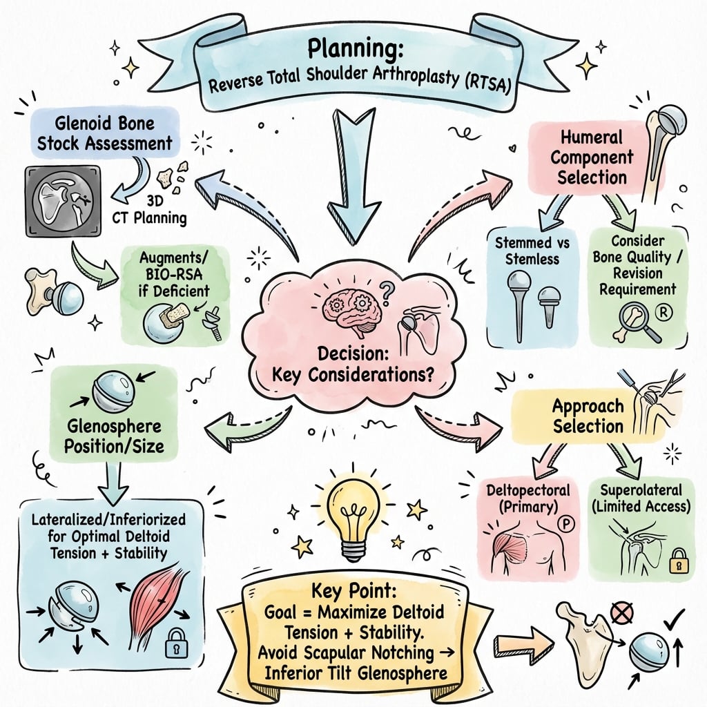
Imaging Protocol for RTSA Planning
Standard shoulder series: AP (neutral, IR, ER), scapular Y, axillary lateral
Key findings to assess:
- Acromiohumeral interval (less than 7mm indicates massive tear)
- Superior migration of humeral head
- Femoralization of humeral head (rounding)
- Acetabularization of acromion and glenoid
- Glenohumeral arthritis severity
- Acromioclavicular joint arthritis
Critical for surgical planning
Glenoid assessment:
- Bone stock: anterior, posterior, inferior
- Version: excessive retroversion (over 15 degrees) may need augmentation
- Erosion pattern: centered vs decentered (Seebauer)
- Vault depth and screw trajectory planning
- Prior hardware if revision
Not essential if diagnosis clear, but useful for:
- Teres minor integrity (predicts postoperative ER)
- Fatty infiltration grading (Goutallier)
- Subscapularis status (repair vs leave)
- Biceps pathology
- Exclude infection if revision
CT scan with 3D reconstruction is essential for RTSA planning. It allows assessment of glenoid version, bone stock, and optimal baseplate trajectory. Excessive retroversion (over 15 degrees) may require bony increased offset (BIO) augmentation or posterior augmented baseplate. The inferior screw trajectory must be planned to avoid scapular spine.
Management Algorithm
RTSA Indications
- Rotator cuff tear arthropathy (CTA): Hamada Grade 3-5 with pseudoparalysis
- Massive irreparable rotator cuff tear: With pseudoparalysis, without arthritis
- Proximal humerus fracture: Elderly (over 70), comminuted, poor bone quality
- Failed hemiarthroplasty: With cuff deficiency or tuberosity non-union
- Revision of failed anatomic TSA with cuff deficiency
- Primary rheumatoid arthritis with massive cuff tear
- Tumor reconstruction (proximal humerus)
- Fracture sequelae (malunion, nonunion)
All indications require intact deltoid function - this is non-negotiable.
Surgical Technique
Patient Positioning
Beach chair position:
- Upright 60-70 degrees, head secured
- Arm free-draped for full access
- Ensure ability to maximally extend arm for humeral preparation
- Radiolucent table for intraoperative imaging
Surgical Approach
Deltopectoral approach (most common):
Deltopectoral Approach Steps
Start from coracoid, extend distally over deltopectoral groove (approximately 15cm). Identify cephalic vein and retract laterally (protects deltoid blood supply).
Develop deltopectoral interval. Identify conjoint tendon (medial), coracoid. Release clavipectoral fascia lateral to conjoint. Identify subscapularis and biceps.
Options: tenotomy, lesser tuberosity osteotomy, or peel. Lesser tuberosity osteotomy provides best healing in RTSA. Tenotomy also acceptable given cuff already deficient.
Dislocate shoulder anteriorly. Use cutting guide or freehand. Resection level and version critical. Many systems use 20-30 degrees retroversion. Preserve deltoid attachment.
Deltopectoral approach preserves deltoid origin and is preferred for RTSA.
Complications
- Incidence
- 20-50 percent
- Prevention
- Inferior tilt, large glenosphere, lateralized design
- Management
- Observation unless symptomatic/progressive
- Incidence
- 2-10 percent
- Prevention
- Appropriate tensioning, avoid excessive retroversion
- Management
- Revision with larger glenosphere, humeral insert
- Incidence
- 2-7 percent
- Prevention
- Avoid excessive arm lengthening
- Management
- Brace, limited abduction, rarely ORIF
- Incidence
- 1-4 percent
- Prevention
- Antibiotics, sterile technique
- Management
- Debridement or staged revision
- Incidence
- 1-2 percent (usually neurapraxia)
- Prevention
- Careful retraction, avoid overtensioning
- Management
- Observation, usually recovers
- Incidence
- 1-3 percent
- Prevention
- Careful technique, assess bone quality
- Management
- ORIF or revision
Acromial fractures are unique to RTSA due to increased deltoid tension. Risk factors include: excessive arm lengthening, osteoporotic bone, female sex, and rheumatoid arthritis. Present with sudden pain and weakness. Management is often conservative with limited abduction and sling immobilization. May catastrophically affect outcome if displaced.
Instability Patterns:
- Anterior: Most common, excessive retroversion, subscapularis failure
- Posterior: Rare, excessive anteversion
- Superior: Very rare, deltoid dysfunction
Postoperative Care
RTSA Rehabilitation Protocol
- Sling immobilization for 4-6 weeks
- No active shoulder motion
- Elbow, wrist, hand exercises permitted
- Pendulum exercises (gravity-assisted) from week 2
- Avoid combined abduction and external rotation (instability position)
- Begin active-assisted ROM exercises
- Progress to active ROM as tolerated
- Forward flexion and abduction focus
- Gentle external rotation (often limited, do not force)
- Periscapular strengthening
- Progressive deltoid strengthening
- Isometric to isotonic exercises
- Functional activities as tolerated
- Avoid heavy lifting (greater than 10kg) long-term
- Full activities of daily living expected
- Ongoing home exercise program
- Lifelong activity modifications (no contact sports, heavy lifting)
- Annual follow-up with radiographs
Activity Restrictions (Long-term):
- Avoid lifting greater than 10-15kg (risk of acromial stress fracture, instability)
- No contact sports or high-impact activities
- Swimming, golf, and tennis often possible with low demand
Outcomes and Prognosis
- Forward elevation: Improves from mean 50 degrees to 120-140 degrees
- External rotation: Often limited postoperatively (0-30 degrees average)
- Internal rotation: Variable, often to sacrum/buttock level
- Pain relief: Excellent in over 90 percent of patients
- 92-95 percent 10-year implant survivorship in modern series
- Revision rates higher in younger patients (under 60)
- Glenoid loosening is long-term concern with medialized designs
- Lateralized designs may have improved long-term outcomes (under investigation)
- Intact teres minor (better external rotation)
- Adequate deltoid function preoperatively
- Patient understanding of activity restrictions
- Primary indication (CTA) vs complex revision
- Surgeon experience with RTSA technique
Restoring Active External Rotation: Combined Reverse and Tendon Transfer
The topic repeatedly notes that RTSA restores elevation but rarely restores active external rotation, and the Exam Warning hints that "latissimus dorsi transfer can improve ER in select cases" - this section develops that under-stated point. When the posterior cuff (infraspinatus and teres minor) is absent or fatty-infiltrated, a reverse alone leaves the patient with a combined loss of active elevation and external rotation (CLEER): pseudoparalysis plus a positive Hornblower sign and external-rotation lag, so the forearm swings uncontrollably into the trunk when they try to reach forward or hold an object away from the body.
The established solution is to combine the reverse with a modified L'Episcopo transfer - detaching the latissimus dorsi and teres major, rerouting them around the humerus and reattaching them posterolaterally so that these internal rotators are converted into active external rotators. Boileau popularised performing both the reverse and the transfer through a single deltopectoral approach in the beach-chair position, avoiding the classic two-incision posterior dissection.
Key selection points:
- CLEER (irreparable postero-superior cuff plus teres minor loss, in cuff tear arthropathy or tumour reconstruction) is the indication for the combined reverse plus transfer.
- Isolated loss of external rotation (ILER) with preserved elevation can be treated by the transfer alone, without a reverse.
- The transfer restores the ability to control the spatial position of the arm (external rotation with the arm at the side), which is the main driver of patient satisfaction, rather than adding elevation.
Modified L'Episcopo Transfer With or Without Reverse (Boileau)
- 15 patients (mean age 63) had a combined latissimus dorsi and teres major transfer through a single deltopectoral approach, minimum 1-year follow-up
- Transfer alone in 7 with isolated loss of external rotation (ILER); combined with a reverse prosthesis in 8 with combined loss of active elevation and external rotation (CLEER)
- Active external rotation gained 27 degrees (ILER) and 28 degrees (CLEER); mean active elevation gained 34.7 degrees; Constant score reached 65.6
- Subjective Shoulder Value improved from 34 to 72 percent; benefit was control of arm position, not extra elevation
A reverse restores elevation but not external rotation. If the patient has combined loss of both (positive Hornblower and a dropping arm from an absent teres minor), consider RTSA plus a modified L'Episcopo latissimus dorsi and teres major transfer through a single deltopectoral approach. If elevation is preserved and only external rotation is lost, the tendon transfer alone may suffice without arthroplasty.
GRAMMONTGRAMMONT - Reverse Shoulder Principles
Hook:Professor Paul GRAMMONT designed the modern reverse shoulder - each letter of his name teaches a principle!
Baseplate Inclination: The RSA Angle and Prosthesis-Scapular Neck Angle
The NOTCH mnemonic and the Sirveaux and Simovitch data tell you to give the baseplate "inferior tilt" and to implant the glenosphere low, but examiners expect you to know how that tilt is measured and why it matters - a point the topic names (the Simovitch card cites the "prosthesis-scapular neck angle" and glenosphere height) but does not define. Two radiographic parameters quantify it:
- RSA angle (Boileau): the inclination of the glenoid baseplate relative to the floor of the supraspinatus fossa (the scapular reference line). Cuff tear arthropathy glenoids are frequently superiorly inclined, and leaving that superior tilt is a leading driver of notching. The target is a neutral inclination - an RSA angle under 5 degrees - obtained by inferior reaming, an inferiorly-tilted or augmented baseplate, and lowering the baseplate on the glenoid.
- Prosthesis-scapular neck angle (PSNA) (Simovitch): the angle between the glenosphere and the scapular neck. Together with the height of glenosphere implantation it predicts inferior notching, and glenosphere height carries roughly eight times more influence than the neck angle - so implant the sphere low.
Combine neutral or inferior inclination with inferior glenosphere overhang of more than 5 mm and a low baseplate position. Persistent superior inclination and a high or flush glenosphere are the malpositions most strongly linked to notching in long-term data.
Baseplate Inclination and Notching - Long-Term BIO-RSA (Boileau)
- 143 shoulders treated with bony increased-offset RSA, mean 75-month (5 to 10 year) follow-up; revision-free survival 96 percent
- Severe (grade 3 to 4) inferior scapular notching in 18 percent; graft incorporated fully in 96 percent
- Notching correlated with superior glenosphere inclination, high or flush glenosphere position, and low body mass index
- Authors confirm implanting the baseplate at neutral inclination (RSA angle under 5 degrees) with inferior glenosphere overhang over 5 mm to prevent notching
"Inferior tilt" is not a vague instruction - it is measured. Target a neutral RSA angle (under 5 degrees) and inferior glenosphere overhang greater than 5 mm, with the glenosphere implanted low on the glenoid. Glenosphere height influences notching roughly eight times more than the prosthesis-scapular neck angle, so lowering the sphere is the single most powerful preventive step.
NOTCHNOTCH - Scapular Notching Prevention
Hook:To prevent scapular NOTCH, remember all five prevention strategies!
Guidelines, Registries & Global Practice
Global Epidemiology and Utilization:
- RTSA utilization has grown several-fold over the past two decades and is now the most common shoulder arthroplasty in many high-income health systems
- It overtook anatomic TSA in the US around the late 2010s and continues to expand into fracture and revision indications
- Mean age at surgery is typically 70-75 years with a female predominance (around 60-65 percent), mirroring cuff tear arthropathy demographics
- Position on RTSA
- RTSA for cuff tear arthropathy with pseudoparalysis and intact deltoid
- Notable Emphasis
- Strongest evidence for pain relief and elevation; cautious in young patients
- Position on RTSA
- RTSA for cuff-deficient arthritis and selected complex fractures
- Notable Emphasis
- Emphasis on shared decision-making and surgeon volume
- Position on RTSA
- RTSA preferred over hemiarthroplasty for non-reconstructable PHF in older patients
- Notable Emphasis
- Removes dependence on tuberosity healing
- Position on RTSA
- Supports expanding indications with appropriate selection
- Notable Emphasis
- Highlights design choice (lateralization) and notching avoidance
- National registries (AOANJRR in Australia, NJR in the UK, the Nordic registries and emerging US data via AJRR) consistently confirm rising RTSA volumes and document higher revision rates in younger patients
- Registries show that fracture and revision indications carry higher early revision than primary cuff tear arthropathy
- Implant survival around 90 percent at 10 years is reproduced across registry and cohort data, broadly matching the Guery/Favard survivorship findings
- In well-resourced systems, preoperative CT (often with 3D planning or patient-specific guides) and a wide range of lateralized implants are standard
- In limited-resource settings, plain radiographs may guide planning, implant choice is narrower, and hemiarthroplasty or non-operative management remains more common for fractures and cuff arthropathy
- Access, surgeon volume and implant availability - not biology - drive much of the global variation in who receives an RTSA
Controversies and Areas of Uncertainty
RTSA is a young procedure with rapidly evolving design philosophy. Examiners reward candidates who can discuss genuine controversy rather than recite dogma.
Classic Grammont medialization minimizes glenoid torque but causes high notching, poor rotation and loss of shoulder contour. Lateralized designs (bony BIO-RSA, metallic lateral glenosphere, lateralized/onlay humerus) reduce notching and improve rotation but theoretically increase baseplate stress. No design is proven superior in long-term survivorship; bony lateralization is currently favoured to balance the two.
Early survivorship data led to advice restricting RTSA to low-demand patients over 70. Indications have since expanded to patients in their 50s-60s, but lifetime revision burden, the difficulty of revising a failed reverse, and limited long-term data remain real concerns. Shared decision-making is essential.
Whether to repair the subscapularis in RTSA is unsettled. Repair may reduce anterior instability in medialized designs but can limit rotation; lateralized designs may tolerate non-repair. Practice varies by implant and surgeon.
For irreparable cuff tears without arthritis in younger, active patients, RTSA competes with superior capsular reconstruction, tendon transfers (lower trapezius, latissimus dorsi) and balloon spacers. The optimal first-line strategy in this group is not yet defined.
The mechanism, true incidence and best management of acromial and scapular spine stress fractures after RTSA remain debated. Reported rates vary widely (around 1-10 percent), they are more common with greater deltoid lengthening, and there is no consensus on operative versus non-operative treatment - outcomes are often disappointing whichever route is chosen.
MCQ Practice Points
Q: What is the key biomechanical principle of the Grammont design? A: Medialization of the center of rotation to the glenoid surface, which eliminates eccentric loading on the glenoid component and increases the deltoid moment arm by 30-40 percent.
Q: How do you prevent scapular notching in RTSA? A: Inferior tilt of baseplate (10-15 degrees), larger glenosphere diameter (38-42mm), inferior glenosphere overhang, and consider lateralized (BIO-RSA) designs.
Q: What is the absolute requirement for RTSA? A: Intact deltoid function and axillary nerve. RTSA is deltoid-powered and cannot function without it.
Q: What Hamada grade indicates RTSA is the appropriate treatment? A: Hamada Grade 4 or 5 with pseudoparalysis. Grade 4a/4b show acromiohumeral interval under 5mm with glenohumeral arthritis. Grade 5 shows humeral head collapse.
Q: What predicts external rotation after RTSA? A: Teres minor integrity. If Hornblower's sign is negative (teres minor intact), expect better postoperative external rotation (mean 30 degrees). If positive, expect limited ER (mean 10 degrees).
Q: What complication is unique to RTSA compared to anatomic TSA? A: Acromial stress fractures (2-7 percent) due to increased deltoid tension from arm lengthening. Usually managed conservatively unless displaced.
Exam Viva Scenarios
Practise clinical reasoning and management decisions out loud
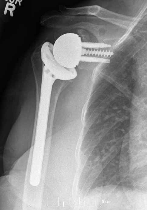
“A 72-year-old female presents with a 2-year history of right shoulder pain and progressive weakness. She cannot lift her arm above 90 degrees. X-ray shows superior migration of the humeral head with acromiohumeral distance of 4mm.”
“You are discussing consent for RTSA. The patient asks about the main complications.”
“A 68-year-old male is 6 weeks post-RTSA and presents with sudden pain and inability to lift his arm. He denies trauma. X-ray shows an acromial fracture.”
Grammont Principles
- Medialize center of rotation to glenoid surface
- Distalize humerus to tension deltoid (increase moment arm 30-40 percent)
- Eliminates eccentric glenoid loading
- Deltoid becomes primary arm elevator
Indications
- Rotator cuff tear arthropathy (Hamada 4-5) is primary indication
- Massive irreparable RC tear with pseudoparalysis
- Complex proximal humerus fracture in elderly (over 70)
- Failed anatomic TSA or hemiarthroplasty with cuff deficiency
Complications
- Scapular notching (20-50 percent) - prevent with inferior tilt and larger glenosphere
- Acromial fractures (2-7 percent) - unique to RTSA
- Instability (2-10 percent) - anterior most common
- Limited external rotation - expected with posterior cuff deficiency
Exam Traps
- Deltoid MUST be intact - absolute requirement for RTSA
- Anatomic TSA contraindicated in CTA (cuff deficient)
- Teres minor integrity predicts postoperative ER
- Counsel on lifelong activity restrictions
Evidence Base
Reverse Prosthesis for Cuff-Deficient Arthritis (Frankle)
- 60 shoulders with glenohumeral arthritis and severe cuff deficiency, mean follow-up 33 months
- Mean ASES score improved from 34.3 to 68.2; forward flexion 55 to 105 degrees, abduction 41 to 102 degrees
- 13 complications in 10 patients (17 percent); 12 percent required revision
- Established the lateralized-glenoid reverse prosthesis as a viable option in North America
Grammont Reverse Prosthesis - Neer Award (Boileau)
- 45 Grammont reverse prostheses for cuff tear arthritis, fracture sequelae and revision arthroplasty
- Active elevation improved 55 to 121 degrees and Constant score 17 to 58, but active external rotation essentially unchanged (7 to 11 degrees)
- Scapular notching in 68 percent; complications far higher in revision (47 percent) than in CTA (5 percent)
- Atrophy or fatty infiltration of teres minor predicted worse external rotation (15 vs 0 degrees) and lower Constant score (66 vs 46)
Ten-Year Survivorship of RTSA (Guery / Favard)
- Multicentre survivorship of 80 reverse prostheses, minimum 5-year (mean 70-month) follow-up
- Survival 91 percent at 120 months with revision as endpoint, 84 percent with glenoid loosening as endpoint
- Survival fell to 58 percent when an absolute Constant score under 30 was used as endpoint (progressive functional decline after ~6 years)
- Cuff tear arthropathy fared significantly better than other aetiologies
Scapular Notching - Incidence and Consequences (Levigne)
- 461 Grammont-type reverse shoulders, mean follow-up 51 months
- Notching occurred in 68 percent, appeared early and generally progressed
- Notching was associated with lower strength, lower elevation and with humeral and glenoid radiolucent lines
- Preoperative superior glenoid erosion predicted notching - avoid cranial baseplate position and superior tilt
Predictors of Scapular Notching (Simovitch)
- 77 Delta III reverse shoulders, minimum 24-month follow-up; inferior notching in 44 percent
- Craniocaudal glenosphere height and the prosthesis-scapular neck angle were strongly correlated with notching
- Glenosphere height had roughly eight times more influence than the neck angle
- Notching was associated with significantly poorer clinical outcome
BIO-RSA: Bony Increased-Offset (Boileau)
- 42 patients with an autologous humeral-head bone graft between glenoid and baseplate, minimum 2-year follow-up
- Graft incorporated in 98 percent; no graft resorption, glenoid loosening or instability
- Inferior scapular notching in only 19 percent; Constant score improved 31 to 67
- Bony lateralization keeps the centre of rotation at the bone-implant interface, avoiding the extra torque of metallic lateralization
RTSA vs Hemiarthroplasty for Acute PHF - RCT (Sebastia-Forcada)
- Blinded RCT of 62 patients over 70 with acute proximal humeral fracture: RTSA vs hemiarthroplasty
- RTSA superior at mean 28 months: Constant 56.1 vs 40.0, UCLA 29.1 vs 21.1, forward elevation 120 vs 80 degrees
- RTSA function was independent of tuberosity healing; 6 hemiarthroplasties required revision to RTSA for proximal migration
- Notching seen in only 1 RTSA patient at short follow-up
AAOS / Society Guidance - Cuff Tear Arthropathy
- RTSA is the recommended arthroplasty for symptomatic cuff tear arthropathy with pseudoparalysis and an intact deltoid
- Anatomic TSA is contraindicated when the rotator cuff is irreparable (eccentric glenoid loading, rocking-horse loosening)
- Deltoid and axillary nerve integrity must be confirmed before surgery; preoperative CT for glenoid version and bone stock is advised
- Society guidance increasingly endorses RTSA over hemiarthroplasty for complex fractures in physiologically older patients