Deltopectoral Approach | Neurovascular Structures at Risk | Rotator Cuff Relationships | Glenoid Exposure
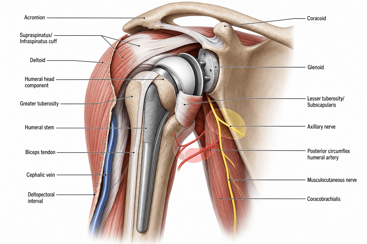
- Axillary nerve lies 5-7 cm inferior to acromion, vulnerable during inferior capsule release
- Musculocutaneous nerve enters coracobrachialis a mean of 56 mm below the coracoid tip (range 31-82 mm) - no distance is a reliable safe zone
- Cephalic vein runs in deltopectoral groove - usually retracted laterally with deltoid (more feeders are deltoid-side); medial retraction is an alternative
- Subscapularis can be released via tenotomy, peel, or lesser tuberosity osteotomy
- Posterior glenoid exposure requires complete anterior capsule release and careful retraction
- “Three critical nerves: axillary (inferior), musculocutaneous (medial), suprascapular (posterior)
- “Deltopectoral interval is internervous and intermuscular - theoretically atraumatic
- “Ascending branch of anterior humeral circumflex artery requires ligation during approach
- “Humeral version averages 30 degrees retroversion relative to transepicondylar axis
Overview and Clinical Significance
Shoulder arthroplasty concentrates critical neurovascular structures in a limited surgical field. In hip arthroplasty the sciatic nerve is the primary concern; shoulder replacement puts four major nerves at risk, the axillary, musculocutaneous, suprascapular and radial, and works around a complex rotator cuff that must be preserved or repaired. The deltopectoral approach gives excellent exposure but passes through multiple tissue planes.
What goes wrong. Neurological complications are reported in 1-4% of cases in most series, the axillary nerve most often, and mastery of this anatomy reduces them and improves functional outcomes. Subscapularis failure causes anterior instability. Native glenoid and humeral version must be understood to position the components: excessive glenoid retroversion leads to posterior instability, and restoring version is critical for implant longevity.
What the exposure has to achieve. Every step of the approach serves one of these goals:
- Safe extensile exposure through an internervous plane
- Protection of the axillary nerve during capsule release
- Subscapularis preservation or secure repair
- Glenoid visualisation without nerve traction injury
- Humeral canal preparation without radial nerve injury
- Restoration of anatomic version relationships
Bony Anatomy and Landmarks
The Glenoid
Shape. The glenoid is a shallow, pear-shaped socket, wider inferiorly, 25-30 mm in diameter (about 35 mm high and 25 mm wide). Its articular arc is 75% of the humeral head's. It sits in 5° of superior tilt, and its morphology changes with disease: the erosion patterns classified below guide component selection.
Fixation depends on bone stock. The subchondral bone is critical for fixation of a glenoid component. The central glenoid vault, 15-25 mm deep, holds the bone stock on which baseplate fixation depends, and its depth is assessed on CT to choose baseplate screw length.
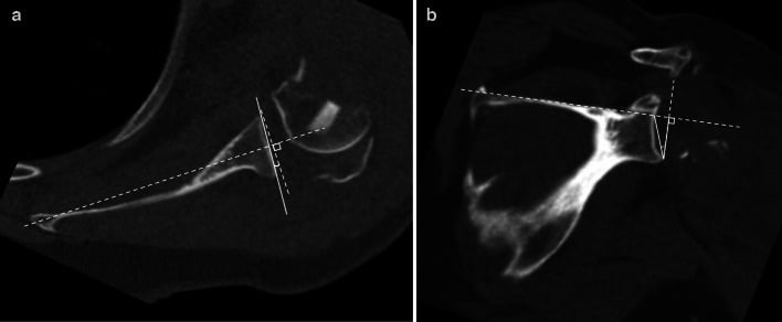
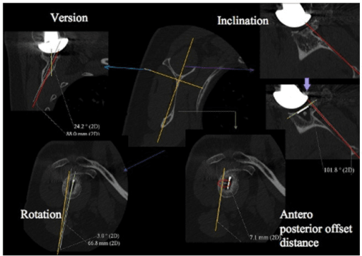
Version. Normal glenoid version is 0-5° of retroversion, measured on CT by the Friedman or scapular body method. Retroversion over 10° is pathological; it occurs with posterior glenoid wear from cuff tear arthropathy or longstanding posterior instability. Neutral version must then be restored by eccentric reaming (which corrects up to 10-15°), an augmented component or bone grafting, because a glenoid left in excessive retroversion leads to component failure and posterior instability.
Glenoid Inclination
The coronal-plane partner of version. Version is the anterior-to-posterior tilt of the glenoid articular surface in the axial plane; inclination is its superior-to-inferior tilt in the scapular (coronal) plane. Glenoid planning therefore needs two CT angles, not one. The native glenoid sits in a few degrees of superior tilt, and that inclination alters the position of a reverse glenosphere.
Measuring it: the beta angle (Maurer). On a true coronal CT reconstruction or a Grashey AP, draw one line along the floor of the supraspinatus fossa, a reproducible bony reference along the scapular spine, and a second across the glenoid fossa. A beta angle near 90° is neutral inclination; a smaller angle indicates superior tilt and a larger one inferior tilt. The reverse shoulder angle is a related measure used specifically for reverse-arthroplasty planning.
Superior tilt is poorly tolerated by both implants.
- Anatomic TSA: a glenoid component left in superior inclination promotes superior migration and a rocking-horse loosening pattern with eccentric loading, so the aim is to ream toward neutral and not retain superior tilt
- Reverse TSA: baseplate superior tilt is a leading risk for inferior scapular notching and instability, so the baseplate is placed in neutral-to-inferior tilt with inferior glenosphere overhang (see Surgical Technique)
The Coracoid
The coracoid projects anterolaterally from the superior scapula, about 25 mm medial to the glenoid rim. It is the key medial landmark and central reference of the deltopectoral approach, used for nerve protection and retractor placement. The conjoint tendon (short head of biceps and coracobrachialis) arises from it, as do the coracoacromial, coracoclavicular and coracohumeral ligaments.
The Humerus
The humeral head is 40-50 mm in diameter. Its retroversion decreases with age, and the relationships that govern component position are set out below.
- Key Feature
- Articular surface
- Measurement
- 130-145 degree neck-shaft angle
- Clinical Significance
- Component position affects ROM and stability
- Key Feature
- Head relative to epicondyles
- Measurement
- 20-40 degrees (average 30)
- Clinical Significance
- Restore native version to prevent instability
- Key Feature
- Supraspinatus, infraspinatus, teres minor insertion
- Measurement
- 5-8 mm superior to articular margin
- Clinical Significance
- Tuberosity malposition causes impingement
- Key Feature
- Subscapularis insertion
- Measurement
- 1 cm medial width
- Clinical Significance
- Osteotomy site preserves tendon-bone healing
- Key Feature
- Long head biceps
- Measurement
- Between tuberosities
- Clinical Significance
- Landmark for version, protect during exposure
Bone quality. Osteoporotic bone increases the risk of intraoperative fracture; the precautions are in the Complications section.
Rotator Cuff Anatomy
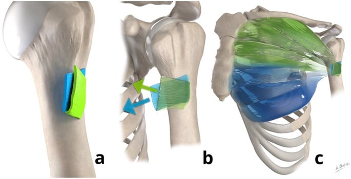
Subscapularis. From the subscapular fossa to the lesser tuberosity, supplied by the upper and lower subscapular nerves (C5-C6). It is the most important muscle for anterior stability and internal rotation, its insertion is critical for TSA stability, and it must be released or osteotomised to expose the humerus. Its upper third runs superiorly, its middle third horizontally and its lower third inferiorly.
The subscapularis footprint. The insertion on the lesser tuberosity has four facets. The first lies most superior and lateral, the second is separated from it by a bony ridge, and the broader third and fourth extend progressively inferiorly and medially. This footprint explains why limited upper-border tears can preserve substantial muscle attachment.
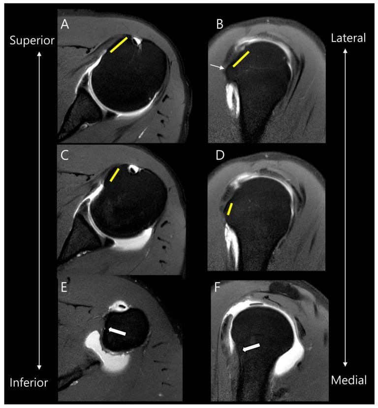
Supraspinatus. From the supraspinatus fossa, under the coracoacromial arch, to the superior facet of the greater tuberosity, supplied by the suprascapular nerve (C5-C6). It initiates abduction (the first 15°) and depresses the humeral head. It is rarely released during arthroplasty.
Posterior cuff. Infraspinatus runs from the infraspinatus fossa to the middle facet of the greater tuberosity (middle and superior greater tuberosity), supplied by the suprascapular nerve; teres minor runs from the lateral scapular border to the inferior facet, supplied by the axillary nerve. Together they externally rotate the humerus and stabilise it posteriorly. The posterior cuff rarely requires release but can be split for a posterior approach.
Rotator interval. The interval lies between the superior border of subscapularis and the anterior edge of supraspinatus, floored by the anterosuperior capsule and containing the coracohumeral ligament, the superior glenohumeral ligament and the long head of biceps. It is the key to safe joint access: it is opened first in the deltopectoral approach, and releasing it improves external rotation in stiff shoulders.
Neurovascular Anatomy - Critical Structures
Axillary Nerve - Highest Risk During Arthroplasty
Course. The axillary nerve arises from the posterior cord (C5-C6) and crosses anterior to subscapularis. It leaves the axilla through the quadrangular space with the posterior humeral circumflex artery, then wraps posteriorly around the surgical neck of the humerus, 5-7 cm inferior to the acromion at rest and closer with the arm abducted. The quadrangular space is bounded by:
- Superiorly, teres minor
- Inferiorly, teres major
- Medially, the long head of triceps
- Laterally, the surgical neck of the humerus
Branches. An anterior branch to the anterior and middle deltoid, a posterior branch to the posterior deltoid and teres minor, and the superior lateral cutaneous nerve of the arm.
When it is injured. The nerve is most at risk during inferior capsule release and glenoid retraction. Injury paralyses the deltoid, with loss of abduction beyond 15°, weakens external rotation through teres minor, and leaves sensory loss over the lateral shoulder (the regimental badge area). It is usually a neurapraxia that recovers in 3-6 months.
Musculocutaneous Nerve - Medial Retraction Risk
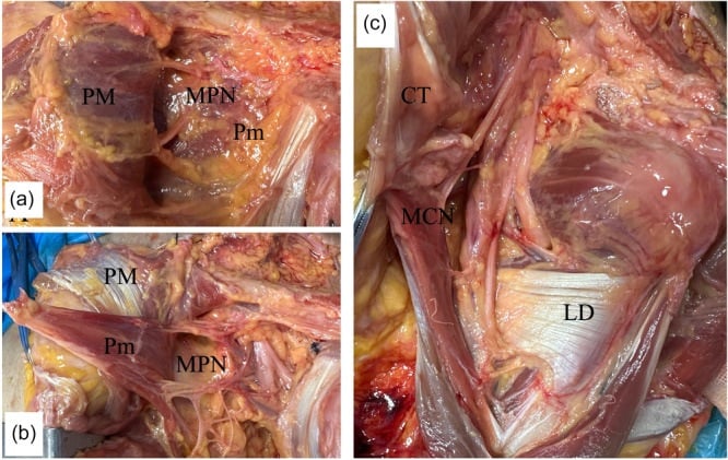
Course. From the lateral cord (C5-C7), the nerve passes between pectoralis minor and subscapularis and enters coracobrachialis a mean of 56 mm below the coracoid tip (range 31-82 mm), with proximal muscular twigs entering as close as 17 mm. It supplies coracobrachialis, biceps brachii and brachialis, and ends at the elbow as the lateral antebrachial cutaneous nerve.
There is no reliable safe zone. The frequently quoted "5-8 cm below the coracoid" fails because 29% of main trunks enter proximal to 5 cm, 74% once the proximal twigs are counted. Identify and protect the nerve rather than trusting a distance.
When it is injured. Excessive medial retraction of the conjoint tendon, a subscapularis release carried too far medially, and coracoid fracture or transfer procedures all threaten it. Injury weakens elbow flexion and supination and causes sensory loss over the lateral forearm; it usually recovers with conservative management.
Suprascapular Nerve - Posterior Glenoid Exposure Risk
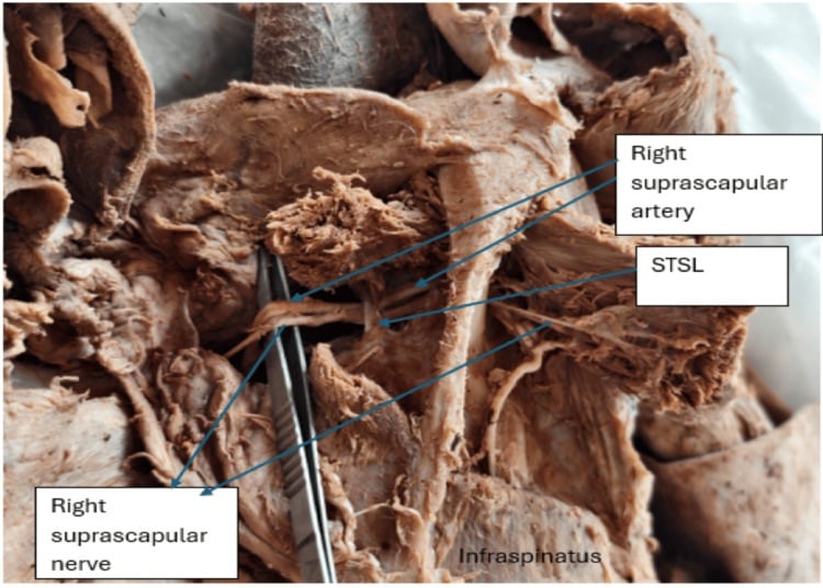
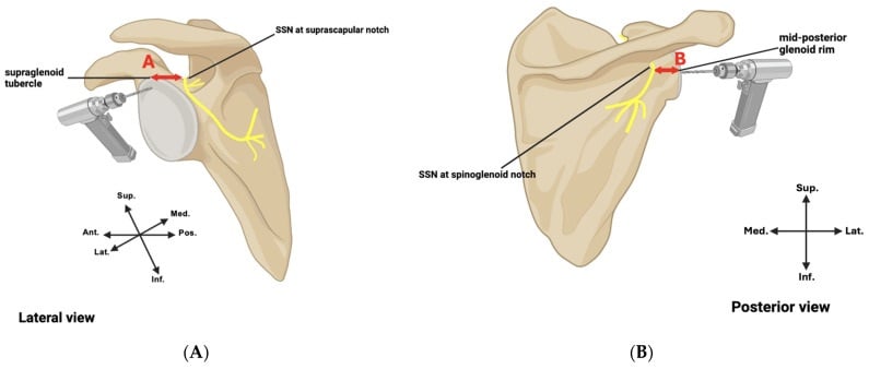
Course. From the upper trunk (C5-C6), the nerve passes through the suprascapular notch under the superior transverse scapular ligament, along the floor of the supraspinatus fossa, and around the spine of the scapula through the spinoglenoid notch, posterior to the glenoid, to the infraspinatus fossa. It supplies supraspinatus and infraspinatus and has no sensory component. The two notches are its fixed points, where it is vulnerable to traction.
When it is injured. Posterior glenoid retraction, a superior-posterior retractor, excessive traction on the posterior capsule and long superior screws all put it at risk. Injury causes supraspinatus and infraspinatus atrophy with weakness of abduction and external rotation, and can mimic a rotator cuff tear after surgery.
Radial Nerve - Humeral Canal Preparation Risk
Course. From the posterior cord (C5-T1), the radial nerve runs behind the humerus in the spiral groove on the posterior mid-shaft, 10-14 cm distal to the surgical neck and 20 cm from the acromion. It supplies triceps, brachioradialis and the wrist and finger extensors, with sensation to the posterior arm and forearm.
When it is injured. Reamers and broaches during canal preparation, perforation of the anterior cortex as a stemmed component goes in, and fracture while broaching osteoporotic bone. Injury produces wrist drop and sensory loss over the dorsum of the hand, and a complete palsy requires nerve exploration.
- Axillary nerve: release the inferior capsule under direct vision, dissect bluntly inferior to subscapularis, never place inferior retractors blindly, limit inferior translation during glenoid exposure, and stay more than 5 cm from the acromion inferiorly
- Musculocutaneous nerve: limit medial retraction of the conjoint tendon, stay superior to the inferior border of subscapularis and the conjoint tendon, avoid dissection more than 5 cm medial to the coracoid, and use self-retaining retractors carefully
- Suprascapular nerve: minimise posterior retraction duration and force, avoid a superior-posterior retractor at the glenoid rim, release the posterior capsule fully before retracting, and recognise anatomical variation at the notches
- Radial nerve: stay centred in the humeral canal, using the line of the shaft as a guide, protect the anterior cortex, broach gently in osteoporotic bone, and consider a cementless short stem to avoid violating the distal canal
Vascular Anatomy
Axillary artery. Medial to the surgical field, behind pectoralis minor. It is the main blood supply to the arm and rarely at risk; avoid excessive medial dissection deep to the conjoint tendon.
Anterior humeral circumflex artery. Ascends in the bicipital groove. Its ascending branch is met at the superior border of pectoralis major during the deltopectoral approach and requires ligation there.
Posterior humeral circumflex artery. Travels with the axillary nerve through the quadrangular space and is at risk during inferior capsule release; protecting the nerve protects the artery.
Deltopectoral Approach - Step by Step
Landmarks and Incision
Surface landmarks. Palpate and mark:
- Coracoid process: anteromedial, 2-3 cm medial and inferior to the AC joint
- Acromion: the lateral shoulder prominence
- Clavicle: the superior reference
- Deltopectoral groove: a visible depression running from the clavicle toward the deltoid insertion
- Axillary fold: the inferior limit of the exposure
Incision. Start at the coracoid and run obliquely toward the lateral edge of the acromion, then distally along the deltopectoral groove, extending to the deltoid insertion if needed: 10-15 cm in total for a standard arthroplasty. A gentle curve following the natural skin lines gives a better scar. Incise skin and subcutaneous fat down to the deltopectoral fascia.
The cephalic vein. The vein marks the interval, and it is visible in the groove in 60-70% of cases. There are two schools, with no significant difference in outcome between them; choose on surgical preference and vein quality, and if the vein is damaged during dissection, ligate it completely.
- Retract laterally with deltoid (the more common choice): coagulate or ligate the medial tributaries from pectoralis major. In a cadaveric study of 40 shoulders the vein had more lateral (deltoid-side) than medial feeder vessels in most specimens, so taking it with the deltoid divides fewer branches (Radkowski 2006)
- Retract medially with pectoralis major: ligate the lateral branches entering deltoid, which in most shoulders means dividing more branches
The Interval
A true internervous plane. Deltoid is supplied by the axillary nerve (C5-C6) and pectoralis major by the medial and lateral pectoral nerves (C5-T1), so the deltopectoral interval is both internervous and intermuscular, and extensile.
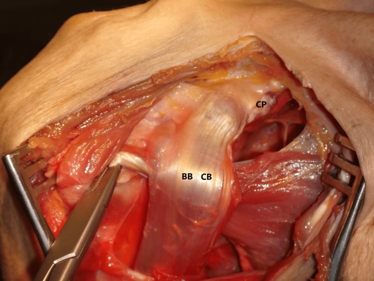
Deep dissection. Release the clavipectoral fascia deep to pectoralis major along its lateral border, which preserves the medial pectoral neurovascular bundle. The ascending branch of the anterior humeral circumflex artery is encountered at the superior border of pectoralis major and is ligated. Retracting pectoralis medially and deltoid laterally then exposes the coracoid, the conjoint tendon (the medial border of the exposure), subscapularis and the anterior capsule.
Retractor placement.
- A self-retaining retractor between deltoid and pectoralis
- The medial retractor superior to the inferior border of the conjoint tendon, protecting the musculocutaneous nerve
- The lateral retractor on deltoid without excessive tension
- No inferior retractors until the capsule has been released, to protect the axillary nerve
Deep Exposure
Rotator interval. Identify the space between supraspinatus superiorly and subscapularis inferiorly and incise the interval tissues, preserving the long head of biceps initially. Opening it improves external rotation and gives access to the anterior capsule.
Long head of biceps. Inspect it for degeneration, subluxation and tearing. Most surgeons perform a routine tenotomy during arthroplasty.
- Preserve it if healthy (infrequent)
- Tenotomy at the origin, released close to the labrum (most common)
- Tenodesis to the bicipital groove or conjoint tendon in a young, active patient
Subscapularis. Released by tenotomy, lesser tuberosity osteotomy or peel; the options and their repair are in Subscapularis Management below.
Capsule. The anterior capsule attaches to the glenoid labrum and provides anterior stability. Incise it vertically along its humeral insertion, then release the inferior capsule carefully, under direct vision and staying on the capsule, because this is where the axillary nerve is at risk. Posterior capsule release improves glenoid exposure, and a humeral head retractor displaces the head posteriorly.
Glenoid Exposure
Sequence.
- Position the arm in extension, external rotation and adduction, translating the humeral head posteriorly
- Place a posterior humeral head retractor (Fukuda-type)
- Release the anterior capsule off the glenoid rim
- Release the inferior capsule to the 6 o'clock position under direct vision
- Place blunt retractors inferiorly, and superiorly only if needed
Capsular release. Complete anterior release, and inferior release to 6 o'clock while protecting the axillary nerve. Posterior release improves exposure of the posterior glenoid, where excessive retraction puts the suprascapular nerve at risk. Superior release is less critical. Complete anterior capsule release and careful retraction are what make the posterior glenoid accessible.
Retractors on the glenoid.
- Anterior retractor on the anterior glenoid rim
- Inferior blunt retractor on the inferior scapular neck, NOT on the inferior capsule where the axillary nerve travels
- Posterior retractor on the posterior glenoid rim, limiting force and duration to protect the suprascapular nerve
- Use blunt retractors on the rim, avoiding damage to the suprascapular nerve; a superior retractor is rarely needed
Labrum. Excise it completely for glenoid preparation, preserve it if needed for anchor placement (reverse shoulder), and send it for culture if infection is a concern.
When exposure is poor.
- Complete the subscapularis release
- Complete the capsular release
- Externally rotate the arm
- Extend the inferior release, with caution
- Consider making the humeral head cut first to decompress the joint
Alternative Approaches
The deltopectoral approach remains the gold standard for most cases: it is extensile, internervous and familiar, and can be extended proximally or distally as needed.
- Indication
- Most shoulder arthroplasty cases
- Advantages
- Extensile, internervous, familiar anatomy, lower infection
- Disadvantages
- Subscapularis must be released/repaired
- Indication
- Irreparable subscapularis deficiency, revision with subscapularis failure, or reverse arthroplasty where subscapularis function is less critical
- Advantages
- Preserves subscapularis, good superior exposure
- Disadvantages
- Risk to axillary nerve, limited inferior exposure
- Indication
- Selected reverse arthroplasty cases: primary reverse arthroplasty in the hands of an experienced surgeon, or a desire to preserve anterior structures
- Advantages
- Preserves anterior and posterior cuff
- Disadvantages
- Limited exposure, supraspinatus release required, steep learning curve
- Indication
- Posterior instability repair (posterior bone block, posterior glenoid bone grafting) or revision with posterior component removal
- Advantages
- Direct posterior access
- Disadvantages
- Requires prone position, limited anterior exposure
Deltoid-Splitting Approaches and the Axillary Nerve Safe Zone
A different axillary nerve risk. The deltopectoral interval passes between deltoid and pectoralis and spares the deltoid, so its axillary nerve danger is deep and inferior: the main trunk at the inferior surgical neck during inferior capsule release. The anterosuperior (Mackenzie) and superior (Neviaser) approaches split the deltoid fibres instead, and a split carried too far distally divides the nerve's anterior (deltoid) branch.
The anatomy. After leaving the quadrangular space, the anterior branch runs transversely across the deep surface of deltoid to supply its anterior and middle heads, crossing roughly 5-7 cm distal to the lateral acromion. The Yildirim study in the evidence section measured the nerve at a mean of 6.0 cm (range 5.5-6.6 cm) from the anterolateral acromion and proposed about 5.5 cm as a practical limit for the deltoid-splitting approach.
The rule. Limit any deltoid split to within about 5 cm of the acromion, and place a stay (anchoring) suture at the distal apex of the split so that retraction cannot propagate it into the nerve.
The caveat. Nerve position is individually variable, predictable in only about 18% of patients in the Yildirim study, and the nerve migrates closer to the acromion with abduction. The safe zone is a guide, not a guarantee: palpate for the nerve on the deep deltoid surface whenever a split is used.
Classification
Walch Classification of Glenoid Wear
- Description
- Minor central erosion, concentric humeral head
- Implications
- Standard glenoid component, no augment needed
- Description
- Major central erosion, concentric humeral head
- Implications
- May need bone graft for central defect
- Description
- Posterior subluxation, no erosion
- Implications
- Asymmetric reaming, consider RSA
- Description
- Posterior subluxation + biconcave posterior erosion
- Implications
- Posterior augment or asymmetric reaming, RSA preferred
- Description
- Monoconcave posterior erosion (retroversion greater than 15°)
- Implications
- Posterior augment mandatory, bone graft, or RSA
- Description
- Dysplastic glenoid (retroversion greater than 25°)
- Implications
- RSA with augment or custom implant
- Description
- Anterior subluxation/erosion
- Implications
- Rare, anterior augment or RSA
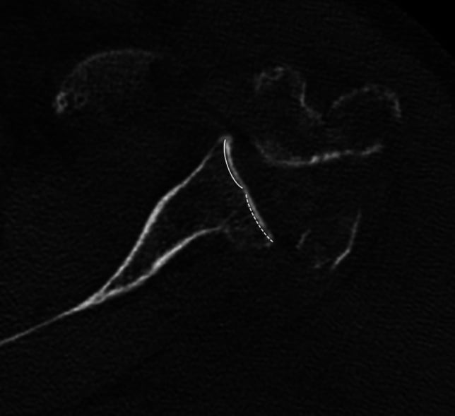
The B2 glenoid is the hardest for TSA. Biconcave erosion with posterior humeral subluxation loads the component eccentrically and brings high glenoid loosening rates. The options are asymmetric reaming, which offers limited correction, a posterior augmented glenoid, or conversion to RSA, which is increasingly preferred.
Hamada Classification of Cuff Tear Arthropathy
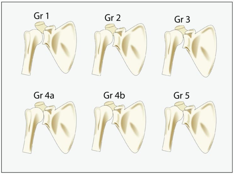
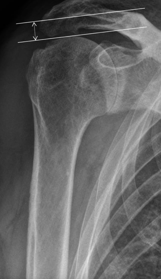
Graded on the acromiohumeral interval (AHI) and the joint changes that follow:
- Grade 1: AHI greater than 6 mm
- Grade 2: AHI 5 mm or less
- Grade 3: acetabularisation of the acromion
- Grade 4A: glenohumeral arthritis without acetabularisation
- Grade 4B: glenohumeral arthritis with acetabularisation
- Grade 5: humeral head collapse
Humeral Bone Loss
- Type 1: minimal bone loss - standard component
- Type 2: metaphyseal deficiency - short metaphyseal stem
- Type 3: diaphyseal extension required - long stem
- Type 4: proximal humerus replacement needed
Investigations
Plain radiographs are essential:
- True AP (Grashey): joint space, humeral head position, glenoid wear
- Axillary lateral: glenoid version, posterior wear, subluxation
- Scapular Y: acromion morphology, os acromiale
- Full-length humerus: stem sizing in revision
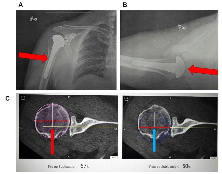
CT with 3D reconstruction is mandatory for preoperative planning, and CT-based planning reduces component malposition and improves outcomes. It provides:
- Glenoid version and inclination
- Subluxation, as the percentage of the humeral head posterior to the glenoid centre line
- The Walch type
- Bone stock for baseplate fixation, vault depth and screw trajectories
- The correction strategy, eccentric reaming or augment
- Virtual templating of implant size, position and screw trajectories in 3D planning software
MRI answers the cuff question, and with it the choice of implant.
- Rotator cuff integrity determines TSA or RSA
- Fatty infiltration (Goutallier) and muscle atrophy: Goutallier 3-4 is irreversible and favours RSA
- A subscapularis tear may need repair or an alternative approach
- Deltoid detachment is a contraindication to RSA
- Metal artefact reduction sequences are used if there is prior hardware
Other tests. Ultrasound gives a dynamic assessment of the subscapularis, and nuclear medicine is used if infection is suspected.
Subscapularis Management
Release
Three techniques. The choice is the most critical decision of the approach.
- Tenotomy: divide the tendon 1-2 cm medial to its lesser tuberosity insertion, preserving a 5-10 mm cuff of tissue on the tuberosity for repair. The release continues inferiorly to the 6 o'clock position, identifying and protecting the axillary nerve below
- Lesser tuberosity osteotomy (LTO): mark 5-8 mm medial to the articular margin and use an osteotome or saw to lift a 5-10 mm (about 1 cm) thick wafer of bone carrying the subscapularis insertion
- Peel: a partial-thickness release from the tuberosity that leaves the deeper capsular fibres attached; less common, and its healing biology is unclear
- Method
- Release 1-2 cm medial to insertion, preserve cuff on bone
- Advantages
- Easier exposure, faster, allows repair to bone
- Disadvantages
- Potential weakness, 5-10 percent failure rate
- Method
- Osteotomise tuberosity with subscapularis attached
- Advantages
- Bone-bone healing, strongest repair, lower failure
- Disadvantages
- Technical demand, tuberosity malunion/nonunion risk
- Method
- Partial thickness peel from tuberosity, leave capsule
- Advantages
- Preserves some insertion, middle ground
- Disadvantages
- Unclear biomechanics, healing variable
Which heals best. Osteotomy gives bone-to-bone healing, stronger and more reliable than tendon-to-bone, preserves the native subscapularis anatomy, and fails less often: 2-3% against 5-10% after tenotomy. It has the best healing rates of the three; the peel has a higher failure rate, with healing put at 95% after osteotomy against 70% after a peel. LTO is the preferred option for TSA.
What the trials add. The randomised trial and network meta-analysis in the evidence section found no difference in motion or clinical outcome scores between the techniques; osteotomy heals more predictably but takes longer, and the choice can be guided by tissue quality, surgeon experience and preference.
Repair After Tenotomy
Preparation. Mobilise the tendon medially until it reaches the lesser tuberosity without tension, clear soft tissue from the tuberosity to leave a bleeding bony bed, and reduce the trial components to assess tension.
Fixation. Suture anchors are the most common: 3-4 anchors in the lesser tuberosity footprint, knotless or traditional, with sutures passed through the tendon as modified Mason-Allen or simple stitches. Tie them with the arm in neutral rotation; external rotation creates excessive tension. Transosseous tunnels are the alternative, drilled from the lesser tuberosity to the lateral cortex, with non-absorbable sutures through tendon and tunnels tied over a lateral bone bridge. Repair strength depends on bone quality and anchor fixation.
Augmentation. Consider a pectoralis major transfer if tissue quality is poor, and a dermal allograft or synthetic patch for large defects (uncommon).
Testing. The belly press should hold resistance at 20-30° of external rotation, and the lag sign should be negative with an intact repair.
Repair After Lesser Tuberosity Osteotomy
Osteotomy repair provides the strongest biomechanical construct but requires technical precision.
- Screws (biomechanically strongest): 2-3 cortical screws of 3.5 or 4.0 mm through the fragment into the medial humeral shaft, heads countersunk to avoid impingement, with washers in osteoporotic bone
- Suture anchors: 2-3 anchors in the medial trough, with sutures through drill holes in the fragment secured as horizontal mattress sutures
- Heavy sutures alone: #5 non-absorbable suture through the tuberosity via bone tunnels and around the humeral neck; less rigid than screws but adequate in good bone
Risks of the osteotomy. A proud or medialised tuberosity causes impingement or weakness, and prominent screws cause impingement. Nonunion occurs in 2-5%, with inadequate fixation or osteoporotic bone.
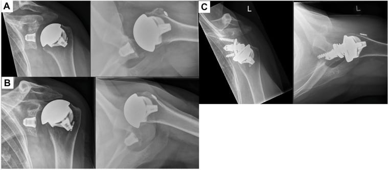
The postoperative protection of the repair is in Postoperative Care.
Management
Choosing the prosthesis. Cuff status and glenoid morphology decide it.
- Recommended Approach
- Anatomic TSA
- Rationale
- Restores normal biomechanics, best ROM
- Recommended Approach
- Reverse TSA (RSA)
- Rationale
- Deltoid-powered elevation, bypasses cuff
- Recommended Approach
- RSA preferred over TSA
- Rationale
- Lower glenoid loosening with baseplate vs pegged glenoid
- Recommended Approach
- RSA preferred
- Rationale
- No tuberosity healing required, reliable outcomes
Correcting glenoid version. The degree of retroversion sets the strategy:
- Less than 15°: asymmetric reaming alone
- 15-25°: posterior augmented glenoid or bone graft
- Greater than 25°: custom implant or RSA with augment
The limit of reaming. Asymmetric reaming can safely correct up to 10-15° of retroversion; beyond this, bone graft or augmented glenoid components are required. Overcorrection by reaming leads to anterior perforation and medialisation.
Bone defects.
- Central defects: autograft from the humeral head, impaction grafting
- Posterior defects (B2): asymmetric reaming, within its 10-15° limit
- Severe bone loss: structural allograft, metal augment or custom implant
Soft tissues. Subscapularis is managed by osteotomy, peel or tenotomy (see Subscapularis Management). A contracted capsule needs circumferential release for exposure, and after previous surgery scar tissue is released and subscapularis mobilised.
Surgical Technique
Humeral Preparation
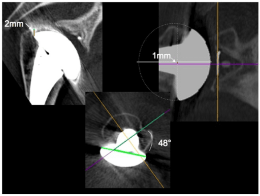
Identify the anatomical neck to set the cut level and preserve bone stock, and match the resection angle to the native neck-shaft angle (130-145°). Set version to the native version or 20-30° of retroversion, using the forearm axis or the transepicondylar axis as the reference. Avoid varus positioning, which risks stress shielding and subsidence.
Glenoid Preparation
Remove peripheral osteophytes first, for orientation, before central reaming. Correct version by asymmetric (posterior) reaming or an augment, avoid breaching the anterior cortex when preparing the pegs, and pressurise the cement into cancellous bone.
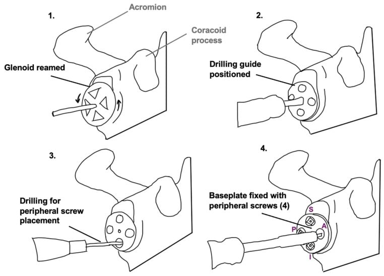
Reverse Arthroplasty: The Baseplate
Position. Centre the baseplate on the glenoid face in neutral to 10-15° of inferior tilt, with inferior glenosphere offset to reduce notching.
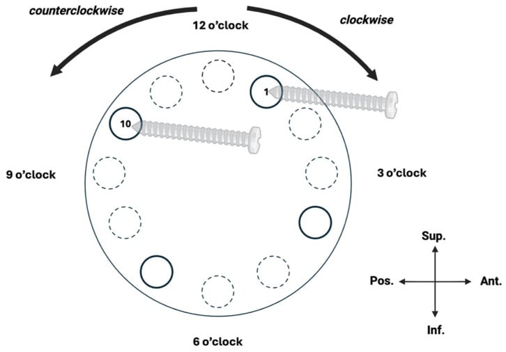
Screws. Use 4 screws at minimum, with the longest in the superior and posterior positions. To protect the suprascapular nerve, limit superior-posterior screw length to under 30 mm.
Complications and Clinical Correlations
Nerve, Tendon and Vessel Injury
- Incidence
- 0.6-4%
- Cause
- Inferior capsule release, retraction, traction
- Prevention
- Careful inferior release, avoid blind retractors, limit traction
- Management
- Observation 3-6 months (most recover), EMG at 6 weeks, consider exploration if no recovery
- Incidence
- 0.3-1%
- Cause
- Excessive medial retraction of conjoint tendon
- Prevention
- Limit medial retraction, stay above inferior subscapularis
- Management
- Usually recovers spontaneously, splint elbow in flexion initially
- Incidence
- Less than 1%
- Cause
- Posterior glenoid retraction, superior retractor
- Prevention
- Limit posterior retraction force and duration, avoid superior-posterior retractors
- Management
- Observation, consider decompression at notch if no recovery 6-12 months
- Incidence
- 2-10%
- Cause
- Poor tissue quality, inadequate repair, excessive tension, noncompliance
- Prevention
- Secure repair technique, protect postoperatively, patient education
- Management
- Revision repair if symptomatic instability, consider pectoralis major transfer
- Incidence
- Less than 0.1%
- Cause
- Excessive medial dissection, fracture, cerclage wires
- Prevention
- Stay lateral, careful medial dissection, avoid cerclage
- Management
- Immediate vascular surgery consultation, repair or graft
Test nerve function in the recovery room before the regional anaesthetic wears off:
- Axillary nerve: deltoid contraction (palpate the muscle during attempted abduction) and sensation over the lateral shoulder
- Musculocutaneous nerve: biceps contraction with elbow flexion
- Radial nerve: wrist and finger extension
- Suprascapular nerve: cannot be tested acutely (motor only)
Document the findings and compare them with a baseline preoperative examination. If there is a new deficit, determine whether it is complete or partial. Most neurapraxias recover with observation, but early recognition guides patient counselling and follow-up planning.
Fractures and Other Complications
Intraoperative and periprosthetic fracture. The greater tuberosity fractures in 3-5% and the shaft in 1-2%. Aggressive reaming or malpositioning fractures the glenoid, and periprosthetic fracture follows postoperative trauma or stress risers. Prevention:
- In osteoporotic bone, gentle preparation, and consider cemented fixation
- On the humerus, avoid excessive torque and ream sequentially
- On the glenoid, get adequate exposure before reaming
Soft tissues. Rotator cuff tear progression reaches 5-10% by 10 years. Deltoid detachment is rare but catastrophic for RSA.
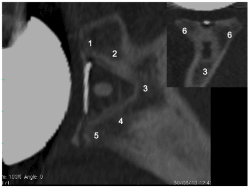
Differential Diagnosis of the Weak or Painful Shoulder After Arthroplasty
When a patient has weakness, restricted motion or pain after shoulder arthroplasty, identify the anatomical structure responsible, because management differs sharply. The distinction is between a nerve injury, a tendon (subscapularis) failure, and a mechanical or septic cause.
- Distinguishing features
- Deltoid weakness (abduction beyond 15 degrees lost), lateral shoulder (regimental badge) sensory loss
- Key test / investigation
- Deltoid palpation on abduction, sensation testing, EMG/NCS at ~6 weeks
- Initial management
- Observation (most are neurapraxias), maintain passive ROM; explore if no recovery by 3-6 months
- Distinguishing features
- Elbow flexion / supination weakness, lateral forearm sensory loss; deltoid intact
- Key test / investigation
- Biceps testing, lateral antebrachial sensation, EMG/NCS
- Initial management
- Usually spontaneous recovery; document, splint, maintain ROM
- Distinguishing features
- Excess passive external rotation, weak internal rotation, anterior instability; neurology intact
- Key test / investigation
- Positive belly-press and lift-off tests; ultrasound or MRI of subscapularis
- Initial management
- Revision repair if symptomatic; consider tendon transfer for irreparable tear
- Distinguishing features
- Supraspinatus/infraspinatus weakness mimicking cuff tear; no sensory loss
- Key test / investigation
- Cannot test acutely (motor only); EMG/NCS, imaging to exclude cuff tear
- Initial management
- Observation; decompression at the notch if no recovery
- Distinguishing features
- Mechanical symptoms, apprehension, malposition or fracture on imaging; neurology and cuff intact
- Key test / investigation
- Plain radiographs and CT for version/position; examine for instability
- Initial management
- Address the structural cause - bracing, revision or fixation as indicated
- Distinguishing features
- Persistent pain, stiffness, effusion, warmth; can present indolently (Cutibacterium)
- Key test / investigation
- Inflammatory markers, aspiration with prolonged culture, tissue sampling
- Initial management
- Treat per infection protocol - debridement or staged revision with targeted antibiotics
Postoperative Care
The sling. The standard protocol uses a sling for 2-4 weeks after TSA and 4-6 weeks after RSA, in internal rotation with slight abduction on a pillow. It comes off for elbow, wrist and hand exercises and for hygiene.
- Timeframe
- 0-4 weeks
- Goals
- Protect repair, control inflammation
- Exercises
- Pendulums, elbow/wrist ROM, isometrics
- Timeframe
- 4-8 weeks
- Goals
- Restore passive ROM
- Exercises
- Passive forward flexion, external rotation
- Timeframe
- 8-12 weeks
- Goals
- Active ROM, light function
- Exercises
- Active assisted to active ROM
- Timeframe
- 12-16 weeks
- Goals
- Progressive strength
- Exercises
- Rotator cuff, deltoid, scapular
After subscapularis repair. Protecting the repair is critical. The protection protocol keeps the sling on for 4-6 weeks, in neutral to slight internal rotation, removed only for hygiene and gentle pendulum exercises. There is no active or passive external rotation and no active internal rotation for 6 weeks, and combined extension and external rotation is avoided.
- Phase 1 (0-6 weeks): sling, passive forward elevation only, gentle pendulums, elbow, wrist and hand motion, and no external rotation
- Phase 2 (6-12 weeks): wean from the sling, active-assisted motion, external rotation gradually to neutral (0°) by 8 weeks, light activities of daily living
- Phase 3 (12+ weeks): progressive external rotation, full by 12-16 weeks, a strengthening programme, and unrestricted activities by 4-6 months
Checking the repair. Test the belly press at 8-12 weeks. A negative belly press by 12 weeks indicates a healed repair; one still positive beyond 12 weeks suggests failure and warrants MRI. Subscapularis failure causes anterior instability and loss of internal rotation.
After reverse arthroplasty. Earlier active motion is allowed, with deltoid-based elevation. External rotation is often limited (subscapularis repair), and full active external rotation may never be achieved.
Return to activity.
- Driving: 6-8 weeks, when comfortable with an emergency manoeuvre
- Swimming: 4-6 months
- Golf: 4-6 months, avoiding power strokes
- Contact sports: generally not recommended
Outcomes
TSA against RSA. TSA gives better range of motion, especially external rotation, but requires an intact rotator cuff. RSA gives reliable pain relief and elevation regardless of cuff status but sacrifices external rotation. The choice depends on cuff integrity, age and activity level.
- TSA
- 140-160°
- RSA
- 120-140°
- TSA
- 40-60°
- RSA
- 20-40°
- TSA
- L1-T12
- RSA
- Sacrum-L3
- TSA
- 85-95%
- RSA
- 85-90%
- TSA
- 90-95%
- RSA
- 85-90%
Anatomy that predicts outcome.
- Glenoid morphology: B2 and B3 glenoids have higher loosening rates, and an uncorrected B2 glenoid a 2-3 times higher loosening rate
- Version: retroversion greater than 15° is associated with increased loosening in TSA; RSA is less sensitive to version than TSA
- Rotator cuff: an intact cuff improves TSA outcomes
- Bone quality: osteoporosis increases fracture and loosening risk
Long-term. Polyethylene wear is a 10-15 year concern for TSA. Scapular notching is seen radiographically in 44-96% but is rarely clinically significant. Late subscapularis failure can turn a TSA into an indication for RSA.
Registry-tracked failure modes. National joint registries (NJR, AJRR, AOANJRR, NZJR, Nordic) track implant survival and revision. Reported revision rates vary by registry, implant and era, so quote the specific registry and implant when citing figures. Glenoid component loosening is a leading cause of anatomic TSA revision, and instability a leading cause of reverse TSA revision.
Guidelines, Registries & Global Practice
Shoulder arthroplasty is one of the fastest-growing joint replacements worldwide, driven mainly by reverse TSA. Wherever it is performed, the deltopectoral approach and its neurovascular relationships are identical, so the surgical anatomy is genuinely a global, exam-universal core. National registries and society guidelines converge on the same anatomical safety principles, differing mainly in implant case-mix and perioperative protocols.
Global Epidemiology
US nationwide data on more than 508,000 primary procedures (Best et al., J Shoulder Elbow Surg 2021, DOI) showed reverse TSA incidence rising from 7.3 to 19.3 per 100,000 persons and anatomic TSA from 9.5 to 12.5 per 100,000 between 2012 and 2017, while hemiarthroplasty fell from 3.7 to 1.5 per 100,000. The largest growth was reverse TSA in men and in patients aged 50-64 years. This rising, increasingly reverse-dominant case-mix means more patients are exposed to the deltopectoral neurovascular risks discussed above, even though reverse TSA may use the same or an anterosuperior approach.
Guidelines Side by Side
- Relevant recommendation
- Glenohumeral OA CPG and the 2009/2010 VTE consensus underpin routine chemoprophylaxis after TSA, associated with falling VTE rates over the following decade
- Evidence basis
- Clinical practice guideline / consensus; registry-linked observational support
- Relevant recommendation
- Joint-replacement standards emphasise consent for nerve injury, infection and revision, surgical-site infection bundles and PROMs capture; no UK guideline mandates a specific shoulder approach
- Evidence basis
- National standards / BOAST principles
- Relevant recommendation
- Teaching materials standardise the deltopectoral internervous interval, axillary-nerve protection during inferior release and structured subscapularis repair
- Evidence basis
- Expert consensus / educational standard
- Relevant recommendation
- Track implant survival, revision rate and the anatomic-versus-reverse case-mix; nerve injury is generally not a discrete registry endpoint
- Evidence basis
- Prospective registry / Level II-III
Registry Evidence and Practice Variation
Joint registries (the National Joint Registry for England, Wales, Northern Ireland and the Isle of Man; the American Joint Replacement Registry; the Australian AOANJRR; the New Zealand Joint Registry; and the Nordic registries) consistently document the global shift from anatomic TSA and hemiarthroplasty toward reverse TSA, and they benchmark implant survival and revision. Neurological injury is rarely a discrete registry field, so its incidence is best taken from clinical series rather than registries. Practice variation is mostly in case-mix and perioperative care rather than in the surgical anatomy: high-resource systems increasingly favour reverse TSA with CT-based planning, whereas in limited-resource settings hemiarthroplasty and standard instrumentation remain more common, and a sound understanding of the deltopectoral approach and axillary-nerve safe zone matters more where navigation and patient-specific guides are unavailable.
Across guidelines and health systems, the same anatomy-driven safety principles recur:
- Surgical-site infection bundle: weight-based intravenous antibiotic prophylaxis within 60 minutes of incision (a first-generation cephalosporin, with a glycopeptide alternative for severe beta-lactam allergy), skin antisepsis and normothermia. Follow the local/national antimicrobial formulary for exact agent and dose.
- VTE prophylaxis for joint-replacement patients, individualised to bleeding and thrombotic risk (AAOS consensus, DOI).
- Documented pre- and immediate post-operative neurological examination to detect axillary, musculocutaneous and radial nerve deficits early, given that injury is rarely captured by registries.
- Shared decision-making and PROMs capture for quality improvement and benchmarking against registry data.
MCQ Practice Points
Q: What is the average distance from the tip of the coracoid process to the point where the musculocutaneous nerve enters the coracobrachialis muscle?
A: In the classic cadaveric study by Flatow et al. (Clin Orthop Relat Res, 1989), the main musculocutaneous nerve trunk entered coracobrachialis at a mean of 56 mm (range 31-82 mm) below the coracoid, and small proximal twigs entered as close as 17 mm. Importantly, the often-quoted "5-8 cm safe zone" is unreliable, because 29 percent of main trunks entered proximal to 5 cm (74 percent when proximal twigs are counted). This anatomic relationship is critical during the deltopectoral approach: excessive medial retraction of the conjoint tendon risks musculocutaneous nerve injury by traction or direct trauma. Protection strategies include limiting medial retraction and staying superior to the inferior border of the subscapularis and conjoint tendon during deep dissection.
Q: What are the boundaries of the quadrangular space, and what structures pass through it?
A: Boundaries: Superior = teres minor, Inferior = teres major, Medial = long head of triceps, Lateral = surgical neck of humerus. Structures: Axillary nerve and posterior humeral circumflex artery. The quadrangular space is clinically important as the axillary nerve is vulnerable during inferior capsule release and humeral retraction for glenoid exposure. The nerve lies approximately 5-7 cm inferior to the acromion. Injury causes deltoid paralysis (loss of active abduction beyond 15 degrees), teres minor weakness, and sensory loss over the lateral shoulder (regimental badge area).
Q: What is the internervous plane for the deltopectoral approach to the shoulder?
A: Deltoid (innervated by axillary nerve from C5-C6) and pectoralis major (innervated by medial and lateral pectoral nerves from C5-T1). This is a true internervous and intermuscular plane, making it theoretically atraumatic to muscles. The interval is marked by the cephalic vein running in the deltopectoral groove. The deep layer requires release of the clavipectoral fascia and ligation of the ascending branch of the anterior humeral circumflex artery. The internervous nature is advantageous but does NOT protect deeper structures like the axillary nerve, musculocutaneous nerve, and suprascapular nerve which remain at risk during capsule release and glenoid retraction.
Q: Compare the biomechanical strength and clinical outcomes of subscapularis tenotomy versus lesser tuberosity osteotomy repair after shoulder arthroplasty.
A: Lesser tuberosity osteotomy provides stronger healing with bone-to-bone healing interface and lower failure rates (2-3 percent) compared to subscapularis tenotomy (5-10 percent failure rate). Osteotomy can be repaired with cortical screws (biomechanically strongest), suture anchors, or heavy sutures. Tenotomy repair uses suture anchors securing tendon to bone at the lesser tuberosity footprint. However, osteotomy carries risks of tuberosity malposition (causing impingement or weakness), nonunion (2-5 percent), and increased surgical complexity. In clinical practice, both techniques can achieve good outcomes with proper technique and postoperative protection. The choice depends on tissue quality, patient factors (younger patients may benefit from osteotomy), revision versus primary surgery, and surgeon experience and preference.
Q: What is normal glenoid version, how is it measured, and what are the implications of pathologic retroversion for shoulder arthroplasty component positioning?
A: Normal glenoid version is 0-5 degrees retroversion (slight posterior tilt of glenoid face). It is measured on axial CT using either the Friedman method (line perpendicular to glenoid face compared to scapular body line) or the scapular body method. Pathologic retroversion (over 10 degrees) occurs with posterior glenoid wear from rotator cuff tear arthropathy, chronic posterior instability, or osteoarthritis. Excessive retroversion leads to posterior humeral subluxation, glenoid component edge loading, loosening, and instability. Correction strategies include: eccentric reaming (can correct up to 10-15 degrees but limited by bone stock), augmented glenoid components (metal or polyethylene wedge on posterior), posterior bone grafting (for severe defects), or accepting retroversion and using posterior augmented component. Failure to address pathologic version results in high rates of posterior instability and glenoid component failure. Preoperative CT planning is mandatory to identify and plan for version abnormalities.
Q: A patient develops weakness in elbow flexion and sensory loss over the lateral forearm after shoulder arthroplasty. What nerve is likely injured, what is the mechanism, and how should this be managed?
A: This presentation suggests musculocutaneous nerve injury. The musculocutaneous nerve (C5-C7 from lateral cord) innervates coracobrachialis, biceps brachii, and brachialis (elbow flexion and forearm supination) and provides sensory innervation as the lateral antebrachial cutaneous nerve (lateral forearm sensation). During shoulder arthroplasty via deltopectoral approach, the nerve is at risk from excessive medial retraction of the conjoint tendon (short head biceps and coracobrachialis). The nerve enters the coracobrachialis a mean of 56 mm (range 31-82 mm) from the coracoid tip, with proximal twigs entering closer, so no fixed safe distance can be relied upon and aggressive medial retraction causes traction injury or direct trauma. Management: Most musculocutaneous nerve injuries are neurapraxias that recover spontaneously in 3-6 months. Initial management includes documentation of deficit, EMG/NCS at 6 weeks for baseline and prognosis, elbow splinting in flexion initially to reduce tension, and range of motion exercises to prevent stiffness. Patient counseling about expected recovery timeline is important. If no recovery by 6 months, consider nerve exploration, although outcomes of late exploration are poor. Prevention is critical: limit medial retraction, stay superior to the inferior border of subscapularis, and avoid dissection greater than 5 cm medial to coracoid tip.
Exam Viva Scenarios
Practise clinical reasoning and management decisions out loud
“You are performing shoulder arthroplasty via deltopectoral approach. Describe the key anatomical landmarks, the surgical intervals you will develop, and the nerves at risk during your dissection.”
“Walk me through your subscapularis management during shoulder arthroplasty. What are the different release techniques, how do you decide which to use, and what are the repair principles for each?”
“During shoulder arthroplasty glenoid exposure, you are concerned you may have injured the axillary nerve. Describe the anatomy of the axillary nerve, how you would assess for injury intraoperatively and postoperatively, and your management if injury is confirmed.”
Key Anatomical Relationships
- Axillary nerve: 5-7 cm inferior to acromion, quadrangular space, at risk during inferior capsule release
- Musculocutaneous nerve: Enters coracobrachialis a mean of 56 mm (range 31-82 mm) from coracoid, twigs closer; no fixed safe zone, at risk with medial retraction
- Suprascapular nerve: Spinoglenoid notch posteriorly, at risk during posterior glenoid retraction
- Radial nerve: Spiral groove at mid-humerus (20 cm from acromion), at risk during humeral canal preparation
- Cephalic vein: Marks deltopectoral interval, usually retracted laterally with deltoid (medial retraction an alternative)
- Ascending branch anterior humeral circumflex: Requires ligation at superior border pectoralis major
Deltopectoral Approach Steps
- Internervous plane: Deltoid (axillary nerve) and pectoralis major (pectoral nerves)
- Incision: Coracoid to lateral acromion, along deltopectoral groove distally
- Identify cephalic vein (marks interval), coagulate medial tributaries and retract laterally with deltoid
- Release clavipectoral fascia, ligate ascending branch anterior humeral circumflex
- Open rotator interval (between supraspinatus and subscapularis)
- Release subscapularis: Tenotomy (1 cm medial to insertion) OR lesser tuberosity osteotomy
Nerve Protection Strategies
- Axillary: Release inferior capsule under direct vision, avoid blind inferior retractors, limit inferior humeral translation
- Musculocutaneous: Limit medial retraction, stay above inferior border subscapularis/conjoint tendon, avoid dissection greater than 5 cm from coracoid
- Suprascapular: Minimize posterior retraction force and duration, avoid superior-posterior retractor placement
- Radial: Stay centered in humeral canal during reaming/broaching, protect anterior cortex, gentle technique in osteoporotic bone
Subscapularis Management
- Tenotomy: Release 1-2 cm medial to insertion, repair with 3-4 suture anchors, 5-10 percent failure rate
- Osteotomy: 1 cm bone wafer with subscapularis attached, repair with screws or anchors, 2-3 percent failure, stronger healing
- Repair: Arm in neutral rotation (not external rotation), secure fixation, avoid excessive tension
- Postoperative: Sling 4-6 weeks, no external rotation, gradual ROM progression, belly press test at 12 weeks
Glenoid and Humeral Anatomy
- Normal glenoid version: 0-5 degrees retroversion, measured on CT, pathologic if over 10 degrees
- Humeral retroversion: 20-40 degrees (average 30 degrees) relative to transepicondylar axis
- Glenoid: Pear-shaped, 25-30 mm diameter, shallow socket (25-30 percent of sphere)
- Quadrangular space: Teres minor (superior), teres major (inferior), triceps long head (medial), surgical neck (lateral)
- Contains: Axillary nerve and posterior humeral circumflex artery
Key Evidence and Complications
- Overall neurologic complication rate: 1-4 percent in shoulder arthroplasty
- Axillary nerve most common (0.6-4 percent), most are neurapraxias recovering in 3-6 months
- Subscapularis failure: 2-10 percent, causes anterior instability and internal rotation weakness
- Immediate postoperative nerve exam before regional anesthesia wears off - document findings
- EMG/NCS at 6 weeks if nerve injury suspected, observation for 3-6 months (most recover)
- Registries (NJR, AJRR, AOANJRR, NZJR, Nordic) track revision and the rising reverse-TSA case-mix; nerve injury is not a discrete registry endpoint, so use clinical series for its incidence
Evidence Base and Key Studies
Neurologic Complications After Total Shoulder Arthroplasty
- 417 total shoulder arthroplasties in 368 patients (1975-1989); 18 shoulders (17 patients) developed a postoperative neurologic deficit
- Most deficits localised to the brachial plexus, with the upper and middle trunks most commonly affected (13 shoulders)
- Presumed mechanism in most cases was intraoperative traction on the plexus rather than direct nerve laceration
- The long deltopectoral approach (deltoid left attached to clavicle/acromion) was significant (p = 0.003), as was methotrexate use (p < 0.0001)
- Operative time also correlated with injury (p = 0.02) - but in the direction OPPOSITE to intuition: SHORTER operative times were associated with more neurologic complications, so speed is not protective here
- Prognosis for recovery was generally good (good in 11 shoulders, fair in 5 at one year), and injury did not compromise long-term arthroplasty outcome
Loss of Subscapularis Function After Total Shoulder Replacement
- Retrospective review of 41 patients after total shoulder replacement with the subscapularis repaired anatomically (9) or through bone tunnels (32)
- Despite meticulous repair, the lift-off test was abnormal in 25 of 37 shoulders (67.5 percent) and the belly-press test abnormal in 24 of 36 (66.6 percent)
- Of patients with an abnormal lift-off finding, 92 percent reported reduced subscapularis function
- Suboptimal return of internal rotation and subscapularis function was a frequent, under-recognised problem
- Subscapularis dysfunction limited activities of daily living such as tucking in a shirt
An Anatomic Study of the Musculocutaneous Nerve and Its Relationship to the Coracoid Process
- The distance from the coracoid to where the main musculocutaneous nerve trunk entered coracobrachialis ranged from 31 to 82 mm (mean 56 mm)
- Small nerve twigs entered the muscle as close as 17 mm below the coracoid (mean 31 mm)
- The frequently quoted 5-8 cm safe zone is unreliable: 29 percent of main trunks entered proximal to 5 cm below the coracoid (74 percent if proximal twigs are counted)
- Excessive medial retraction or coracoid mobilisation is the principal mechanism of injury
- If coracoid mobilisation is needed the nerve and its twigs should be identified and protected directly