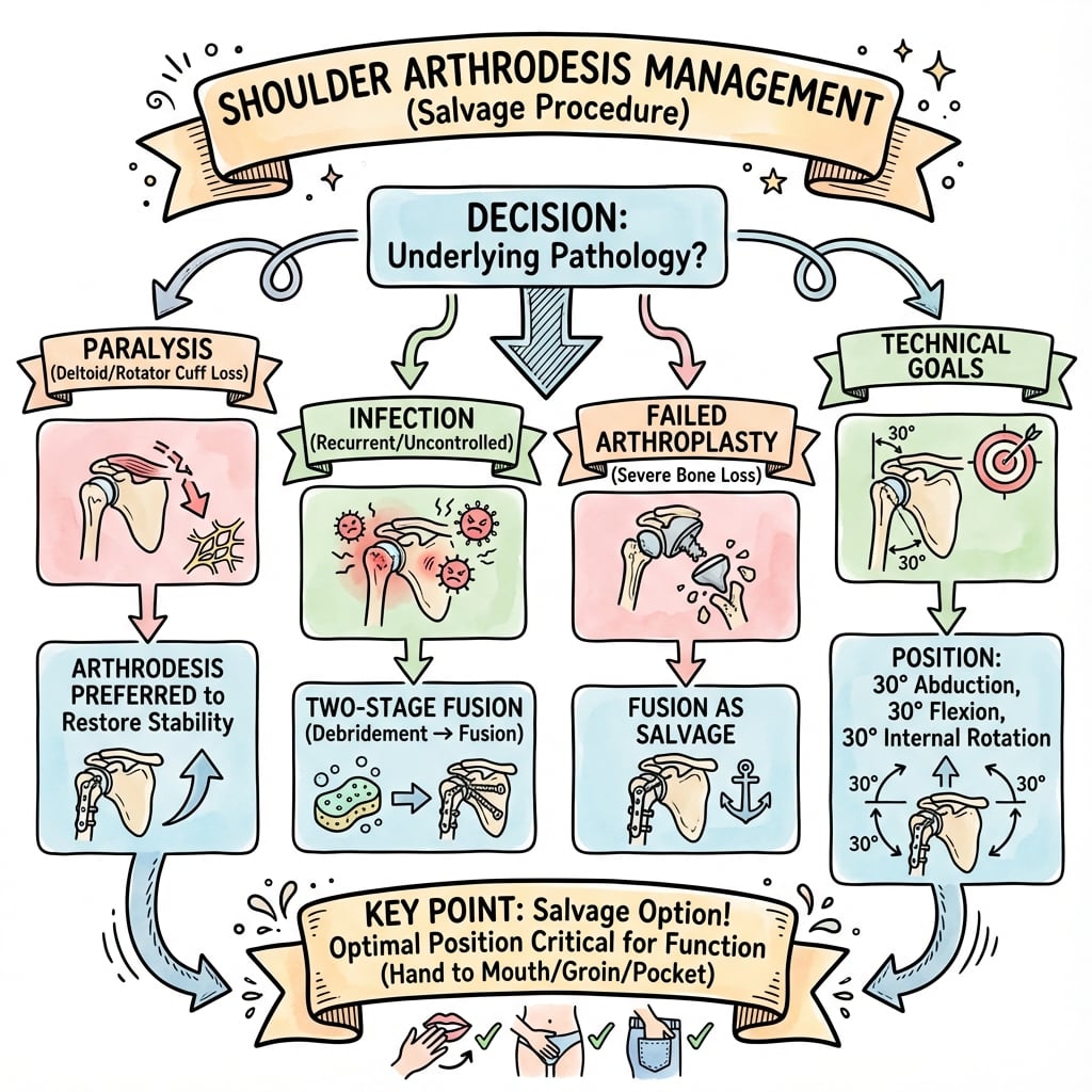
Salvage Procedure | C5-C6 Must Be Intact | Fusion Position Critical | Rarely Performed Now
- C5-C6 MUST be intact - needed for scapular stabilization (trapezius, serratus anterior)
- Fusion position: 20-30° abduction, 20-30° flexion, 30-40° internal rotation (hand-to-mouth position)
- Deltoid is only remaining motor - must preserve and repair meticulously
- Rarely performed now - reverse shoulder arthroplasty preferred for most indications
- Non-union rate: 5-15%, higher in revision or infection cases
- “Fusion position critical: test hand to mouth, opposite axilla, perineum BEFORE fixing
- “Axillary nerve 5-7cm below acromion - must protect during deltoid splitting
- “Superior plate from scapular spine to humeral shaft with minimum 3 screws in scapula
- “Modern alternative: reverse TSR provides better function even without rotator cuff
Absolute requirement: C5-C6 nerve roots must be functional to innervate trapezius and serratus anterior for scapular stabilization. Without scapular stabilization, the patient cannot elevate the fused shoulder unit. This is an absolute contraindication if C5-C6 are deficient.
Optimal position: 20-30° abduction, 20-30° flexion, 30-40° internal rotation. This is the hand-to-mouth position. Must test clinically: hand to mouth, hand to opposite axilla, hand to perineum BEFORE definitive fixation. Malposition cannot be revised.
Deltoid is the ONLY remaining shoulder motor after arthrodesis. Must preserve during approach (axillary nerve 5-7cm below acromion) and repair meticulously if detached. Deltoid failure results in complete loss of shoulder elevation.
Reverse shoulder arthroplasty has largely replaced arthrodesis for most indications. Reverse TSR provides better function (active motion vs passive scapulothoracic), better cosmesis, and acceptable outcomes even without rotator cuff. Arthrodesis now reserved for: young patients with infection, massive bone loss, or when arthroplasty not possible.
- Indication
- C5-C6 intact, C7-T1 deficient
- Alternative
- Arthrodesis if deltoid functional
- Key Consideration
- Must test C5-C6 function preoperatively
- Indication
- Massive soft tissue loss, bone loss
- Alternative
- Arthrodesis or amputation
- Key Consideration
- Reverse TSR may still be possible with bone graft
- Indication
- After infection eradication
- Alternative
- Staged arthrodesis
- Key Consideration
- Must wait 6-12 months after infection clear
- Indication
- Pseudoparalysis, deltoid intact
- Alternative
- Reverse TSR (preferred) or arthrodesis
- Key Consideration
- Reverse TSR now standard - arthrodesis rarely indicated
C5-C6Nerve Requirements
Hook:C5-C6 together provide scapular stabilization (trapezius, serratus) and deltoid function - both essential for arthrodesis success.
Overview and Epidemiology
Shoulder arthrodesis is a salvage procedure that fuses the glenohumeral joint, converting the shoulder into a fixed unit. Function depends on scapulothoracic motion and requires intact C5-C6 nerve roots for scapular stabilizers (trapezius and serratus anterior).
Shoulder arthrodesis was once the primary treatment for severe shoulder dysfunction, particularly in cases of brachial plexus injury and failed arthroplasty. However, the advent of reverse shoulder arthroplasty has dramatically reduced the indications for arthrodesis. Reverse TSR now provides superior outcomes for most conditions that previously required arthrodesis.
- Brachial plexus injury with C5-C6 intact (most common current indication)
- Failed shoulder arthroplasty with massive soft tissue deficiency and bone loss
- Chronic infection with bone loss after infection eradication
- Severe deltoid insufficiency with irreparable rotator cuff (rare - reverse TSR usually preferred)
- Frequency: Rarely performed now (less than 1% of shoulder procedures)
- Age: Typically younger patients (under 60) with brachial plexus injury or infection
- Gender: Male predominance due to higher rates of trauma and brachial plexus injury
- Trend: Declining frequency due to reverse shoulder arthroplasty success
Reverse shoulder arthroplasty has revolutionized treatment of rotator cuff arthropathy and failed arthroplasty. Reverse TSR provides better function (active motion vs passive scapulothoracic only), better cosmesis, and acceptable outcomes even without rotator cuff. Arthrodesis is now reserved for: young patients with infection, massive bone loss, or when arthroplasty is not possible. Brachial plexus injury with C5-C6 intact remains a valid indication.
Indications and Contraindications
Primary Indications
1. Brachial Plexus Injury (Most Common Current Indication)
- C5-C6 intact, C7-T1 deficient
- Deltoid functional (C5-C6 innervation)
- Patient accepts functional limitations
- Rationale: Arthrodesis provides stable base for hand function
2. Failed Shoulder Arthroplasty
- Massive soft tissue deficiency
- Significant bone loss
- Multiple failed revisions
- Rationale: When reverse TSR not possible due to bone loss
3. Chronic Infection with Bone Loss
- After infection eradication (6-12 months)
- Significant glenoid or humeral bone loss
- Rationale: Staged fusion after infection clearance
4. Severe Deltoid Insufficiency (Rare)
- Irreparable rotator cuff with pseudoparalysis
- Deltoid intact but cuff deficient
- Note: Reverse TSR now preferred for this indication
Understanding these indications helps guide patient selection and surgical decision-making.
Anatomy and Biomechanics
- C5-C6 nerve roots: Must be intact
- C5: Innervates deltoid, supraspinatus, upper trapezius
- C6: Innervates serratus anterior, biceps, middle trapezius
- Function: Scapular stabilization (trapezius and serratus) and deltoid power
- Why critical: Without scapular stabilization, patient cannot elevate fused shoulder unit
- Location: 5-7cm below acromion
- Course: Wraps around surgical neck of humerus
- Innervation: Deltoid (critical for post-fusion function) and teres minor
- Protection: Must stay medial to nerve during deltoid splitting approach
- Function: Only remaining shoulder motor after arthrodesis
- Origin: Clavicle, acromion, scapular spine
- Insertion: Deltoid tuberosity of humerus
- Critical: Must preserve and repair meticulously if detached
- Trapezius: Upper fibers (C5-C6), elevates and rotates scapula
- Serratus anterior: C5-C6-C7, protracts and stabilizes scapula
- Function: After arthrodesis, scapulothoracic motion provides arm elevation
- Glenohumeral motion: Eliminated (fused)
- Scapulothoracic motion: Becomes primary mechanism for arm elevation
- Functional elevation: 40-60° achievable through scapular rotation
- Deltoid function: Provides force for scapular elevation (if intact)
Classification Systems
Classification by Primary Indication
Type I: Brachial Plexus Injury
- C5-C6 intact, C7-T1 deficient
- Most common current indication
- Deltoid functional
- Outcome: Good if hand function preserved
Type II: Failed Arthroplasty
- Massive soft tissue deficiency
- Significant bone loss
- Multiple failed revisions
- Outcome: Variable, higher non-union risk
Type III: Chronic Infection
- After infection eradication
- Bone loss from infection
- Staged approach required
- Outcome: Lower fusion rates (60-70%)
Type IV: Severe Deltoid Insufficiency
- Irreparable rotator cuff
- Pseudoparalysis
- Note: Reverse TSR now preferred
This classification helps guide treatment approach and predict outcomes.
Clinical Assessment
- Mechanism of injury (brachial plexus, trauma, infection)
- Previous surgeries and outcomes
- Functional goals and expectations
- Hand dominance and occupation
Physical Examination
- C5-C6 function: Test trapezius (shoulder shrug), serratus anterior (wall push), deltoid (abduction)
- C7-T1 function: Test hand function, finger extension, intrinsic muscles
- Axillary nerve: Test deltoid strength, sensation over deltoid
- EMG/NCS: Confirm C5-C6 integrity if clinical exam equivocal
- Active and passive range of motion
- Deltoid strength (critical)
- Scapular motion and stability
- Hand function (determines overall utility)
- X-rays: AP, axillary, scapular Y views
- CT scan: Assess bone stock, glenoid version, humeral head condition
- MRI: Evaluate soft tissue, rotator cuff, deltoid integrity
- Demonstrate fusion position preoperatively
- Test hand to mouth, hand to opposite axilla, hand to perineum
- Patient must approve position before surgery
Investigations
- AP shoulder: Assess glenohumeral joint, bone stock
- Axillary view: Glenoid version, humeral head condition
- Scapular Y view: Scapular anatomy, acromion shape
- 3D reconstruction: Essential for planning
- Bone stock assessment: Glenoid width, humeral head condition
- Version analysis: Glenoid version, humeral retroversion
- Bone loss quantification: Critical for fixation planning
- Soft tissue evaluation: Deltoid integrity, rotator cuff status
- Nerve assessment: Brachial plexus, axillary nerve
- Infection evaluation: If history of infection
- EMG/NCS: Confirm C5-C6 integrity
- Axillary nerve function: Critical for deltoid function
- Brachial plexus assessment: Complete evaluation if brachial plexus injury
- Infection markers: ESR, CRP if infection history
- Nutritional status: Albumin, prealbumin (affects fusion)
- Bone health: Vitamin D, calcium if osteoporotic
Management Algorithm
The key decision is whether arthrodesis is truly indicated or if reverse shoulder arthroplasty is possible. Modern reverse TSR can address most conditions that previously required arthrodesis, with better functional outcomes.
Decision Tree
Step 1: Assess Nerve Function
- C5-C6 intact? → Proceed to Step 2
- C5-C6 deficient? → Arthrodesis contraindicated
Step 2: Evaluate Indication
- Brachial plexus injury with C5-C6 intact? → Arthrodesis indicated
- Failed arthroplasty? → Consider reverse TSR first, arthrodesis if not possible
- Chronic infection? → Staged approach after infection clearance
- Irreparable cuff? → Reverse TSR preferred, arthrodesis rarely indicated
Step 3: Assess Bone Stock
- Adequate glenoid and humeral bone? → Standard arthrodesis
- Significant bone loss? → May require bone graft or tibiotalocalcaneal fusion
Step 4: Patient Selection
- Accepts functional limitations? → Proceed
- Unrealistic expectations? → Reconsider or counsel extensively
The goal is stable, pain-free shoulder with hand-to-mouth function through scapulothoracic motion.
Surgical Technique
Pre-operative Planning Steps
- Demonstrate fusion position preoperatively
- Test hand to mouth, opposite axilla, perineum
- Patient must approve position
- CT 3D reconstruction for bone stock assessment
- Plan plate length and screw positions
- Identify bone graft requirements
- Superior locking plate (long enough to span scapula to humerus)
- Locking screws (3.5mm or 4.5mm)
- Compression screws for supplemental fixation
- Bone graft (autograft from iliac crest or local)
- Fluoroscopy for position confirmation
- Re-examine C5-C6 function on day of surgery
- Confirm deltoid strength
- Document baseline function
Proper planning ensures optimal positioning and fixation.
SPLITSurgical Approach Steps
Hook:SPLIT the deltoid carefully, protecting the axillary nerve, and test position before final fixation.
Complications
- Incidence
- 5-15%
- Risk Factors
- Inadequate fixation, smoking, malnutrition, infection, revision cases
- Management
- Revision with better fixation, bone graft, address risk factors, dual plate construct
- Incidence
- 5-10%
- Risk Factors
- Incorrect fusion position, inadequate intraoperative testing
- Management
- Functional impairment - cannot reach mouth/perineum, may require revision if severe
- Incidence
- 2-5%
- Risk Factors
- Inadequate repair, axillary nerve injury, excessive retraction
- Management
- Complete loss of shoulder function - may require revision or accept limitations
- Incidence
- 2-5%
- Risk Factors
- Aggressive deltoid splitting, dissection more than 7cm below acromion
- Management
- Functional disaster - observe for recovery, may be permanent
- Incidence
- 10-15%
- Risk Factors
- Superior plate location, thin skin, osteoporotic bone
- Management
- Removal if symptomatic after fusion, may require revision fixation
- Incidence
- 20-30%
- Risk Factors
- Compensatory overuse, poor scapular mechanics
- Management
- Physical therapy, scapular stabilization exercises
- Incidence
- 3-5%
- Risk Factors
- Higher in salvage cases, previous infection, poor soft tissue
- Management
- Debridement, antibiotics, may require hardware removal
- Incidence
- 2-3%
- Risk Factors
- Osteoporotic bone, excessive stress, hardware failure
- Management
- ORIF with longer plate, bone graft
- Incidence
- 15-20%
- Risk Factors
- AC joint, elbow, wrist overuse from compensatory motion
- Management
- Symptomatic management, may require additional procedures
Non-union at the glenohumeral junction is the most common complication (5-15%). Risk factors include inadequate fixation (especially scapular side - poor bone quality), smoking, malnutrition, infection, and inadequate surface preparation. Treatment requires revision with better fixation, bone graft, addressing risk factors, and often dual plate construct for revision cases.
Postoperative Care and Rehabilitation
Early Postoperative Period (0-6 weeks)
- Sling or shoulder spica in fusion position
- Maintain fusion position for 6 weeks minimum
- Avoid any stress on fusion site
- No active deltoid use
- Monitor for infection
- Drain removal when output less than 30ml/day
- Suture removal at 10-14 days
- X-rays at 2 weeks: Check hardware position
- X-rays at 6 weeks: Assess early fusion signs
- Multimodal analgesia
- Avoid NSAIDs (may affect fusion)
- Regional anesthesia for first 48 hours
Proper immobilization is essential for fusion success.
Outcomes and Prognosis
- Primary arthrodesis: 85-90% union rate with optimal technique
- Revision arthrodesis: 70-80% union rate (higher non-union risk)
- Infection cases: 60-70% union rate (staged approach)
- Hand to mouth: Achievable in 90%+ of successful fusions
- Functional elevation: 40-60° through scapulothoracic motion
- Pain relief: 80-90% achieve good to excellent pain control
- Patient satisfaction: 70-80% satisfied with outcome
- Adjacent joint problems: 15-20% develop AC joint, elbow, or wrist issues
- Hardware issues: 10-15% require hardware removal for prominence
- Scapulothoracic dysfunction: 20-30% develop pain or dysfunction
- Return to work: 6-12 months for manual labor
Good outcomes are associated with: optimal fusion position (tested preoperatively), stable fixation (rigid construct), intact C5-C6 function, functional deltoid, adequate bone stock, and patient acceptance of limitations. Poor outcomes are associated with: malposition, non-union, deltoid failure, C5-C6 deficiency, and unrealistic patient expectations.
Choosing Between Salvage Options
When a shoulder is unreconstructable by conventional means, the decision is essentially a differential between competing salvage strategies. The table below contrasts the realistic options.
- Best Indication
- Intact C5-C6 periscapular muscles; infection, young/high-demand, cuff and deltoid both deficient
- Active Motion
- Passive scapulothoracic only (40-60° elevation)
- Key Limitation
- Irreversible position; high complication rate; no rotation
- Best Indication
- Cuff arthropathy / pseudoparalysis with functional deltoid and axillary nerve
- Active Motion
- Active elevation restored (deltoid-driven)
- Key Limitation
- Implant longevity, notching, not viable in active infection or absent deltoid
- Best Indication
- Recalcitrant periprosthetic infection with massive bone loss
- Active Motion
- Poor (flail); abduction usually under 90°
- Key Limitation
- Instability and weakness; pain relief inconsistent
- Best Indication
- Isolated motor deficit with reconstructable plexus, within timeframe
- Active Motion
- Restores selected active motion
- Key Limitation
- Requires donor function and microsurgical capacity
- Best Indication
- Failed reconstruction with intractable pain or non-functional flail limb
- Active Motion
- None at shoulder
- Key Limitation
- Psychological impact; prosthetic dependence
What Actually Powers a Fused Shoulder: the Scapulothoracic Mechanism and the Rest of the Limb
The topic states that "scapulothoracic motion becomes the primary mechanism", lists "severe scapulothoracic dysfunction" as a relative contraindication and "scapulothoracic pain/dysfunction (20-30%)" as a complication, cites the requirement for periscapular muscle strength, and names "ipsilateral elbow arthrodesis" as a relative contraindication - but never brings these together into the single governing principle: a fused shoulder only works if the whole kinetic chain that positions the hand works.
- Assess the scapulothoracic engine before you fuse. Because the glenohumeral joint is abolished, ALL post-fusion elevation comes from the scapula rotating on the chest wall, driven by the periscapular muscles. So the true determinant of success is not the fusion itself but the periscapular reserve: grade the trapezius, serratus anterior and rhomboids (the modern screw-arthrodesis literature makes scapular muscle strength below about grade 4 / under ~75% of normal an explicit contraindication), and confirm adequate passive scapulothoracic excursion (a stiff, previously-operated or radiated scapulothoracic articulation will not deliver motion). This is why intact C5-C6 matters - it is the innervation of that engine.
- Know the failure mode. If the scapulothoracic mechanism is inadequate (or fails later), the patient is left with a stiff, poorly-elevating, and often painful fused unit - the fusion succeeded but the limb does not function, and the compensatory overuse produces the 20-30% scapulothoracic pain and periscapular fatigue. This is a functional, not a bony, failure and cannot be fixed by "more fixation".
- The fused shoulder borrows the elbow and hand - never fuse two joints in series. Hand-to-mouth, hand-to-perineum and hand-to-axilla are only achievable because a mobile ipsilateral elbow and a functioning hand reposition the hand once the shoulder is fixed. Therefore a stiff or fused ipsilateral elbow, or a non-functional hand, negates the benefit - a shoulder fusion combined with an elbow fusion in the same limb leaves the hand unable to reach the mouth or perineum. Preserving elbow and hand motion (and honest appraisal of hand function) is a prerequisite, which is exactly why hand function ultimately decides whether a brachial-plexus arthrodesis is worthwhile.
Q: What determines whether a glenohumeral fusion will actually function? A: Not the fusion - the rest of the kinetic chain. Post-fusion elevation is entirely scapulothoracic, so you must confirm periscapular muscle strength (≥ about grade 4; the screw-arthrodesis literature makes weaker than grade 4 a contraindication) and adequate passive scapulothoracic excursion (intact C5-C6 is what innervates this). And because the fixed shoulder relies on a mobile ipsilateral elbow and a working hand to place the hand in space, you must preserve elbow/hand motion and never fuse the shoulder and elbow in the same limb - and be honest that poor hand function makes the whole exercise futile.
Taking Down a Fusion: Conversion of Shoulder Arthrodesis to Reverse Arthroplasty
The topic states repeatedly that arthrodesis is "irreversible", that "malposition cannot be revised", and that reverse arthroplasty "has largely replaced" fusion - which naturally raises the question it never addresses: can a fusion be taken down and converted to a reverse total shoulder arthroplasty to restore motion? This is a real, if uncommon and demanding, salvage that examiners increasingly probe.
- Why and when it is considered. A patient fused years earlier (often for a now-recovered brachial plexus injury, or a malpositioned/painful fusion, or simply a young patient dissatisfied with the functional loss) may request restoration of active motion. Conversion to a reverse arthroplasty is the only construct that could give active elevation, because it makes the deltoid drive the arm - so the prerequisite is a viable, innervated deltoid (intact axillary nerve); without it, takedown offers nothing and the fusion should be left alone.
- It is a major undertaking, not a routine revision. Taking down a solid fusion means osteotomising through the fusion mass to recreate a glenohumeral interval, dealing with distorted anatomy, retained hardware, and frequently deficient glenoid and proximal humeral bone stock (from the original disease and the fusion), often needing bone graft/augments and a long-stem or reconstruction implant. The deltoid is typically scarred and its excursion uncertain.
- Expect high complications and temper expectations. Reported conversions restore some active elevation and can relieve a malpositioned fusion, but carry high rates of instability, deltoid dysfunction, infection, fracture and reoperation, with function inferior to a primary reverse arthroplasty. It is therefore reserved for the motivated patient with a viable deltoid and reconstructable bone, counselled that the realistic goal is modest active motion, not a normal shoulder - and that a well-functioning, painless fusion is usually best left undisturbed.
Q: Can a shoulder arthrodesis be reversed, and what does it require? A: Yes - a fusion can be taken down and converted to a reverse total shoulder arthroplasty to restore active, deltoid-driven elevation, but only with a viable, innervated deltoid (intact axillary nerve) and reconstructable glenoid/humeral bone. It requires osteotomy through the fusion mass, managing distorted anatomy/retained hardware/bone loss (graft, augments, long-stem/reconstruction implants), and carries high complication and reoperation rates with outcomes inferior to a primary reverse. Reserve it for the motivated patient with a working deltoid; leave a painless, well-functioning fusion alone.
HANDFusion Position - Hand to Mouth
Hook:HAND position allows hand to mouth function - the primary goal of shoulder arthrodesis.
Guidelines, Registries & Global Practice
Global Epidemiology and Trends:
- Shoulder arthrodesis is now a rare salvage procedure worldwide (well under 1% of shoulder surgery), having declined sharply since the broad adoption of reverse total shoulder arthroplasty
- Most contemporary cases cluster in tertiary upper-limb and brachial-plexus units; many general units see fewer than 1-2 cases per year
- The dominant remaining indications globally are post-traumatic brachial plexus injury (with intact C5-C6 periscapular function), failed/infected arthroplasty with bone loss, and tumour reconstruction
- AAOS / ASES (US) and BESS-BOA (UK) position reverse arthroplasty as first-line for cuff arthropathy and pseudoparalysis, reserving arthrodesis for the young, high-demand, infected or unreconstructable shoulder
- AO Foundation describes plate-and-screw glenohumeral fusion (incorporating the acromion) as the reference fixation technique
- EFORT / European consensus echoes a salvage-only role and stresses intact periscapular muscle and scapulothoracic motion as prerequisites
- Arthrodesis is not separately tracked in major arthroplasty registries (NJR, AOANJRR, AJRR, SHAR); registry value here is indirect - documenting the strong shift toward reverse arthroplasty volumes
- In brachial-plexus practice, arthrodesis is integrated with nerve transfer, free functional muscle transfer and tendon/trapezius transfer rather than used in isolation
- Best fusion and function data come from high-volume single-centre series rather than registries
- Reverse arthroplasty, nerve transfers and free functional muscle transfer are readily available, so arthrodesis is genuinely a last-resort salvage
- Locking plates, intraoperative fluoroscopy and 3D CT planning are standard
- Tumour and complex revision cases handled in specialist centres
- Arthrodesis retains a larger role where arthroplasty implants, revision capacity or microsurgical nerve reconstruction are unavailable
- Screw-only or single-plate constructs are durable, low-cost and avoid implant dependence
- A pain-free, stable, fused shoulder providing hand-to-mouth function can be a highly cost-effective outcome for a manual labourer
Controversies and Areas of Uncertainty
The classic Cofield series fused at ~45° abduction, yet modern practice favours lower abduction (around 20-30°) to limit periscapular fatigue and pain. Wagner et al found a fusion position of 25° or more of abduction gave better function. There is no single proven-optimal position - a tested, patient-approved range is more defensible than a fixed figure.
Cofield reported high fusion rates with screws alone; adding a plate avoids prolonged casting. There are no high-level comparative trials defining the best construct, and choice remains guided by bone quality and revision status rather than randomised evidence.
For irreparable cuff with pseudoparalysis (deltoid intact), reverse arthroplasty has largely displaced arthrodesis, but durability in young, high-demand patients is uncertain - some argue arthrodesis remains preferable in heavy manual workers despite poorer function.
Whether to pursue staged arthrodesis, permanent resection arthroplasty, or a two-stage reimplantation after periprosthetic infection is unsettled; fusion rates fall in infected and revision settings and the threshold for accepting a resection arthroplasty endpoint varies by centre.
MCQ Practice Points
Q: What nerve roots must be intact for functional shoulder arthrodesis? A: C5-C6 must be intact. These innervate trapezius and serratus anterior for scapular stabilization, and deltoid for elevation force. Without C5-C6, the patient cannot elevate the fused shoulder unit.
Q: What is the optimal fusion position for shoulder arthrodesis? A: 20-30° abduction, 20-30° flexion, 30-40° internal rotation. This is the hand-to-mouth position. Must test clinically: hand to mouth, opposite axilla, perineum before fixing.
Q: What is the most common complication after shoulder arthrodesis? A: Non-union at the glenohumeral junction (5-15% incidence). Risk factors include inadequate fixation (especially scapular side), smoking, malnutrition, infection, and poor bone quality.
Q: Why is shoulder arthrodesis rarely performed now? A: Reverse shoulder arthroplasty has largely replaced arthrodesis for most indications. Reverse TSR provides better function (active motion vs passive scapulothoracic), better cosmesis, and acceptable outcomes even without rotator cuff. Arthrodesis now reserved for: young patients with infection, massive bone loss, or when arthroplasty not possible.
Q: Where is the axillary nerve located during deltoid-splitting approach? A: 5-7cm below the acromion. Must stay medial to the nerve during deltoid splitting. The deltoid is the only remaining shoulder motor after arthrodesis, so axillary nerve injury is a functional disaster.
Exam Viva Scenarios
Practise clinical reasoning and management decisions out loud
“A 35-year-old construction worker presents with brachial plexus injury following a motorcycle accident. Examination shows intact C5-C6 function with deltoid strength 4/5, but C7-T1 are deficient with hand weakness. What are your treatment options and what is your recommendation?”
“You are performing a shoulder arthrodesis for brachial plexus injury. Walk me through how you determine the optimal fusion position and how you achieve it.”
“A patient presents 6 months after shoulder arthrodesis with inability to elevate the arm and X-rays show non-union at the glenohumeral junction. How do you manage this?”
Key Anatomy
- C5-C6 must be intact: innervate trapezius, serratus, deltoid
- Axillary nerve: 5-7cm below acromion - must protect
- Deltoid: only remaining shoulder motor after fusion
- Scapular stabilizers: trapezius and serratus (C5-C6) essential
Fusion Position
- 20-30° abduction: allows clearance and reach to perineum
- 20-30° flexion: part of hand-to-mouth position
- 30-40° internal rotation: completes hand-to-mouth position
- Test: hand to mouth, opposite axilla, perineum before fixing
Indications
- Brachial plexus injury: C5-C6 intact, C7-T1 deficient
- Failed arthroplasty: massive soft tissue loss, bone loss
- Chronic infection: after eradication, bone loss
- Rare now: reverse TSR preferred for most
Surgical Technique
- Approach: deltoid-splitting, sabre incision along scapular spine
- Protect axillary nerve: stay medial, less than 7cm below acromion
- Fixation: superior plate scapula to humerus, 3+ screws scapula, 4-6 humerus
- Deltoid repair: critical - only remaining motor
Complications
- Non-union: 5-15% (most common), higher in revision
- Malunion: functional impairment if position wrong
- Deltoid failure: complete loss of function
- Axillary nerve injury: functional disaster
Evidence Base and Key Trials
Glenohumeral Arthrodesis: Operative and Long-Term Functional Results
- Landmark series of 71 shoulders (70 patients), mean follow-up 9.5 years, all with internal fixation
- Solid fusion after a single operation in 68 of 71 shoulders; 3 needed a second arthrodesis
- Average fused position was 45° abduction, 25° flexion, and 21° rotation - the position of fusion had little effect on the result
- Three-quarters of patients could reach the trunk and obtained adequate pain relief; 82% felt they had benefited
Long-Term Outcomes of Glenohumeral Arthrodesis
- 29 primary arthrodeses (neurologic injury 12, cuff arthropathy/pseudoparalysis 7, infection 3, instability 3, tumour 4), mean 12-year follow-up
- High complication rate of 41%: 7 nonunions, 6 periprosthetic fractures, 3 infections; 38% needed further surgery
- Reasonable pain relief and stability but marked functional limitation (mean DASH 58, Subjective Shoulder Value 35)
- Patients with neurologic injuries had worse function; a fusion position of 25° or more of abduction gave better functional outcomes
Shoulder Arthrodesis and Resection Arthroplasty (Instructional Course)
- Defines the acceptable position range: 20-45° abduction, 20-45° flexion, 30-60° internal rotation
- Fusion achievable in 90-95% of patients with screws; adding a plate avoids prolonged postoperative casting
- Intra-articular fusion incorporating the acromion is the favoured technique
- Pain is usually but not always relieved; function is limited, especially for rotation-dependent tasks
Salvage Procedures of the Shoulder: Glenohumeral Arthrodesis and Resection Arthroplasty
- Contemporary systematic review of indications, technique and outcomes for glenohumeral arthrodesis and resection arthroplasty
- Indications: brachial plexus injury, tumour resection, chronic infection, failed arthroplasty, refractory instability, combined irreparable cuff and deltoid loss
- Open arthrodesis remains the gold-standard technique but still carries a high complication rate
- Arthrodesis gives good stability and pain relief with function relying mainly on the scapulothoracic joint
Screw Arthrodesis of the Shoulder
- Compression arthrodesis using three glenohumeral and three acromiohumeral screws, fusing both the glenohumeral and acromiohumeral joints
- Target position 20° abduction, 30° flexion (anteversion), 40° internal rotation - preserving scapulothoracic motion
- Explicit contraindication: scapular (periscapular) muscle strength below grade 4 (under 75% of normal) or insufficient passive scapulothoracic motion
- Primary fusion of all surfaces in all 4 prospective patients; mean 60° abduction and 40° flexion achieved
Reconstructive Operations for the Upper Limb After Brachial Plexus Palsy
- 109 patients with brachial plexus lesions underwent 144 reconstructive procedures including 23 shoulder arthrodeses
- Arthrodesis was integrated with trapezius transfer, rotational osteotomy and distal tendon transfers within a staged plan
- Shoulder function improved in 100% and shoulder stability in over 90% of patients
- Procedure choice was tailored to the individual neuromuscular deficit, passive joint function and bony deformity
Shoulder Arthroplasty: Evolving Techniques and Indications
- Reverse (Grammont) prosthesis is a validated option for massive irreparable cuff tears with pseudoparalysis and for cuff arthropathy
- Reported 10-year reverse-prosthesis survival of 91% overall and 94% for osteoarthritis with head migration
- Provides active elevation rather than the passive scapulothoracic motion of arthrodesis
- Has expanded into indications (failed cuff repair, tumour resection of cuff insertions) once managed by arthrodesis
Management of Adult Brachial Plexus Injuries
- Comprehensive review placing shoulder arthrodesis within the brachial-plexus reconstruction algorithm alongside nerve transfer, free functional muscle transfer and tendon transfer
- Outcomes are favourable for shoulder and elbow function when nerve reconstruction is performed within 6 months
- Arthrodesis and amputation remain late-stage options when neurologic reconstruction fails or is not feasible
- Restoration of hand function remains the key unsolved challenge in complete plexus injury