Anatomic vs Reverse | Glenoid Component Selection | Outcomes and Complications
- Rotator cuff integrity determines anatomic vs reverse TSA selection
- Glenoid version greater than 15° retroversion may require bone grafting or augmented component
- Reverse TSA relies on deltoid function - deltoid must be intact
- Subscapularis repair critical for anatomic TSA stability and outcomes
- Periprosthetic infection is devastating - 1-2% incidence, chronic infection requires staged revision
- “Walch classification of glenoid morphology guides component selection (A1, A2, B1, B2, C in the original 1999 paper; B3 and D were added by the later modification)
- “Lateralised glenoid designs reduce scapular notching in reverse TSA
- “Metal-backed glenoid components have higher failure rates - all-polyethylene preferred
- “Deltopectoral approach is standard - preserves axillary nerve, allows subscapularis repair
Overview and Epidemiology
Total shoulder arthroplasty is the definitive treatment for end-stage glenohumeral arthritis, in two forms: the anatomic replacement for a shoulder with an intact rotator cuff, and the reverse for a cuff-deficient one. Rotator cuff integrity is what decides between them. Glenoid component selection and fixation remain the weak link, and glenoid loosening is the most common long-term failure mode.
Indications. The joint is replaced for:
- Glenohumeral osteoarthritis, primary or secondary
- Cuff tear arthropathy (reverse TSA)
- Inflammatory arthritis (RA) with severe joint destruction
- Osteonecrosis (AVN) of the humeral head
- Acute proximal humerus fracture (reverse TSA, over 70 years old)
- Failed prior surgery (hemiarthroplasty, fixation)
The rise of the reverse. Reverse TSA has revolutionised the treatment of cuff tear arthropathy and of proximal humerus fractures in the elderly. In the USA National Inpatient Sample, primary shoulder arthroplasty rose 103.7% between 2011 and 2017, driven by reverse TSA, which rose 191.3% to 63,845 procedures in 2017 (Wagner et al 2020). Reverse TSA is now the most commonly implanted primary shoulder arthroplasty (AOANJRR, NJR, AJRR data), exceeding anatomic in most registries, and the AOANJRR reports it as the majority of primary shoulder replacements.
The patient. Primary glenohumeral osteoarthritis has a female predominance, and the mean age is approximately 70 years.
Anatomy and Biomechanics
The bones. The glenoid is pear-shaped, about 39mm high and 29mm wide, and natively sits in 2-10° of retroversion. The humeral head measures 45-55mm across with a 130° head-shaft angle. An anatomic design reproduces the offset centre of rotation, and with it normal kinematics.
The rotator cuff. Each part of the cuff has a role that the arthroplasty depends on:
- Subscapularis: the anterior dynamic stabiliser, which also resists posterior subluxation
- Supraspinatus: superior stabiliser and deltoid synergist
- Infraspinatus and teres minor: posterior stability and external rotation
A massive tear lets the head migrate superiorly and ends in cuff tear arthropathy.
Nerves at risk. The axillary nerve crosses the inferior capsule 5-10mm from the glenoid rim at the 6 o'clock position (average 7mm), which puts it at risk during inferior capsular release. The musculocutaneous nerve enters coracobrachialis 5-8cm distal to the coracoid tip, and aggressive medial retraction puts it at risk.
- Anatomic TSA
- Lateral, anatomic position
- Reverse TSA
- Medialised to glenoid face
- Anatomic TSA
- Rotator cuff muscles
- Reverse TSA
- Deltoid muscle (cuff-independent)
- Anatomic TSA
- Concavity-compression, cuff balance
- Reverse TSA
- Semiconstrained design, inherent stability
- Anatomic TSA
- Intact cuff, concentric OA
- Reverse TSA
- Cuff deficiency, CTA, fracture sequelae
- Anatomic TSA
- Near-normal if cuff intact
- Reverse TSA
- Limited external rotation, good elevation
Classification Systems
Walch's classification describes the glenoid wear pattern and version, and it guides component selection and version correction. The original 1999 paper described A1, A2, B1, B2 and C only. The 2016 modification added B3 (monoconcave, worn posteriorly, retroverted 15° or more, or subluxated 70% or more) and D (glenoid anteversion, or anterior humeral head subluxation), so if you cite B3, cite the modification, not Walch 1999.
- Morphology
- Minor central erosion, concentric
- Version
- Normal (under 10°)
- Treatment Strategy
- Standard all-polyethylene component
- Morphology
- Major central erosion, deep concentric wear
- Version
- Normal
- Treatment Strategy
- May need augmented or thicker component
- Morphology
- Posterior subluxation (under 70%), asymmetric wear
- Version
- 10-15° retroversion
- Treatment Strategy
- Eccentric reaming (up to 10° safe)
- Morphology
- Biconcave glenoid, severe posterior wear
- Version
- 15-25° retroversion
- Treatment Strategy
- Bone graft or augmented component
- Morphology
- Dysplastic, severe retroversion and bone loss
- Version
- Over 25° retroversion
- Treatment Strategy
- Bone graft mandatory or reverse TSA
Maximum 10° correction with eccentric reaming without unacceptable medialisation or glenoid fracture risk. For retroversion greater than 15° (B2 or C), consider bone grafting (autograft or allograft), augmented components, or reverse TSA if cuff-deficient.
Clinical Assessment
History. Establish what the shoulder costs the patient and what they want back from it:
- Pain: location, severity, night pain, functional limitation
- Function: activities of daily living and work affected
- Prior treatment: physiotherapy, injections, previous surgery
- Expectations: activity demands, occupation, goals
- Comorbidities: diabetes, smoking, immunosuppression
Examination. The examination is aimed at the cuff and the deltoid, because they decide the implant.
- Active against passive range of motion: pseudoparalysis suggests a cuff tear
- Cuff strength: lag signs, drop arm, external rotation weakness
- Instability: load and shift, sulcus sign
- AC joint: tenderness, cross-body adduction pain
- Neurovascular: axillary nerve (deltoid sensation), radial pulse
- Distinguishing Features
- Gradual pain/stiffness, intact cuff, posterior joint-line tenderness
- Key Imaging
- Joint-space loss, inferior osteophyte, posterior glenoid wear (Walch)
- Implication for TSA
- Classic indication for anatomic TSA if cuff intact
- Distinguishing Features
- Weakness/pseudoparalysis, superior migration, anterosuperior escape
- Key Imaging
- Acromiohumeral distance under 7mm, acetabularised acromion
- Implication for TSA
- Indication for reverse TSA, not anatomic
- Distinguishing Features
- Polyarticular, morning stiffness, serology positive, soft-tissue thinning
- Key Imaging
- Symmetric/central erosion, osteopenia, frequent cuff attrition
- Implication for TSA
- Often reverse TSA if cuff deficient; bone quality affects fixation
- Distinguishing Features
- Steroid/alcohol/sickle history, preserved joint space early
- Key Imaging
- Crescent sign, subchondral collapse, intact glenoid early
- Implication for TSA
- Hemiarthroplasty or anatomic TSA depending on glenoid involvement
- Distinguishing Features
- Global loss of active AND passive ROM, especially external rotation; normal radiographs
- Key Imaging
- Plain films normal; no arthritis
- Implication for TSA
- Not a TSA indication - treat non-operatively
- Distinguishing Features
- Acute, systemic signs, raised CRP/ESR, exquisite pain
- Key Imaging
- Effusion, rapid erosion; aspirate positive
- Implication for TSA
- Absolute contraindication until eradicated
Investigations
Radiographs are essential: a true AP, a scapular Y and an axillary lateral, with the contralateral shoulder for comparison. They show the arthritis, the glenoid version and wear pattern, the bone stock and any superior migration: an acromiohumeral distance of less than 7mm suggests a massive cuff tear.
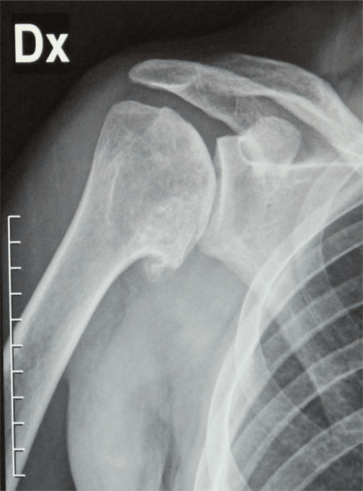
CT with 3D reconstruction is standard. It measures glenoid version, bone stock and the Walch wear pattern, and it is the study on which component position is planned and the need for a bone graft or augmented component decided.
MRI is added when cuff status is unclear. It shows cuff integrity and fatty infiltration (Goutallier classification): stage 3-4 infiltration suggests an irreparable tear, and reverse TSA should be considered. It also assesses the subscapularis, whose integrity is critical for an anatomic replacement.
Management Algorithm
The decision. Rotator cuff integrity is the key. An anatomic TSA needs an intact cuff, especially the subscapularis and supraspinatus; a reverse TSA is for cuff deficiency, massive tears or cuff tear arthropathy, so preoperative imaging must assess the cuff.
Absolute. Active infection, deltoid paralysis (axillary nerve palsy for reverse TSA), Charcot arthropathy.
Relative. Young age (under 50 for anatomic), high activity demands, uncorrectable glenoid bone loss, severe osteoporosis, a noncompliant patient. For reverse TSA specifically: glenoid bone loss requiring bone grafting, a young active patient (under 65), and an intact cuff, where anatomic TSA is preferred.
Who. Age under 75, an intact rotator cuff with no fatty infiltration, concentric or correctable glenoid wear (Walch A or B1), good bone stock and realistic expectations. The goal is to restore anatomy, preserve cuff function and maximise longevity, and in the active patient under 65 with primary OA and an intact cuff it gives the best function and longevity.
The glenoid. Walch A glenoids are the best candidates, with excellent outcomes from a standard anatomic TSA. A B1 glenoid can be corrected by eccentric reaming without a bone graft. A B2 glenoid requires version correction with a graft or an augmented component.
Anatomic versus Reverse: The Modern Selection Debate
Intact cuff to anatomic, deficient cuff to reverse is the starting rule, and it does not settle every case. The B2 and C glenoid already carry reverse TSA as an option, and a 75-year-old with a B2 glenoid should consider a reverse.
Reverse for the difficult glenoid. Reverse TSA is increasingly chosen for primary glenohumeral OA even when the cuff is intact, if the glenoid is severely retroverted or biconcave (Walch B2/B3) or has posterior bone loss that an anatomic TSA cannot reliably reconstruct. The dominant long-term failure of anatomic TSA is glenoid loosening, driven by eccentric posterior loading on a retroverted or biconcave glenoid. A reverse baseplate, fixed with a central post and peripheral screws into the scapular body, tolerates posterior bone loss and version far better than a cemented anatomic glenoid and removes that failure mode.
The trade. The reverse exchanges glenoid loosening for its own profile: scapular notching, acromial and scapular-spine stress fracture, limited external rotation, and non-anatomic, medialised kinematics.
Who gets which. In the difficult glenoid the call turns on glenoid bone stock plus age and demand, not cuff status alone. The older, lower-demand patient with a severely deformed or bone-deficient glenoid is well served by a reverse, which needs no perfect version correction. The younger patient with a correctable glenoid and an intact cuff is still better served by an anatomic TSA with version correction.
Why the reverse is not the default. Registries show reverse TSA carries a lower cumulative revision rate than anatomic TSA at comparable follow-up, in the early and mid term. A lower revision rate does not make it the better operation for every patient.
- With an intact cuff, anatomic TSA restores near-normal kinematics and external rotation. The reverse sacrifices external rotation and imposes a medialised, non-anatomic centre of rotation
- Reverse-specific problems accumulate: notching, acromial and scapular-spine stress fractures, and glenosphere and baseplate wear accrue over time. There is little long-term (15-20 year) outcome data for reverse in younger, higher-demand patients, which is why it is controversial in the young and a relative contraindication under about 65
- A failed anatomic TSA, typically from glenoid loosening or cuff failure, can usually be revised to a reverse. A failed reverse is a harder, more bone-consuming revision, so choosing anatomic in the younger intact-cuff patient keeps the reverse in reserve
Select on cuff status + age/demand + glenoid bone stock + the revision pathway. An intact cuff makes anatomic TSA possible, not obligatory: anatomic remains preferred for the younger intact-cuff patient, with reverse reserved for cuff deficiency, the irreconcilable glenoid and the older, lower-demand patient.
Surgical Technique
Position. Beach chair is preferred for TSA: anatomic orientation is easier, the glenoid is better reached, and range of motion can be assessed during surgery. Lateral decubitus is used for arthroscopy or by surgeon preference, but glenoid exposure is more difficult.
Setup Checklist
Beach chair, 30-45° upright, on a specialised shoulder positioning system. Secure the head in neutral alignment on a padded headrest and pad the contralateral shoulder against pressure injury.
- Pad the medial elbow, avoiding direct pressure on the ulnar nerve
- Protect the brachial plexus by avoiding excessive head rotation or lateral flexion
- Pad the sacrum, heels and occiput
- Place the arm adducted and internally rotated on a mobile arm positioner
- Expose the sternoclavicular joint medially, the AC joint and the deltoid insertion laterally
- Free-drape the arm to allow full range of motion for trial reduction
- Position for C-arm AP and axillary views if needed
Deltopectoral Approach
From the coracoid to the deltoid insertion, approximately 15cm, in Langer lines for cosmesis. Identify the cephalic vein in the deltopectoral interval through the subcutaneous tissue.
The deltopectoral interval is an internervous plane. Retract the cephalic vein laterally with the deltoid, which protects it from thrombosis, and develop the areolar tissue bluntly from the coracoid superiorly to the pectoralis major insertion inferiorly.
Incise the clavipectoral fascia lateral to the conjoint tendon. Divide the superior 1-2cm of the pectoralis major tendon if exposure needs it, and tag it for later repair.
Palpate the bicipital groove: the long head of biceps is the landmark for the rotator interval. Tenotomy or tenodesis is surgeon preference; tenotomy is faster, and tenodesis reduces cramping but adds time.
During inferior capsular release, stay on bone and retract with a Fukuda or similar retractor to protect the axillary nerve. Avoid aggressive medial retraction, which endangers the musculocutaneous nerve.
Reverse Total Shoulder Arthroplasty Technique
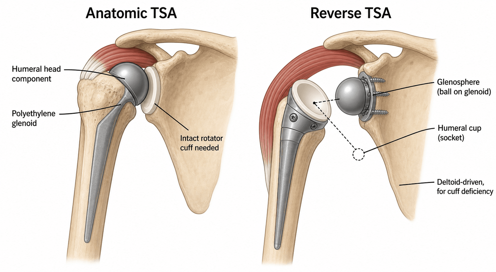
How the reverse works. The anatomic replacement puts a metal head on the humeral stem against a polyethylene glenoid. The reverse swaps ball and socket: a metal glenosphere is fixed to the glenoid and a concave polyethylene cup sits on the humeral component. This medialises and lowers the centre of rotation, which tensions the deltoid and lets it elevate the arm without a cuff. The design features, and what each buys, are set out below.
- Effect
- Increases deltoid tension and moment arm
- Clinical Benefit
- Compensates for absent cuff, improves elevation
- Effect
- Inherent stability without cuff
- Clinical Benefit
- Allows function despite massive cuff tear
- Effect
- Increases contact arc, reduces notching
- Clinical Benefit
- Better ROM, lower impingement
- Effect
- Moves COR laterally, reduces notching
- Clinical Benefit
- Improves external rotation, reduces scapular impingement
Complications
- Incidence
- 10-25% at 10 years
- Risk Factors
- Eccentric loading, excessive retroversion, poor cementation
- Management
- Observation if asymptomatic, revision to reverse TSA if symptomatic
- Incidence
- 1-2% (up to 5% in revision)
- Risk Factors
- Diabetes, immunosuppression, prior surgery, long operating time
- Management
- I&D and component retention if acute (under 4 weeks), staged revision if chronic
- Incidence
- 2-5%
- Risk Factors
- Subscapularis failure, component malposition, cuff deficiency
- Management
- Revision subscapularis repair, liner exchange, convert to reverse TSA
- Incidence
- 62-68%
- Risk Factors
- Medialised glenosphere, small glenosphere, high or superiorly tilted baseplate
- Management
- Prevention: lateralisation, larger glenosphere, inferior tilt. Observation if asymptomatic.
- Incidence
- 1-3%
- Risk Factors
- Excessive retraction, inferior capsular release, traction
- Management
- Observation, EMG at 6 weeks, consider nerve exploration if no recovery at 3-6 months
- Incidence
- 1-2% (higher in osteoporotic bone)
- Risk Factors
- Osteoporosis, trauma, loose component, press-fit stems
- Management
- ORIF if stable component, revision to longer stem if loose, cerclage if intraoperative
- Incidence
- 2-4%
- Risk Factors
- Excessive deltoid tension, osteoporosis, trauma
- Management
- Observation if nondisplaced, ORIF if displaced with functional deficit
- Incidence
- 5-10% (higher with tenotomy)
- Risk Factors
- Poor repair, excessive tension, osteoporotic bone, early mobilisation
- Management
- Revision repair if acute, latissimus transfer or convert to reverse if chronic
Shoulder infection is its own disease. It is not hip or knee infection with a different incision. The dominant pathogen is Cutibacterium acnes (formerly Propionibacterium acnes), an anaerobic commensal of the sebaceous glands that are dense around the shoulder girdle, which is also why infection is substantially commoner in men.
How it presents. C. acnes is low-virulence and indolent. There is often no sinus, no erythema and no fever, CRP and ESR are frequently normal, and the presentation is simply a stiff or painful arthroplasty, or unexplained early loosening. It is the reason a shoulder revision that "looks aseptic" so often is not: all three deep infections in the Neer Award series were in the revision group, and its authors advise vigilance for low-grade infection when revising.
This changes the microbiology request, and it is the single most practical fact in the topic. Standard cultures are held for around 5 days, and Cutibacterium frequently will not have grown by then, so a routine result reads "no growth" and the infection is missed. Ask explicitly for prolonged anaerobic culture held for 14 days, and send multiple separate tissue samples rather than a swab: a single positive can be a contaminant, while two or more matching samples are meaningful. Sonication of the explant raises the yield further.
Treatment by timing.
- Acute, under 4 weeks. I&D with component retention, exchange of modular parts and long-term suppressive antibiotics, with a success rate of 50-70%
- Chronic, over 4 weeks. Two-stage revision is the gold standard: component removal, an antibiotic spacer for 6-12 weeks, then reimplantation, with a success rate of 80-90%
- Culture-negative. In the shoulder, treat "culture-negative" as a hypothesis about the culture rather than a conclusion about the joint. Biofilm and insufficient incubation are the usual explanations
Postoperative Care and Rehabilitation
Anatomic TSA Rehabilitation Timeline
Interscalene block (wears off in 12-24 hours) with multimodal analgesia (opioids, paracetamol, NSAIDs). Sling with the arm in neutral rotation. Pendulum exercises from day 1 if pain is controlled. Chemical (enoxaparin or aspirin) and mechanical DVT prophylaxis.
Sling continuously for 4-6 weeks, off only for exercises. Passive range of motion only, to 90° of forward elevation and 30° of external rotation in the scapular plane; no active motion (protecting the subscapularis repair), and no lifting, pushing or pulling. Physiotherapy 2-3 times a week for passive stretching.
Wean the sling at 6 weeks if subscapularis healing is confirmed (examination, imaging if concerned). Begin active-assisted exercises (pulleys, cane) and gentle isometric work for the scapular stabilisers, aiming for forward elevation of 120-140°, external rotation of 40-50° and internal rotation to L5.
Progressive resistance with Therabands and light weights (1-5 lbs), and functional activities of daily living and light work. Full range is forward elevation 140-160°, external rotation 50-60° and internal rotation to L3. Swimming returns at 3-4 months and golf at 4-6 months.
Active internal rotation is prohibited for 6 weeks after anatomic TSA to protect the subscapularis repair. Failure to protect it leads to a 5-10% repair failure rate and anterior instability. Educate the patient (no lifting, pushing or reaching behind the back); sling compliance improves healing.
Activities. The elbow and hand are unrestricted from the start.
- No lifting over 5 lbs for 12 weeks
- Driving at 6-8 weeks, if off opioids with adequate range of motion
- Return to work at 4-6 weeks for sedentary work, 3-6 months for manual labour
- Contact sports avoided permanently, for the risk of dislocation or fracture
Follow-up.
- 2 weeks: wound check, sutures or staples out, range of motion assessed
- 6 weeks: radiographs (AP, axillary, scapular Y), progress to active motion
- 12 weeks: clinical and radiographic assessment, full range expected
- 6 months, 1 year and 2 years: surveillance for loosening and wear
- Annually thereafter if asymptomatic
Outcomes and Prognosis
- Anatomic TSA
- Excellent (90-95% significant improvement)
- Reverse TSA
- Excellent (90-95% significant improvement)
- Anatomic TSA
- 140-160° (near-normal if cuff intact)
- Reverse TSA
- 120-140° (deltoid-dependent)
- Anatomic TSA
- 40-60° (requires intact posterior cuff)
- Reverse TSA
- 20-40° (limited by absent cuff)
- Anatomic TSA
- 90-95%
- Reverse TSA
- 90-95%
- Anatomic TSA
- 70-90% (glenoid loosening main failure)
- Reverse TSA
- 85-95% (more durable glenoid fixation)
Scores. Functional scores improve after arthroplasty:
- ASES score from 30-40 before surgery to 75-85 after
- Constant score from 20-30 to 60-70
- Pain VAS from 7-8/10 to 1-2/10
- Forward elevation improves by 50-80°; external rotation is variable
- 70-80% return to golf or swimming
Registries. The common indications for revision are instability or dislocation, infection, and glenoid component loosening.
- Anatomic TSA. Unrecognised rotator cuff tear (leads to early failure), subscapularis failure (instability), glenoid loosening (excessive retroversion or eccentric loading)
- Reverse TSA. Deltoid dysfunction (axillary nerve injury), infection, scapular notching with pain, acromial fracture
- Both. Periprosthetic infection, nerve injury, patient noncompliance with rehabilitation
Guidelines, Registries & Global Practice
Global Epidemiology
- USA: primary shoulder arthroplasty rose 103.7% from 2011-2017; reverse TSA rose 191.3% with 63,845 reverse procedures in 2017 (Wagner et al 2020)
- Shoulder arthroplasty is growing faster than hip or knee arthroplasty
- Projected 174,810-350,558 annual procedures in the USA by 2025
- Reverse TSA is now the most common shoulder replacement in most national registries (AOANJRR, NJR, AJRR)
- Ageing populations with cuff tear arthropathy and proximal humeral fractures
- Broadening reverse TSA indications (massive cuff tear, fracture, revision, tumour)
- Improved implant longevity and surgeon familiarity
- Shift away from hemiarthroplasty for glenohumeral OA and acute fracture
Guideline and Registry Positions
- Position on TSA
- Supports anatomic TSA for end-stage glenohumeral OA with intact cuff and reverse TSA for cuff-deficient/CTA patterns; emphasises individualised indication
- Evidence basis
- Clinical practice guideline / appropriate-use criteria (moderate to limited evidence)
- Position on TSA
- Shoulder replacement recommended for severe, refractory glenohumeral arthritis; cuff status determines anatomic vs reverse; registry submission encouraged
- Evidence basis
- Guideline plus National Joint Registry (NJR) surveillance
- Position on TSA
- Reverse TSA favoured for displaced 3-4 part proximal humeral fractures in older patients over ORIF/hemiarthroplasty where cuff/tuberosity healing is unreliable
- Evidence basis
- Expert consensus and comparative cohort data
- Position on TSA
- Endorse reverse TSA as standard for CTA and increasingly for fracture; highlight scapular notching and baseplate position as key technical issues
- Evidence basis
- Consensus statements and registry data
- Position on TSA
- Track reverse versus anatomic revision risk; consistently favour all-polyethylene cemented glenoids over metal-backed in anatomic TSA
- Evidence basis
- Population-level observational registry data
National joint registries (AOANJRR, England/Wales NJR, American AJRR) consistently show: (1) reverse TSA is now the most commonly implanted shoulder arthroplasty; (2) metal-backed glenoid components have higher revision rates than cemented all-polyethylene designs in anatomic TSA; and (3) leading revision causes are instability/dislocation, infection and glenoid loosening. Registry data are observational and complement, rather than replace, randomised evidence.
Practice Variation
- Fracture management: higher reverse TSA use for elderly proximal humeral fractures in North America/Europe than in some Asian and lower-resource settings
- Glenoid fixation: near-universal cemented all-polyethylene in anatomic TSA; uncemented/hybrid use varies by region and implant availability
- Augments vs bone graft for B2/B3 glenoids varies with access to patient-specific instrumentation and augmented components
- Quantified risks to discuss: infection (1-2%), nerve injury (1-3%), instability (2-5% anatomic), glenoid loosening (radiographic 10-25% at 10 years anatomic), revision (single-digit percent at 10 years)
- Document: rotator cuff integrity (MRI), glenoid morphology (Walch on CT), and component-selection rationale
Key documentation requirements: Preoperative imaging assessment (XR, CT with version measurement, MRI for cuff assessment), informed consent with specific complications discussed (infection, nerve injury, loosening, instability, limited ROM especially external rotation in reverse), component selection rationale (anatomic vs reverse, glenoid design, version correction strategy), intraoperative findings (cuff integrity, bone quality, component positioning), postoperative instructions (sling compliance, ROM restrictions, PT protocol). Common litigation issues: Unrecognized rotator cuff tear leading to early anatomic TSA failure (failure to obtain MRI preoperatively), nerve injury from excessive retraction, infection from inadequate prophylaxis or contamination, subscapularis failure from inadequate repair or early mobilization, wrong implant selection (anatomic when reverse indicated). Protective measures: Thorough preoperative assessment with appropriate imaging, detailed consent process, meticulous surgical technique with nerve protection, careful component selection based on cuff integrity and glenoid morphology, clear postoperative instructions and compliance monitoring.
MCQ Practice Points
Q: What is the Walch classification system for glenoid morphology in primary glenohumeral osteoarthritis? A: Walch classification assesses glenoid wear pattern and version to guide component selection. A1: Minor concentric erosion, normal version (best candidate for standard component). A2: Major concentric erosion (may need thicker component). B1: Posterior subluxation with asymmetric wear, 10-15° retroversion (eccentric reaming up to 10° safe). B2: Biconcave glenoid with severe posterior wear, 15-25° retroversion (requires bone graft or augmented component). C: Dysplastic with over 25° retroversion (bone graft mandatory or consider reverse TSA).
Q: What are the key biomechanical principles of reverse total shoulder arthroplasty? A: Reverse TSA creates a semiconstrained ball-and-socket design with inverted anatomy (glenosphere on glenoid, concave cup on humerus). Medialized center of rotation increases deltoid tension and moment arm, allowing elevation despite absent rotator cuff. Deltoid becomes prime mover replacing cuff function. Design is inherently stable without requiring cuff balance. Lateralization (lateralized glenosphere or humeral offset) reduces scapular notching and improves external rotation. Functioning deltoid (axillary nerve intact) is essential - deltoid paralysis is absolute contraindication.
Q: What is the preferred glenoid component design for anatomic TSA and why? A: All-polyethylene cemented glenoid components are gold standard with 70-90% survivorship at 10-20 years. Metal-backed glenoid components have significantly higher failure rates due to polyethylene dissociation, increased wear from thin polyethylene, and osteolysis - they are no longer recommended. Cemented fixation with keel or pegged designs provides durable fixation. Keel designs easier to insert, pegged designs may offer better cement interdigitation. Pressurization of cement into cancellous bone is critical for fixation strength.
Q: Compare lesser tuberosity osteotomy versus subscapularis tenotomy for anatomic TSA. A: Lesser tuberosity osteotomy: Preserves subscapularis insertion on bone fragment, allows bone-to-bone healing (6-8 weeks), lower failure rate (under 5%), better strength restoration. More technically demanding, risk of fracture or nonunion. Subscapularis tenotomy: 5mm medial to insertion, faster, easier exposure and repair. Tendon-to-bone healing (variable), higher failure rate (5-10%), longer protection (12 weeks). Subscapularis failure leads to anterior instability and poor outcomes after anatomic TSA, making secure repair critical.
Q: What is the most common long-term complication of anatomic TSA and how is it managed? A: Glenoid loosening occurs in 10-25% of patients at 10 years (radiographic), with 5-10% requiring revision. Caused by eccentric loading, excessive retroversion, poor cementation, or component malposition. Presents with recurrent pain and functional decline. Management: Observation if asymptomatic with stable component, revision to new glenoid component if symptomatic and bone stock adequate, conversion to reverse TSA if bone loss or rotator cuff failure develops. Prevention through proper version correction, cemented all-polyethylene components, and balanced soft tissues.
Q: What are the key findings from the Australian Orthopaedic Association National Joint Replacement Registry (AOANJRR) regarding shoulder arthroplasty? A: AOANJRR data show that reverse TSA is now the most commonly implanted primary shoulder arthroplasty and carries a lower cumulative revision rate than anatomic TSA at comparable follow-up. The leading revision indications are instability/dislocation, infection and glenoid component loosening. Cemented all-polyethylene glenoid components have lower revision rates than metal-backed designs in anatomic TSA. Registry data are observational and guide implant selection and patient counselling.
Exam Viva Scenarios
Practise clinical reasoning and management decisions out loud
“A 72-year-old retired teacher presents with 2-year history of progressive right shoulder pain and weakness. Unable to lift arm above shoulder height. Examination: forward elevation 70° active, 110° passive. Positive lag signs for supraspinatus and infraspinatus. XR shows superior migration of humeral head with acromiohumeral distance 4mm, glenohumeral joint space narrowing. MRI shows massive retracted rotator cuff tear with stage 3 Goutallier fatty infiltration of supraspinatus and infraspinatus. What is your diagnosis and management?”
“You are planning an anatomic TSA for a 65-year-old with primary glenohumeral OA. Preoperative CT shows Walch B2 glenoid morphology with 18° of retroversion and biconcave wear pattern. MRI confirms intact rotator cuff. Walk me through your approach to managing this glenoid morphology.”
“A 68-year-old presents 6 months after anatomic TSA with 3 weeks of increasing shoulder pain, swelling, and low-grade fever. Wound healed primarily. Exam shows warmth, effusion, painful ROM. ESR 65, CRP 45. Aspiration grows coagulase-negative Staph. How do you manage this patient?”
Key Decision Points
- Rotator cuff integrity determines anatomic (intact cuff) vs reverse TSA (cuff deficiency)
- Walch B2/C glenoid (retroversion over 15°) requires bone graft or augmented component
- Deltoid must be intact for reverse TSA - axillary nerve palsy is absolute contraindication
- All-polyethylene cemented glenoid is gold standard (metal-backed abandoned)
Walch Glenoid Classification
- A1 = minor concentric erosion, normal version (best candidate)
- A2 = major concentric erosion (thicker component)
- B1 = posterior subluxation 10-15° retroversion (eccentric reaming safe)
- B2 = biconcave 15-25° retroversion (bone graft or augment needed)
- C = dysplastic over 25° retroversion (graft mandatory or reverse TSA)
Surgical Approach Pearls
- Deltopectoral approach: retract cephalic vein laterally with deltoid
- Axillary nerve 5-10mm from glenoid rim at 6 o'clock - protect during capsular release
- Lesser tuberosity osteotomy has lower failure rate (under 5%) vs tenotomy (5-10%)
- Eccentric reaming safe to 10° correction - beyond requires bone graft or augment
Reverse TSA Specifics
- Semiconstrained design with medialized center of rotation - deltoid powered
- Lateralized glenosphere reduces scapular notching (62-68% with Grammont-style designs)
- External rotation limited 20-40° due to absent posterior cuff - counsel preop
- Earlier mobilization safe than anatomic (no cuff repair to protect)
Complications and Management
- Glenoid loosening (10-25% at 10 years anatomic) - observation vs revision to reverse
- Periprosthetic infection (1-2%): acute (under 4 weeks) = I&D, chronic = staged revision
- Subscapularis failure (5-10%) leads to instability - lesser tuberosity osteotomy preferred
- Scapular notching (62-68% in the series cited here) often asymptomatic, and no glenoid loosening was seen - prevent with lateralization and a low, inferiorly tilted baseplate
Key Evidence and Outcomes
- 10-year survival: 70-90% anatomic TSA, 85-95% reverse TSA
- 90-95% patient satisfaction and pain relief for both types
- Reverse TSA is now the most common shoulder arthroplasty (AOANJRR registry data)
- Scapular notching linked to high baseplate position/superior tilt; place glenosphere low (Levigne/Walch multicentre)
Evidence Base and Key Trials
Walch Classification: Morphologic Study of the Glenoid in Primary Glenohumeral OA
- Serial CT analysis of 113 osteoarthritic shoulders defined three glenoid types: A, B and C
- Type A (centred head, symmetric erosion) 59 percent; Type B (posterior subluxation, asymmetric/posterior wear) 32 percent; Type C (retroversion over 25 degrees, dysplastic) 9 percent
- Posterior subluxation of the humeral head drives asymmetric load and posterior glenoid wear in Type B
- Classification discriminates dysplastic retroversion (Type C) from acquired posterior erosion (Type B)
Grammont Reverse Prosthesis: Design, Rationale and Biomechanics
- Grammont design innovations: large neckless glenosphere and a 155-degree humeral cup medialising and lowering the centre of rotation
- Medialisation minimises torque on the glenoid component and recruits more deltoid fibres as abductors
- Lowering the humerus restores and increases deltoid tension, allowing the deltoid to compensate for a deficient cuff
- External and internal rotation are often not restored; scapular notching and polyethylene wear from cup-on-neck impingement are key concerns
Neer Award 2005: Grammont Reverse Prosthesis in CTA, Fracture Sequelae and Revision
- 45 reverse prostheses (21 cuff tear arthropathy, 5 fracture sequelae, 19 failed arthroplasty), mean follow-up 40 months
- Active elevation improved from 55 to 121 degrees and Constant score from 17 to 58; active external rotation essentially unchanged (7 to 11 degrees)
- Complications far higher in revision than primary CTA (47 percent vs 5 percent); 22 percent required further surgery
- Scapular notching in 68 percent; teres minor atrophy/fatty infiltration predicted poorer external rotation and function