Gold Standard for OA | Intact Cuff Required | 95% Survival
- Requires INTACT rotator cuff (contraindicated if torn)
- Walch B2 (biconcave) is the most common operative challenge
- Subscapularis management is critical for success
- Axillary nerve is at risk during inferior capsular release
- Glenoid loosening is the main mode of long-term failure
- “External Rotation lag = Cuff tear (Contraindication for aTSA)
- “Pseudoparalysis vs Stiffness differentiation is key
- “Lesser tuberosity osteotomy has highest subscap healing rate
- “Critical Shoulder Angle under 30 degrees associated with OA
Overview and Epidemiology
Definition Anatomic Total Shoulder Arthroplasty (TSA) is a surgical procedure that involves replacing the damaged humeral head with a metal sphere and the glenoid with a polyethylene dish, strictly replicating the native anatomy. The success of the procedure relies entirely on a functioning rotator cuff to compress the head into the glenoid ("concavity compression") and generate rotation.
Epidemiology
- Trends: While Anatomic TSA volume is increasing globally, Reverse TSA (rTSA) volume has grown exponentially and now surpasses aTSA in many registries (including Australia and USA). This is due to expanded indications for Reverse TSA (such as cuff tear arthropathy, fractures, and revision), as well as the desire to avoid late glenoid loosening associated with anatomic implants.
- Demographics: Typically active patients aged 60-75 years with primary osteoarthritis. Patients under 50 present a significant challenge due to concerns about implant longevity—specifically the "polyethylene problem."
- Risk Factors:
- Primary: Osteoarthritis (genetic, age-related).
- Secondary: Previous trauma (Post-traumatic OA), instability surgery (Capsulorraphy Arthropathy—over-tightening leads to posterior wear), Avascular Necrosis (Steroids, Alcohol, Sickle Cell), Inflammatory Arthritis (Rheumatoid), and Hemochromatosis ("Iron fist, iron shoulder").
Pathophysiology of Osteoarthritis Primary OA is characterized by a predictable sequence of joint destruction:
- Cartilage Loss: Early loss of articular cartilage, often starting centrally or posteriorly.
- Posterior Wear: The hallmark of shoulder OA is posterior glenoid wear (retroversion) and posterior humeral subluxation. This eccentric loading creates a "Rocking Horse" phenomenon. The humeral head acts as a fulcrum, levering the glenoid component out of the bone if not corrected.
- Soft Tissue Contracture: The subscapularis and anterior capsule become contracted and scarred, drastically limiting external rotation. The posterior capsule stretches due to chronic subluxation but is rarely the primary problem.
- Osteophyte Formation: Large osteophytes form on the inferior humeral head ("Goat's Beard") which can encroach on the axillary nerve space.
In patients under 50 years, glenoid components have high failure rates due to polyethylene wear and loosening (aseptic loosening is the #1 failure mode). The "Ream and Run" procedure (Hemiarthroplasty with concentric glenoid reaming) is an option for high-demand patients willing to undergo extended rehab. Reverse TSA is generally avoided due to finite lifespan and salvage difficulties.
Anatomy and Biomechanics
Normal Anatomy
- Glenoid Version: Typically retroverted 2-8° relative to the axis of the scapula body. However, the scapula itself is anteverted 30° on the thorax. The net result is a glenoid face that is slightly retroverted relative to the body axis.
- Inclination: Superior inclination is typically 0-5°.
- Critical Shoulder Angle (CSA): The angle between the glenoid inclination and the lateral acromion edge.
- CSA under 30°: Associated with Osteoarthritis. The deltoid force vector compresses the joint, leading to cartilage wear.
- CSA over 35°: Associated with Cuff Tears. The deltoid force vector shears superiorly, pulling the head into the supraspinatus.
Biomechanics of Concavity Compression The shoulder is inherently an unstable joint (golf ball on a tee). Stability in aTSA is dynamic, provided by the rotator cuff.
- Concavity Compression: The rotator cuff muscles pull the humeral head into the glenoid concavity, creating a stable fulcrum.
- Force Couples:
- Coronal Plane: Deltoid (upward force) vs Supraspinatus/Infraspinatus (compressive/downward force).
- Axial Plane: Subscapularis (anterior) vs Infraspinatus/Teres Minor (posterior).
- Implication: Any deficiency in the cuff leads to eccentric loading (edge loading) of the glenoid component, causing the "Rocking Horse" phenomenon and early loosening. This is why an intact cuff is non-negotiable for aTSA.
Biomechanical Goals of aTSA
- Restore Version: Correct retroversion to neutral or slight retroversion (within 10°) to prevent eccentric loading. This centers the humeral head on the glenoid, distributing forces evenly across the cement interface.
- Restore Head Height: Reproduce the relationship between tuberosities and articular surface height (typically 8mm above the Greater Tuberosity). Restoring the native Center of Rotation (COR) is vital for cuff mechanics. If the head is placed too high, the cuff is over-tensioned; if too low, the cuff is lax, leading to instability.
- Restore Offset: Lateralization is critical for proper deltoid tensioning and the length-tension relationship of the rotator cuff. Loss of global offset leads to weakness and impingement.
Avoid correcting more than 10 degrees of retroversion with eccentric reaming alone. This removes excessive anterior bone stock, compromising peg fixation. ("Robbing Peter to pay Paul"). For over 10-15 degrees of correction, use augmented glenoids (Wedge) or bone graft to preserve bone stock.
Classification Systems
Walch Classification of Glenoid Morphology
Critical for pre-operative planning and implant selection. Developed to describe glenoid morphology in primary osteoarthritis using CT scans.
- Morphology
- Concentric wear
- Pathology
- A1: Minor erosion A2: Major erosion (Protrusio)
- Treatment Strategy
- Standard Glenoid
- Morphology
- Posterior wear
- Pathology
- B1: Posterior narrowing B2: Biconcave (Paleo/Neo)
- Treatment Strategy
- Correction Required (Augment/Ream)
- Morphology
- Retroversion over 25°
- Pathology
- Developmental dysplasia
- Treatment Strategy
- Complex (Bone Graft vs Reverse)
Clinical Assessment
- Pain: Deep, aching, toothache-like. Worse at night.
- Function: Difficulty with ADLs (esp. hygiene, reaching back, toileting). Females complain about bras; males about wallets.
- Stiffness: Progressive loss of ER and Abduction. "Screwing in a lightbulb" is difficult.
- Crepitus: Audible/palpable grinding ("Ratchet-like").
- Look: Supra/Infraspinatus atrophy (Chronic cuff tear?). Anterior scar?
- Feel: Posterior joint line tenderness. Anterior subluxation of the humeral head.
- Move: Blocked ER (capsular restriction). Scapulothoracic compensation (shrugging).
- Power: MUST test Cuff integrity. ER Lag sign? Belly Press? Lift-off?
Differentiate Pseudoparalysis (Cuff failure = Cannot lift arm actively but Full PASSIVE ROM) from Stiffness (OA = Cannot lift arm actively AND Limited PASSIVE ROM).
Stiff shoulder = Anatomic TSA. Pseudoparalytic shoulder = Reverse TSA.
- Key Distinguishing Feature
- Posterior wear, stiff, full passive ROM lost
- Implant Implication
- Anatomic TSA
- Key Distinguishing Feature
- Superior migration, pseudoparalysis, acetabularisation
- Implant Implication
- Reverse TSA
- Key Distinguishing Feature
- Symmetric erosion, medial wall erosion, soft bone
- Implant Implication
- aTSA if cuff intact; often reverse
- Key Distinguishing Feature
- Preserved glenoid early, crescent sign, head collapse
- Implant Implication
- Hemiarthroplasty or aTSA
- Key Distinguishing Feature
- Pain at rest, raised inflammatory markers, C. acnes
- Implant Implication
- Address infection first
- Key Distinguishing Feature
- Global passive ROM loss, normal joint space
- Implant Implication
- Non-operative; not arthroplasty
Imaging and Investigations
Imaging Protocol
- AP (Grashey): Joint space loss, subchondral sclerosis, osteophytes ("Goat's Beard" on inferior humerus).
- Axillary Lateral: CRITICAL. Assess posterior subluxation (percentage) and version. Look for biconcavity.
- Outlet: Acromial shape and cuff status signs (high riding head).
- Version: Quantify retroversion (Friedman method).
- Bone Stock: Assess glenoid vault depth for peg penetration.
- Planning: Essential for PSI (Patient Specific Instrumentation) guides to execute version correction accurately.
- Indication: If cuff strength is equivocal on exam or history of tear.
- Finding: Fatty infiltration (Goutallier over 2 contraindicates aTSA) and tendon retraction (Patte).

Non-Operative Management
- NSAIDs: First line for pain and inflammation.
- Analgesia: Paracetamol/codeine sparingly. Avoid opioids.
- Injections:
- Corticosteroid: Short term relief. WARNING: Increases risk of infection if performed under 3 months before arthroplasty.
- Hyaluronic Acid: Variable evidence, less risk.
- Maintain ROM: Stretching to prevent secondary adhesive capsulitis.
- Strengthening: Scapular stabilizers and cuff to maintain centering.
- Activity Modification: Avoid heavy overhead loading and impact activities.
Management Algorithm
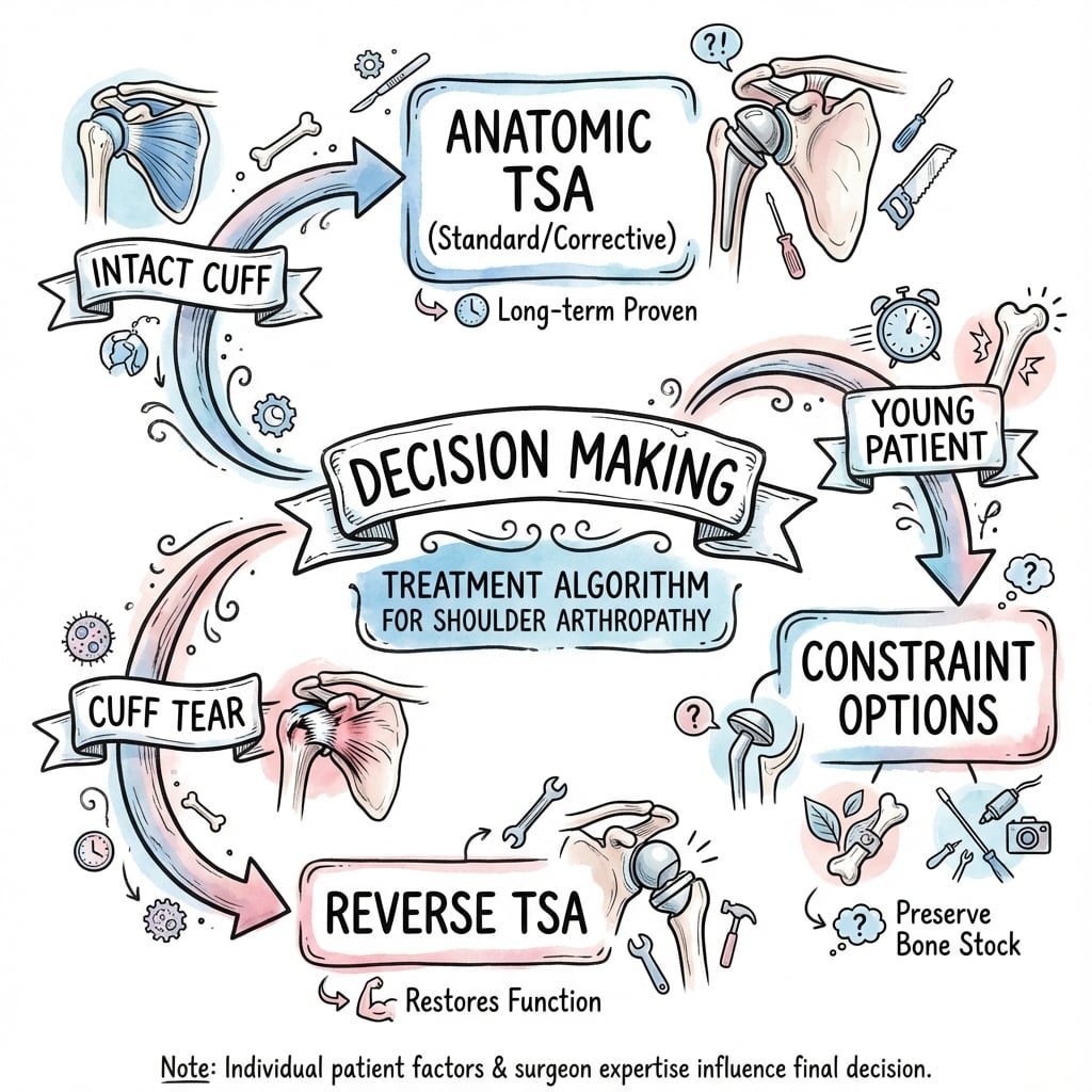
Osteoarthritis with Intact Cuff
Decision Pathway
Standard glenoid component (All-poly pegged). Reaming to expose subchondral bone. Excellent outcomes expected.
- Mild (under 10°): Eccentric reaming (high side down).
- Severe (over 10°): Augmented Glenoid (Wedge) or PSI.
- Goal: Center the head to prevent early loosening. Avoid reaming over 10° to preserve bone.
Treatment Options
- Indication
- Young (under 50), mechanical symptoms
- Pros
- Low risk, buys time
- Cons
- Unpredictable pain relief
- Indication
- Young laborer, insufficient glenoid bone
- Pros
- No glenoid loosening risk
- Cons
- Glenoid erosion pain
- Indication
- Classic OA, Intact cuff
- Pros
- Best ROM, Normal anatomy
- Cons
- Glenoid loosening risk
- Indication
- Cuff tear arthropathy, Elderly
- Pros
- Reliable with no cuff
- Cons
- Limited rotation, contour
Surgical Technique
Deltopectoral Approach
The workhorse approach for TSA. It is an extensile, internervous approach that preserves the deltoid origin, which is crucial for rehabilitation.
Exposure Steps
Incision from coracoid tip to deltoid insertion (~10-15cm). Identify Cephalic vein in the deltopectoral groove. Pearl: Retract Cephalic vein laterally with Deltoid to preserve venous drainage from the arm (most branches come from deltoid). Ligate small feeding branches (The "Delta" vein).
Incise clavipectoral fascia lateral to the conjoined tendon. Identify "Conjoined Tendon" (Coracobrachialis/Short head Biceps). Retract Medially. Danger: Musculocutaneous nerve enters coracobrachialis 5-8cm distal to coracoid. Do not retract heavily here. Use a self-retainer (Kolbel).
External rotate the arm. Identify the "Three Sisters" (anterior circumflex humeral vessels) at the inferior border. Ligate/cauterize. Techniques:
- Tenotomy: 1cm medial to insertion (easier to repair later).
- Peel: Sharply from lesser tuberosity.
- Osteotomy: Lesser tuberosity osteotomy (Highest healing rate).
Release capsule inferiorly and anteriorly. Protect Axillary Nerve! Palpate it in the quadrangular space before releasing. The "Tug Test" ensures safety. Release the inferior capsule off the humeral neck (not the glenoid side yet) to mobilize the humerus.
Use of nerve monitoring is controversial. It does not prevent injury but may alert the surgeon. The "Tug Test" (palpating the nerve) remains the most reliable method of confirmation.
Navigation and PSI In complex B2/B3 glenoids, standard instrumentation is inaccurate (often under-correcting retroversion).
- PSI (Patient Specific Instrumentation): Pre-operative CT planning creates a 3D mold that sits on the glenoid face/coracoid to guide the central pin. Evidence suggests improved accuracy of version correction compared to standard guides. The guides are typically printed in sterile nylon and allow for precise pin placement within 2-3 degrees of the plan. This is particularly useful in "B2" glenoids where the native landmarks are eroded.
- Navigation/Robotics: Real-time feedback on version and inclination. Useful for trainees and difficult morphology. Optical or accelerometer-based systems are available, though cost remains a barrier to widespread adoption in the public system.
Overstuffing and Restoration of Native Humeral Geometry
The word "anatomic" is the operative principle of aTSA: the reconstruction must reproduce the native centre of rotation (COR), head height, radius of curvature and global offset. The topic repeatedly warns against "over-stuffing" the joint, but the concept deserves its own treatment because it is one of the most common technical errors and a frequent cause of a stiff, painful, or failed anatomic replacement.
What overstuffing means Overstuffing is an increase in the effective soft-tissue tension and joint volume beyond native anatomy. It occurs when the composite humeral construct is too big or sits too proud, most often because:
- The humeral head component is too thick or too large in diameter (over-sized), over-tensioning the cuff and capsule.
- Peripheral (inferior "goat's beard") osteophytes are NOT removed before templating, so the anatomic neck is misjudged and an oversized head is selected.
- The neck cut is too low/conservative or the head is seated too proud, lateralising and elevating the COR.
- Global (medial-to-lateral) offset is over-restored, lengthening the deltoid and cuff moment arms excessively.
Consequences
- Loss of motion, particularly external rotation and elevation, because an over-tensioned subscapularis and posterior cuff cannot excurse.
- Increased subscapularis repair tension, raising the risk of early repair failure and anterior escape.
- Eccentric glenoid rim loading and edge loading, accelerating polyethylene wear and the rocking-horse mechanism that drives glenoid loosening.
- Persistent pain and stiffness, and a shoulder that never feels "natural" despite a technically seated implant.
Conversely, under-stuffing (a head that is too small or a neck cut too high) leaves the cuff lax and the joint unstable. The goal is a balanced, tension-neutral reconstruction.
How to avoid it
- Remove osteophytes first, then define the true anatomic neck and template head diameter and thickness to native geometry.
- Match native head size (coverage and radius of curvature); resist the temptation to up-size for "stability".
- Restore, not exceed, offset and head height so the COR is reproduced rather than lateralised.
- Check passive external rotation on the table — the reconstructed shoulder should reach roughly neutral to greater than 30 degrees of ER with the repaired subscapularis under acceptable tension; a shoulder that will not externally rotate past neutral suggests overstuffing.
- Assess the "shuck" / translation — an appropriately balanced head allows about 50 percent posterior translation that reduces spontaneously, indicating neither over- nor under-stuffing.
In anatomic TSA the single unifying goal is to reproduce the native centre of rotation, head height, radius and offset. Remove osteophytes BEFORE sizing the head, then match native geometry. Overstuffing (over-sized/proud head, un-resected osteophytes) over-tensions the cuff, stiffens the shoulder, overloads the subscapularis repair and edge-loads the glenoid; test passive external rotation on the table to detect it before closure.
Complications
- Timing
- Early (under 3mo)
- Cause
- Poor repair/compliance
- Management
- Repair (under 3mo) or Pec Transfer/Reverse
- Timing
- Late (over 5-10y)
- Cause
- Eccentric load/Rocking Horse
- Management
- Revision to Reverse TSA
- Timing
- Acute/Late
- Cause
- C. acnes (slow growing)
- Management
- Debridement or 2-stage Revision
- Timing
- Early
- Cause
- Malversion/Soft tissue imbalance
- Management
- Revision usually required
- Timing
- Intra-op/Late
- Cause
- Reaming/Trauma
- Management
- Cerclage/Plate/Revision Stem
Cutibacterium acnes is a slow-growing anaerobe in shoulder skin flora. Causes indolent infections (pain, loosening) without systemic signs (normal WCC/CRP). Require anaerobic culture holding for 14 days. Prophylaxis includes Benzoyl Peroxide pre-op wash.
Diagnosis of Infection Diagnosing shoulder periprosthetic joint infection (PJI) is notoriously difficult due to the low-virulence nature of C. acnes.
- Serum Markers: CRP and ESR are often NORMAL.
- Aspiration: High rate of dry tap or false negatives.
- Intra-op: Fluid/Tissue cultures are gold standard. Hold cultures for 14 days as C. acnes is slow growing.
- Sonication: Explant sonication increases sensitivity by disrupting the biofilm.
Nerve Injury Nerve injury is a feared complication.
- Axillary Nerve: 1-2% incidence. Traction injury or direct laceration during inferior release. Monitor deltoid function.
- Musculocutaneous Nerve: Retractor injury. Biceps weakness. Do not place retractors deep to conjoined tendon.
- Suprascapular Nerve: Injury during posterior release or retractor placement.
Rotator Cuff Failure Secondary cuff failure leads to superior migration and "Rocking Horse" loosening of the glenoid. This is the main reason for avoiding aTSA in patients with questionable cuff status. If the cuff fails, the mechanics of the joint are destroyed, and conversion to Reverse TSA is required.
Grading Glenoid Radiolucency (Lazarus / Franklin)
Because aseptic glenoid loosening is the leading long-term failure mode of anatomic TSA, an examiner will expect you to interpret peri-glenoid radiolucent lines and to grade them. The topic's viva on the asymptomatic 2 mm peg lucency invokes the "Lazarus lines" scale but never defines it — this section closes that loop.
The scale Franklin originally described a system for radiolucency around keeled glenoid components; Lazarus and colleagues modified it for pegged components (J Bone Joint Surg Am, 2002). Both use an ordinal 0-to-5 scale in which higher grades indicate a greater extent and completeness of lucency around the pegs (or keel):
- Grade 0 — no radiolucency.
- Grades 1-2 — incomplete lucency around one or more pegs (thin, partial).
- Grades 3-4 — complete lucency around one, then progressively all, of the pegs/keel.
- Grade 5 — gross loosening: component tilt, shift, subsidence or migration.
How to use it
- Radiolucent lines are extremely common and often benign. In the Lazarus series, only 20 of 328 glenoids showed NO radiolucency on the immediate postoperative film — so an isolated thin, incomplete line is expected, not alarming.
- Progression matters more than a single film. Serial radiographs (matched AP and axillary) looking for widening lucency, new complete peri-peg lucency, or a change in component position are what distinguish stable fixation from evolving loosening.
- Correlate with symptoms. A low-grade, non-progressive lucency in a pain-free patient is surveillance, not surgery ("treat the patient, not the X-ray"); a rising grade, component shift (grade 5), or new start-up/rest pain triggers an infection work-up (including Cutibacterium acnes) and CT assessment of bone stock before considering revision — typically to a reverse prosthesis.
Glenoid radiolucency is graded 0-5 (Franklin for keels, Lazarus modification for pegs): 0 equals no lucency, 5 equals gross loosening (tilt/shift/migration). Thin incomplete lines are near-universal on the first postoperative film and are usually benign; it is PROGRESSION on serial films and component MIGRATION, correlated with pain, that define clinical loosening.
Postoperative Care
Rehabilitation Protocol
- Sling full time.
- Passive Elevation to 90° only in scapular plane.
- ER restricted to neutral (protect subscap).
- NO Active Internal Rotation (protect repair).
- Goal: Protect the subscapularis repair while preventing adhesive capsulitis.
- Wean sling.
- Active Assisted → Active ROM.
- Hydrotherapy.
- Begin gentle internal rotation.
- Goal: Regain active control and range of motion.
- Cuff strengthening (bands).
- Scapular stabilization.
- Return to golf/swimming ~4-6 months.
- Goal: Functional restoration and strength.
Outcomes and Prognosis
- Pain: 90-95% achieve excellent pain relief. Anatomic TSA is the gold standard for pain relief in OA.
- Function: Range of motion typically superior to Reverse TSA (better rotation). Patients often forget they have a replacement.
- Satisfaction: High patient satisfaction scores (Subjective Shoulder Value ~90%).
- Durability: Young age (under 55) is strongest predictor of revision. Glenoid loosening remains the primary mode of failure.
Guidelines, Registries & Global Practice
Global Epidemiology Primary glenohumeral OA is the dominant indication for anatomic TSA worldwide, typically in patients aged 60-75. Across large national registries (AOANJRR, UK NJR, US AJRR), reverse TSA volume has grown sharply and now exceeds anatomic TSA for many indications, driven by expanded reverse indications and concern over late glenoid loosening. Anatomic TSA remains preferred in younger, higher-demand patients with an intact cuff to maximise rotation.
Society Guidance (Side by Side)
- Position on aTSA
- aTSA for GHOA with functioning cuff
- Emphasis
- Shared decision-making; limited evidence for one implant over another
- Position on aTSA
- aTSA reasonable for OA with intact cuff
- Emphasis
- Cuff integrity and glenoid morphology drive implant choice
- Position on aTSA
- Augmented glenoids / reverse for B2/B3
- Emphasis
- Version correction and bone preservation prioritised
- Position on aTSA
- Correct version, secure glenoid fixation
- Emphasis
- Avoid eccentric loading and the rocking-horse mechanism
Registry Evidence Registries consistently show cemented all-polyethylene glenoids out-survive metal-backed designs, and that younger age is the strongest predictor of revision (aseptic glenoid loosening). Augmented all-poly components are increasingly used for B2/B3 bone loss.
High- vs Limited-Resource Practice In high-resource settings, preoperative CT, 3D planning, patient-specific instrumentation (PSI), and navigation are used to improve glenoid version correction, especially in B2/B3 deformity. In limited-resource settings, plain radiographs with an axillary view guide planning, standard instrumentation and cemented all-poly glenoids predominate, and hemiarthroplasty or non-operative care may be selected where implant cost or revision capacity is constrained.
Controversies & Areas of Uncertainty
Reverse TSA is increasingly offered to older patients with an intact cuff to avoid late glenoid loosening, but at the cost of rotation and a finite lifespan. The aTSA-versus-rTSA question in this group is being tested prospectively (RAPSODI-UK protocol, BMJ Open 2025) and is not yet settled.
Eccentric reaming, posterior augments, bone grafting, and conversion to reverse all have advocates. Augments preserve bone and correct version reliably at short term, but central-peg osteolysis in severe B3 remains a concern.
LTO heals most reliably radiographically, yet RCTs show no clear functional advantage over tenotomy or peel. The "best" technique remains debated.
Equivalent short/mid-term outcomes are reported, but long-term survivorship and behaviour in poor metaphyseal bone are unproven.
Avoids the polyethylene weak link but rehab is demanding and pain relief less predictable than aTSA. Patient selection is contentious.
Improve version-correction accuracy in studies, but cost-effectiveness and whether they change long-term survivorship are unestablished.
Mnemonics
ABCWalch Types
Hook:Easy as ABC: Aligned, Backwards, Crazy retroversion.
MACStructures at Risk
Hook:Big MAC Attack: Beware the nerves during approach.
PINContraindications
Hook:Don't put a PIN/Prosthesis In a Neuropathic joint!
MCQ Practice Points
Q: What is the most common cause of late failure in Anatomic TSA? A: Aseptic loosening of the glenoid component, often due to the 'Rocking Horse' effect from eccentric loading or cuff failure.
Q: A Walch B2 glenoid is characterized by what morphology? A: Biconcavity (Paleoglenoid and Neoglenoid) and posterior subluxation.
Q: A Critical Shoulder Angle (CSA) under 30 degrees is associated with what pathology? A: Osteoarthritis. (Conversely, over 35 degrees is associated with Cuff Tears).
Q: Where is the Axillary nerve at risk during the deltopectoral approach? A: Inferior border of Subscapularis. It must be palpated ('Tug test') or visualized before tenotomy or inferior capsular release.
Q: Why is Deltoid paralysis an absolute contraindication for TSA? A: Powered by Deltoid. Both Anatomic and Reverse rely on the deltoid for elevation. A flail shoulder cannot be salvaged by arthroplasty.
Q: What is the most common organism causing periprosthetic infection in shoulder arthroplasty? A: Cutibacterium acnes (formerly Propionibacterium acnes). It is an indolent, slow-growing anaerobe.
At a Glance
- Anatomic TSA
- OA with Intact Rotator Cuff
- Reverse TSA
- Cuff Tear Arthropathy, Fracture
- Key Pearl
- Cuff status determines choice
- Anatomic TSA
- Restores normal anatomy
- Reverse TSA
- Medializes center of rotation
- Key Pearl
- Anatomic needs cuff; Reverse needs deltoid
- Anatomic TSA
- Better ER/IR (if cuff healthy)
- Reverse TSA
- Limited IR, Good Elevation
- Key Pearl
- Anatomic feels more 'natural'
- Anatomic TSA
- Glenoid loosening (late)
- Reverse TSA
- Notching, Stress fracture (early)
- Key Pearl
- Loosening main fail mode in aTSA
Clinical Decision Scenarios
Practise clinical reasoning and management decisions out loud
“A 45-year-old weightlifter presents with severe primary OA (Walch B2). He wants a replacement. Discuss options.”
“6 months post TSA, patient complains of weakness and anterior pain. On exam, positive belly press. X-rays show anterior subluxation.”
“Routine 5 year follow up. Patient asymptomatic. X-ray shows 2mm lucency around the glenoid peg.”
Key Indications
- OA with Intact Cuff
- AVN
- Inflammatory Arthritis
- Post-traumatic Arthritis
Key Steps
- Deltopectoral Approach
- Subscapularis Management
- Version Correction
- Glenoid Cementing
Complications
- Subscap Failure/Rupture
- Glenoid Loosening (Long term)
- Periprosthetic Fracture
- Infection (C. acnes)
Classification
- Walch A (Centered)
- Walch B (Posterior Subluxation)
- Walch C (Dysplastic)
- Samilson-Prieto (Dislocation AR)
Evidence
- Walch 1999: glenoid morphology classification
- CSA under 30 = OA (Moor 2013)
- LTO heals best; strength equal (Lapner/Levine)
- Pegged glenoids superior to keeled (Lazarus)
Exam Pearls
- 'Intact Cuff' is the key
- 'B2 Glenoid' is the challenge
- Axillary nerve 'Tug Test'
- Young patient = Poly wear risk
Evidence Base
Walch Classification of Glenoid Morphology
- Three glenoid types defined on CT: A (centred, 59%), B (posterior subluxation/asymmetric wear, 32%), C (dysplastic retroversion over 25 degrees, 9%).
- Posterior humeral head subluxation drives the asymmetric posterior wear that characterises type B.
- Type C retroversion is dysplastic in origin and explains early-onset osteoarthritis.
Critical Shoulder Angle (CSA)
- Mean CSA was 33.1 degrees in controls, 38.0 degrees in rotator cuff tears, and 28.1 degrees in primary OA.
- Of shoulders with CSA over 35 degrees, 84% were in the cuff-tear group.
- Of shoulders with CSA under 30 degrees, 93% were in the osteoarthritis group.
Lesser Tuberosity Osteotomy vs Subscapularis Peel
- No significant difference in subscapularis dynamometer strength at 24 months (LTO 4.4 kg vs peel 5.5 kg, p=0.131).
- WOOS and ASES scores were equivalent between groups at all time points.
- Both techniques improved significantly from baseline.
Subscapularis Tenotomy vs LTO (Healing)
- LTO healed more reliably: 27/29 (93.1%) showed bone-to-bone healing on radiograph.
- Tenotomy: 26/30 (86.7%) had no full-thickness subscapularis tear on 3-month ultrasound.
- Clinical outcomes and ROM were equivalent; LTO added operative and repair time.
Stepped Augmented Glenoid for B2/B3
- Posteriorly stepped augmented glenoids corrected pathologic retroversion and posterior subluxation in B2/B3 glenoids.
- Penn Shoulder Score, ROM and version improved significantly at minimum 2 years (p under 0.0001).
- Persistent posterior subluxation correlated with worse teres minor fatty infiltration and residual component retroversion.
Pegged vs Keeled Glenoid Fixation
- Mean radiolucency score was lower for pegged (1.3) than keeled (1.8) components (p=0.0004).
- Pegged components more often achieved 'better cementing' than keeled components (p=0.0028).
- Complete seating was difficult to achieve and surgeon experience was an important variable.
Stemless vs Stemmed Anatomic TSA
- No significant difference in postoperative Constant scores (MD 1.26, p=0.59) or complication rates (OR 1.79, p=0.22).
- Stemless TSA had significantly shorter operative time (MD -15 minutes, p=0.0008).
- Stemless TSA had significantly less intraoperative blood loss (MD -97 mL, p=0.0002).
Joint Registry Evidence
- Reverse TSA volume now exceeds anatomic TSA in most large registries, even for some osteoarthritis indications.
- Aseptic glenoid loosening is the leading cause of anatomic TSA revision; younger age is the strongest predictor of revision.
- Cemented all-polyethylene glenoids continue to show better long-term survival than metal-backed designs.