The Cuff Muscle That Belongs to the Axillary Nerve
- The only rotator cuff muscle supplied by the axillary nerve — posterior branch, C5 and C6.
- Origin: upper two-thirds of the dorsal surface of the lateral border of the scapula, between two aponeurotic laminae.
- Insertion: inferior facet of the greater tuberosity, with the lowest fibres inserting directly onto the surgical neck.
- Forms the superior boundary of both the quadrangular space and the triangular space.
- A fusiform pseudoganglion on the nerve branch to teres minor is a normal anatomical finding, not a tumour.
- “Hornblower's sign localises to teres minor; the drop sign localises to infraspinatus.
- “Teres minor is spared in suprascapular nerve lesions at either notch, which is a useful confirmatory MRI sign.
- “Isolated teres minor fatty atrophy on MRI suggests quadrilateral space syndrome or axillary neuropathy, and is often asymptomatic.
- “Fatty infiltration of teres minor predicts poor external rotation after reverse total shoulder arthroplasty.
Overview
The teres minor is the smallest and most inferior of the four rotator cuff muscles, running obliquely from the lateral border of the scapula to the inferior facet of the greater tuberosity. It is easily dismissed as a minor contributor, and that is a mistake. Three features give it disproportionate surgical weight.
Its nerve. It is the only cuff muscle supplied by the axillary nerve. That makes the interval between it and the infraspinatus a true internervous plane, and makes its selective sparing or selective loss a powerful localising sign.
Its border. It forms the roof of the quadrangular space, so every posterior and inferior exposure of the shoulder is oriented around its inferior border.
Its quality. In the modern era of reverse arthroplasty, teres minor is the single most examined muscle on the pre-operative sagittal MRI, because its condition decides whether external rotation will follow the elevation the prosthesis restores.
Attachments, Innervation and Relations
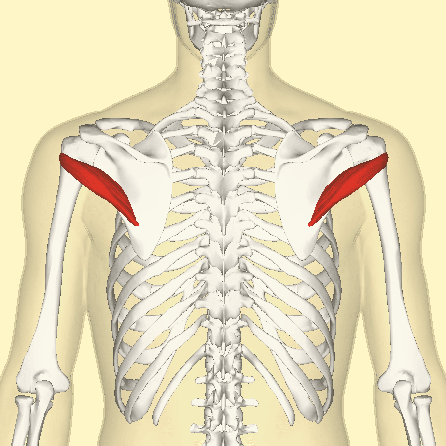
Origin. The muscle arises from the upper two-thirds of the dorsal surface of the lateral (axillary) border of the scapula, on a narrow strip of bone. Its fibres run superolaterally, roughly parallel to the infraspinatus and roughly perpendicular to the teres major beneath it.
The two laminae. Teres minor lies between two aponeurotic laminae, one separating it from the infraspinatus above and the other from the teres major below. Recognising these laminae is how the true intervals are found at operation.
Insertion. It inserts on the inferior facet of the greater tuberosity, the lowest of the three posterior facets, and its lowest fibres insert directly and muscularly onto the surgical neck for approximately 2 cm below the facet. This broad, partly muscular insertion is one reason isolated teres minor tendon tears are rare compared with tears of the tendinous supraspinatus and infraspinatus. The deep surface of the tendon blends with the posteroinferior glenohumeral joint capsule.
Action and Biomechanics
The transverse (axial) force couple. The subscapularis anteriorly is balanced by the infraspinatus and teres minor posteriorly. Teres minor is the inferior part of that posterior arm, and because its line of pull is more inferiorly directed than that of the infraspinatus, it contributes disproportionately to head depression as well as to rotation.
Position dependence. External rotation is its principal action, and how much it contributes depends on the position of the arm. The infraspinatus moment arm is greatest with the arm adducted, so with the arm at the side the infraspinatus supplies most external rotation torque and teres minor plays second fiddle. The teres minor moment arm increases with abduction, and at 90 degrees it becomes a major external rotator, so the shoulder retains external rotation strength across the whole abduction arc. That single fact underwrites the examination: test at the side and you load infraspinatus, test in abduction and you load teres minor.
Other actions. Beyond external rotation, it acts in three ways:
- Head depression: its inferiorly and medially directed vector resists the superior shear of the deltoid, helping to maintain the acromiohumeral interval.
- Posteroinferior stabilisation: it resists posterior and inferior translation of the head, particularly in the abducted externally rotated position, and its capsular blending contributes to the posterior band of the inferior glenohumeral ligament complex.
- Horizontal extension: a minor contribution alongside the posterior deltoid.
Synergists and antagonists. The infraspinatus is its synergist in external rotation, and the posterior deltoid in external rotation in abduction and in horizontal extension. Its antagonists are the subscapularis, pectoralis major, latissimus dorsi and teres major.
Length-tension. The muscle is short with a relatively small excursion, so it works over a narrow part of its length-tension curve. Chronic retraction is poorly tolerated, and a teres minor that has undergone fatty change generates very little force even if the tendon remains in continuity.
Compensation. In a chronic massive posterosuperior tear the teres minor frequently hypertrophies. This is a genuinely favourable finding: a hypertrophied teres minor maintains the posterior arm of the couple, preserves external rotation and predicts better function both with and without surgery.
A reverse total shoulder arthroplasty restores a fulcrum. It medialises and distalises the centre of rotation, which lengthens the deltoid moment arm and recruits more deltoid fibres, so it reliably restores elevation. What it cannot do is generate external rotation, because there is no prosthetic substitute for a posterior rotator.
If the teres minor is intact or hypertrophied, external rotation is usually preserved or improved after reverse arthroplasty. If it is fatty infiltrated, the patient elevates well but cannot externally rotate: Simovitch and colleagues showed this to be an independent predictor of a lower Constant score and markedly worse external rotation.
The clinical consequence is the hornblower posture, in which the patient must abduct the shoulder to shoulder height to bring the hand to the mouth. Patients find this more disabling in daily life than the loss of elevation that the arthroplasty corrected. The solution is a reverse arthroplasty combined with a latissimus dorsi and teres major transfer, converting internal rotators into external rotators at the same sitting.
Surface Anatomy and Examination
Finding the muscle. Teres minor lies deep to the posterior deltoid and is not reliably palpable as a discrete muscle. It is identified indirectly by following the lateral border of the scapula upward from the inferior angle with the arm across the body, which brings the strip of origin subcutaneous in a thin patient.
Finding the quadrangular space. Deep palpation of the posterior soft spot, just lateral to the lateral scapular border and about 2 to 3 cm below the posterior glenohumeral joint line, with the arm abducted and externally rotated, localises the space. Point tenderness here in an overhead athlete supports quadrilateral space syndrome.
Judging wasting. Wasting is best assessed on the sagittal MRI, not by inspection, because the posterior deltoid covers the muscle. This is an important difference from the infraspinatus, whose wasting is visible from across the room.
- How to perform
- Ask the patient to bring the hand to the mouth
- Positive finding
- The patient must abduct the shoulder to 90 degrees to reach the mouth
- What it means
- Irreparable degeneration of teres minor; severe posterior cuff loss
- False positives
- Elbow flexion contracture or a stiff shoulder forcing the same posture
- How to perform
- Arm 90 degrees abduction in the scapular plane, elbow 90 degrees, resist external rotation
- Positive finding
- Weakness against resistance
- What it means
- Posterior cuff weakness in the abducted position, biased toward teres minor
- False positives
- Fatigue on repeated testing; scapular substitution
- How to perform
- Passive full external rotation at 90 degrees abduction, then release
- Positive finding
- The arm drops into internal rotation
- What it means
- Predominantly infraspinatus, with teres minor when severe
- False positives
- Pain inhibition; unsupported arm allowing deltoid substitution
- How to perform
- Elbows tucked in, resist external rotation
- Positive finding
- Weakness
- What it means
- Predominantly infraspinatus; a normal result here with a positive hornblower's sign isolates teres minor
- False positives
- Pain from posterior capsular pathology
- How to perform
- Deep pressure in the posterior soft spot with the arm abducted and externally rotated
- Positive finding
- Reproduction of poorly localised posterolateral shoulder pain, sometimes with paraesthesia
- What it means
- Quadrilateral space syndrome
- False positives
- Posterior capsular tenderness; infraspinatus trigger points
Never interpret hornblower's sign alone. Read it against the drop sign.
- Drop sign positive, hornblower negative: infraspinatus deficient, teres minor intact. External rotation at the side is lost, but hand-to-mouth is preserved.
- Drop sign positive, hornblower positive: infraspinatus and teres minor both deficient. This is combined loss of elevation and external rotation, and it changes the operation.
- Drop sign negative, hornblower positive: uncommon and should prompt a search for an axillary nerve lesion, since teres minor loss with a preserved infraspinatus follows the nerve, not the tendon.
Pitfalls in testing. Support the arm during testing at 90 degrees. An unsupported arm recruits the posterior deltoid, which is also an external rotator in abduction, and the deltoid can substitute so effectively that a patient with an isolated teres minor deficit appears normal until specifically tested in abduction.
Passive range first. Passive external rotation must be full before a lag sign or the drop sign can be interpreted. A stiff shoulder produces a false positive in every lag test.
Complications
Iatrogenic axillary nerve injury. The nerve is injured by dissection or diathermy below the inferior border of teres minor, by a posterior deltoid split extending more than about 5 cm below the scapular spine, or by over-vigorous inferior retraction during posterior glenoid exposure. The result is simultaneous loss of the deltoid, teres minor and lateral arm sensation. That converts a reconstructable shoulder into one for which no reliable salvage exists, since a reverse arthroplasty requires a functioning deltoid.
Denervation from the wrong plane. Splitting the infraspinatus raphe instead of the infraspinatus-teres minor interval denervates the lower infraspinatus. A plane taken too low divides teres minor itself across its motor entry near the inferior border.
Persistent external rotation deficit after arthroplasty. This is the commonest functional disappointment after an otherwise successful reverse arthroplasty. The patient elevates well and is pleased for six weeks, then becomes frustrated at being unable to reach the back of the head, place a hand on the opposite shoulder, or bring food to the mouth without lifting the elbow. Identify it before surgery with hornblower's sign and the sagittal MRI, and either plan a combined transfer or set the expectation explicitly at consent.
Missed or overcalled quadrilateral space pathology. Isolated teres minor atrophy is reported on MRI more often than the clinical syndrome occurs, and operating on the image rather than the patient is the error. Conversely, a genuinely symptomatic vascular quadrilateral space syndrome in a throwing athlete can present with digital ischaemia and be misattributed to a vasospastic disorder; the shoulder is the source.
Failure of an external rotation transfer. A latissimus dorsi and teres major transfer requires an intact, functioning deltoid and adequate passive external rotation. It fails when the deltoid is compromised, when the transfer is tensioned in internal rotation, or when the patient cannot be taught to fire the transferred muscle. Donor morbidity is generally well tolerated but matters in wheelchair users and crutch users, who rely on latissimus dorsi for transfers and for propulsion.
Clinical Relevance
Isolated teres minor atrophy. Read the teres minor against the deltoid and the infraspinatus on the same study, because the combination separates the causes.
- Teres Minor
- Fatty atrophy
- Deltoid
- Normal
- Infraspinatus
- Normal
- Discriminator
- Selective posterior branch compression; often an incidental and asymptomatic MRI finding
- Teres Minor
- Atrophy
- Deltoid
- Atrophy
- Infraspinatus
- Normal
- Discriminator
- History of dislocation, regimental badge sensory change, deltoid weakness
- Teres Minor
- Normal
- Deltoid
- Normal
- Infraspinatus
- Atrophy plus supraspinatus atrophy
- Discriminator
- Teres minor sparing confirms the suprascapular nerve as the culprit
- Teres Minor
- Normal
- Deltoid
- Normal
- Infraspinatus
- Isolated atrophy
- Discriminator
- Paralabral cyst and posterosuperior labral tear on MRI
- Teres Minor
- Hypertrophy or atrophy
- Deltoid
- Normal or hypertrophied
- Infraspinatus
- Atrophy
- Discriminator
- Teres minor hypertrophy is compensatory and favourable; atrophy is a poor prognostic sign
- Teres Minor
- Variable
- Deltoid
- Variable
- Infraspinatus
- Variable
- Discriminator
- Severe prodromal pain then patchy multi-nerve involvement, often crossing nerve territories
Definition and cause. Quadrilateral space syndrome is compression of the posterior branch of the axillary nerve, and sometimes the posterior circumflex humeral artery, within the quadrangular space. The causes are fibrous bands, hypertrophy of the surrounding muscles in throwers and volleyball players, a paralabral cyst tracking inferiorly, or a space-occupying lesion.
The vascular variant. Repeated abduction and external rotation can cause posterior circumflex humeral artery thrombosis with distal digital emboli. Subclavian or brachial angiography in abduction demonstrates this form, and it may require arterial reconstruction.
Presentation. Poorly localised posterolateral shoulder pain, worse in abduction and external rotation, with point tenderness in the quadrangular space. Sensory change over the lateral deltoid is variable and often absent.
Imaging. The MRI hallmark is isolated fatty atrophy of teres minor with a normal deltoid. Cothran and Helms, reviewing a shoulder MRI referral population, found this appearance to be uncommon although not rare, and, the point that matters, rarely an isolated abnormality, usually accompanying other shoulder pathology that better explains the symptoms. It must therefore be correlated clinically before it is treated.
Management. Activity modification and physiotherapy come first. Surgical decompression of the space is reserved for refractory cases with corroborating findings.
The working slice. The sagittal oblique MRI at the level of the scapular spine, medial to the coracoid, shows all four cuff bellies, with teres minor in the inferior posterior quadrant.
Denervation versus disuse. Denervation of teres minor appears first as diffuse T2 hyperintensity of the whole belly, then as uniform fatty replacement. Disuse looks different: the muscle thins but keeps its fascicular architecture. Look at the deltoid on the same images to decide whether the posterior branch alone or the main axillary nerve is involved.
Ultrasound. Teres minor is accessible in the posterior window between the infraspinatus and the posterior deltoid but is difficult to grade for fatty change; MRI remains the standard.
- What it grades
- Fatty infiltration, grades 0 to 4
- Teres minor relevance
- Grade 3 to 4 predicts persistent external rotation loss after reverse arthroplasty and poor response to transfer
- What it grades
- Coronal retraction, stages 1 to 3
- Teres minor relevance
- Rarely applied to teres minor in isolation because the insertion is broad and partly muscular, so retraction is uncommon
- What it grades
- Dropping sign and hornblower's sign
- Teres minor relevance
- Hornblower's sign is the bedside equivalent of a Goutallier 3 to 4 teres minor
- What it grades
- Radiographic stage of cuff tear arthropathy
- Teres minor relevance
- Advanced Hamada stages usually coexist with posterior cuff degeneration including teres minor
Surgical Relevance
Teres minor is the reference structure for three separate surgical decisions.
The interval above it is internervous. Infraspinatus is supplied by the suprascapular nerve and teres minor by the axillary nerve, so the plane between them is a true internervous plane. It is the working interval of the Judet posterior approach, of posterior glenoid exposure for posterior Bankart repair and bone block, and of posterior glenoid osteotomy. Carried medially, it reaches the suprascapular nerve at the spinoglenoid notch, 2 cm medial to the posterior glenoid rim.
The raphe above it is a decoy. The bipennate infraspinatus has an oblique intramuscular raphe that mimics the interval. Find the true plane by following the inferior border of infraspinatus laterally, by identifying both aponeurotic laminae, and by locating the circumflex scapular vessels emerging through the triangular space, where the circumflex scapular artery is itself a structure at risk.
The border below it is the danger line. The inferior border of teres minor is the roof of the quadrangular space, and the axillary nerve and posterior circumflex humeral vessels lie immediately beneath. Any posterior interval carried below teres minor enters the quadrangular space.
- Relationship to teres minor
- Enters at the soft spot between infraspinatus and teres minor
- Structure at risk
- Axillary nerve inferiorly, suprascapular nerve medially
- Distance rule
- 2 cm inferior and 1 cm medial to the posterolateral acromial corner
- Relationship to teres minor
- Passes through or immediately adjacent to teres minor
- Structure at risk
- Axillary nerve
- Distance rule
- Stay close to the glenoid rim; the nerve lies several centimetres inferolateral
- Relationship to teres minor
- Works in the interval directly above teres minor
- Structure at risk
- Suprascapular nerve medially, axillary nerve at the inferior border
- Distance rule
- Do not dissect more than 2 cm medial to the posterior glenoid rim, nor below teres minor
- Relationship to teres minor
- Retracts deltoid to expose teres minor and the interval
- Structure at risk
- Axillary nerve exiting the quadrangular space
- Distance rule
- Split no more than about 5 cm below the scapular spine
- Relationship to teres minor
- Divides fibrous bands at the inferior border of teres minor
- Structure at risk
- The axillary nerve itself and the posterior circumflex humeral vessels
- Distance rule
- Identify the nerve before releasing anything
- Relationship to teres minor
- Teres minor is left undisturbed posteriorly
- Structure at risk
- Its function, not its structure, determines external rotation outcome
- Distance rule
- Assess Goutallier grade pre-operatively rather than intra-operatively
The axillary nerve exits the quadrangular space at the inferior border of teres minor, roughly 1.5 to 2 cm distal to the inferior glenoid rim. Bleeding from the quadrangular space is difficult to control, and in a bloody posterior wound the temptation is to sweep a finger inferiorly and coagulate. Blind diathermy in the depths of a posterior wound is how axillary nerves are burned, and that single manoeuvre accounts for a substantial share of iatrogenic axillary nerve palsies in posterior shoulder surgery. Pack, retract and obtain direct vision before coagulating. Identify the inferior border of teres minor, stop there, and if the exposure demands more, formally identify the nerve first.
Not a donor. Teres minor is not a useful donor, either as a flap or as a transfer: it is small, its excursion is short and its pedicle is shared with the axillary nerve.
A target. The tendon of a latissimus dorsi transfer for an irreparable posterosuperior cuff tear is routinely sutured to the residual infraspinatus and teres minor stumps as well as to the greater tuberosity.
A structure to preserve. In reverse arthroplasty, in posterior approaches and in humeral head fracture surgery, an intact teres minor is the difference between a patient who can externally rotate afterward and one who cannot.
The modified L'Episcopo transfer. In Boileau's modification the latissimus dorsi and teres major are detached from the medial lip of the bicipital groove and transferred posterolaterally to the humerus, converting them from internal to external rotators. It is combined with a reverse arthroplasty when the teres minor is degenerate and hornblower's sign is positive.
- 1Test hand-to-mouthIf the patient must abduct the shoulder to 90 degrees to reach the mouth, hornblower's sign is positive and teres minor is functionally absent.
- 2Grade teres minor on the sagittal MRIGoutallier grade 3 or 4, or clear atrophy relative to the opposite side. Hypertrophy is the opposite finding and is reassuring.
- 3Check the deltoid and axillary nerveTeres minor atrophy with deltoid atrophy indicates an axillary nerve lesion, which contraindicates a reverse arthroplasty rather than modifying it.
- 4Choose the constructIntact teres minor: reverse arthroplasty alone. Degenerate teres minor with a functioning deltoid: reverse arthroplasty combined with latissimus dorsi and teres major transfer.
Guidelines, Registries & Global Practice
Anatomical variation across populations
- Cadaveric series describe variable fusion of teres minor with the infraspinatus, in which the two share a common tendon at the greater tuberosity and no discrete plane exists. Where present, this variant complicates the posterior internervous approach and increases the risk of denervating either muscle.
- The teres minor branch of the axillary nerve may arise before the nerve enters the quadrangular space in a minority of specimens, which alters the pattern of denervation after a proximal injury.
- Fibrous bands within the quadrangular space are described with varying prevalence across dissection series and are the presumed substrate for compressive quadrilateral space syndrome.
- Reported prevalence of isolated teres minor atrophy on shoulder MRI differs substantially between imaging cohorts, reflecting the fact that most of it is incidental.
Side-by-side guidance
- Position Relevant to Teres Minor
- Cuff guidance recognises muscle quality and posterior cuff integrity as determinants of surgical decision-making rather than tear size alone.
- Position Relevant to Teres Minor
- Support reverse arthroplasty for cuff tear arthropathy with pseudoparalysis, with pre-operative assessment of posterior cuff function informing consent about achievable external rotation.
- Position Relevant to Teres Minor
- Emphasises the infraspinatus-teres minor internervous plane in posterior approaches and treats the inferior border of teres minor as the limit of safe dissection.
- Position Relevant to Teres Minor
- Support combining an external rotation transfer with reverse arthroplasty when hornblower's sign is positive and teres minor is degenerate.
Registry and outcome signals
- National joint registries record reverse total shoulder arthroplasty survivorship but do not capture range of motion by plane, so the functional impact of a degenerate teres minor is visible only in institutional series. Those series consistently identify teres minor quality as an independent determinant of external rotation.
- Lateralised reverse designs have been introduced partly to improve external rotation and to reduce scapular notching. Registry data on survivorship of lateralised versus medialised designs continue to accumulate; the functional claim regarding external rotation still rests on cohort studies rather than registry endpoints.
- Combined reverse arthroplasty and external rotation transfer is performed in low volumes, and the evidence base is a series of institutional cohorts rather than randomised trials.
High-resource versus limited-resource practice
- Well-resourced settings: routine sagittal MRI for Goutallier grading of all four cuff muscles, lateralised reverse implants, and combined transfer where indicated.
- Limited-resource settings: hornblower's sign performs the same triage function as the MRI at no cost, reliably identifying the patient in whom a reverse arthroplasty alone will disappoint. Where reverse arthroplasty is unavailable, an isolated latissimus dorsi and teres major transfer in a younger patient with a preserved fulcrum remains a valuable operation.
- The essential clinical skill transfers everywhere: examine hand-to-mouth before promising external rotation.
MCQ Practice Points
Q: Which rotator cuff muscle is not supplied by the suprascapular or subscapular nerves? A: Teres minor, supplied by the posterior branch of the axillary nerve (C5, C6).
Q: Onto which facet of the greater tuberosity does teres minor insert? A: The inferior facet, with the lowest fibres inserting muscularly onto the surgical neck for about 2 cm below it.
Q: What is the fusiform swelling found on the nerve branch to teres minor? A: A pseudoganglion — a normal anatomical structure containing no synapses. It is not a tumour and requires no action.
Q: What are the boundaries and contents of the quadrangular space? A: Teres minor above (subscapularis above anteriorly), teres major below, long head of triceps medially, surgical neck of the humerus laterally. Contents: the axillary nerve and posterior circumflex humeral vessels.
Q: What passes through the triangular space? A: The circumflex scapular artery. Boundaries: teres minor above, teres major below, long head of triceps laterally.
Q: Why does the examination position change which posterior cuff muscle you are testing? A: The infraspinatus external rotation moment arm is greatest with the arm adducted; the teres minor moment arm improves with abduction. So testing at the side loads infraspinatus and testing at 90 degrees loads teres minor.
Q: Which muscle should you grade before promising external rotation after a reverse total shoulder arthroplasty? A: Teres minor. Fatty infiltration predicts a persistent external rotation deficit despite restored elevation.
Q: What does teres minor hypertrophy on MRI signify in a massive posterosuperior cuff tear? A: A favourable compensatory response that maintains the posterior arm of the transverse force couple and preserves external rotation.
Q: Why is teres minor spared in a spinoglenoid notch ganglion? A: Because the lesion affects the suprascapular nerve, whereas teres minor is supplied by the axillary nerve. Preserved teres minor bulk alongside infraspinatus wasting confirms the level of the lesion.
Q: How far distal to the inferior glenoid rim does the axillary nerve lie at the inferior border of teres minor? A: Approximately 1.5 to 2 cm. Treat the inferior border of teres minor as an absolute limit to blind dissection.
Exam Viva Scenarios
Practise clinical reasoning and management decisions out loud
“A 76-year-old man with a massive cuff tear can elevate to only 60 degrees and, when asked to drink from a cup, lifts his elbow to shoulder height to get the cup to his mouth. What is the sign, what does it tell you, and how does it change your plan?”
“A radiologist reports isolated fatty atrophy of the teres minor with a normal deltoid on the MRI of a 28-year-old recreational tennis player whose actual complaint is anterior shoulder pain. What do you make of it?”
“You are planning an open posterior bone block for recurrent posterior instability with glenoid deficiency. Describe your interval, your limits, and the two nerves you are thinking about throughout.”
Attachments
- Origin: upper two-thirds, dorsal surface of the lateral scapular border
- Lies between two aponeurotic laminae
- Insertion: inferior facet of the greater tuberosity
- Lowest fibres insert muscularly onto the surgical neck
- Blends with the posteroinferior capsule
Nerve and Vessels
- Axillary nerve, posterior branch, C5-C6
- Only cuff muscle not supplied by suprascapular or subscapular nerves
- Pseudoganglion on the branch is a normal finding
- Posterior circumflex humeral artery is the main supply
- Circumflex scapular artery emerges below in the triangular space
Spaces
- Quadrangular: teres minor above, teres major below, triceps long head medial, humerus lateral — axillary nerve
- Triangular: teres minor above, teres major below, triceps long head lateral — circumflex scapular artery
- Triangular interval: teres major above, triceps long head medial, humerus lateral — radial nerve
- Axillary nerve exits 1.5 to 2 cm distal to the inferior glenoid rim
Examination
- Hornblower's sign: teres minor
- Drop sign: infraspinatus
- Patte test at 90 degrees biases teres minor
- Support the arm to prevent posterior deltoid substitution
- Wasting is assessed on MRI, not by inspection
Surgical Rules
- Internervous plane above it: infraspinatus versus teres minor
- Never dissect blind below its inferior border
- Posterior deltoid split limited to about 5 cm below the scapular spine
- Grade Goutallier before reverse arthroplasty
- Degenerate teres minor plus hornblower equals reverse plus transfer
Evidence Base
Impact of Fatty Infiltration of the Teres Minor Muscle on the Outcome of Reverse Total Shoulder Arthroplasty
- 42 shoulders in 42 patients, average age 71, treated with a reverse Delta-III arthroplasty for cuff tear arthropathy or irreparable cuff deficiency with pseudoparesis, minimum 24 months follow-up
- Teres minor fatty infiltration graded pre-operatively on MRI by the Goutallier system
- The 30 shoulders with stage 0 to 2 infiltration had significantly better final Constant scores and subjective shoulder values than the 12 with stage 3 or 4
- Relative Constant score rose 41 per cent in the low-grade group against 32 per cent in the high-grade group (p=0.033)
- The SUBJECTIVE shoulder value separates the groups more widely than the Constant does - up 44 per cent against 25 per cent (p=0.018) - so patients felt the difference more than the objective score captured it
- Extremity positioning gained 6.2 points against 5.3 (p=0.033)
- EXTERNAL ROTATION IS THE STARK ONE: the low-grade group gained 9 degrees with the arm at the side while the high-grade group LOST 7 degrees (p less than 0.001) - a net swing of 16 degrees, and the only outcome where the two groups move in opposite directions
The Dropping and Hornblower's Signs in Evaluation of Rotator Cuff Tears
- 54 patients operated on for combined supraspinatus and infraspinatus tears, with the signs correlated against Goutallier stage 3 or 4 fatty degeneration
- Hornblower's sign had 100 per cent sensitivity and 93 per cent specificity for irreparable degeneration of the teres minor
- The dropping sign had 100 per cent sensitivity and 100 per cent specificity for irreparable degeneration of the infraspinatus
- Seven patients had HYPERTROPHY of the teres minor, and the authors note that an intact teres minor can give useful function for activities of daily living - the compensation that makes the muscle worth assessing rather than merely recording
- The reference standard was chosen for a reason the card should state: Goutallier stage 3 or 4 fatty degeneration had ALREADY been correlated with poorer outcome after reconstructive surgery, so the signs are being validated against a marker of irreparability rather than against the tear itself
Anatomical Basis of the Variable Aspects of Injuries of the Axillary Nerve (Excluding the Terminal Branches in the Deltoid Muscle)
- Thirty-two embalmed adult cadaveric shoulders dissected to map the axillary nerve
- The nerve was divided into five segments, from its origin to its intramuscular distribution in the deltoid
- The origins of the branches to teres minor, to the subscapularis and to the shoulder joint varied widely in level, without right-to-left symmetry
- The lateral cutaneous nerve of the arm was absent in four specimens
Reverse Shoulder Arthroplasty Combined with a Modified Latissimus Dorsi and Teres Major Tendon Transfer for Shoulder Pseudoparalysis Associated with Dropping Arm
- 11 consecutive patients, mean age 70, with combined loss of active elevation and external rotation (pseudoparalysis with a dropping arm)
- All had severe cuff tear arthropathy (Hamada stage 3, 4 or 5) with severe atrophy or fatty infiltration of both infraspinatus and teres minor on pre-operative MRI or CT
- Reverse arthroplasty was combined with transfer of latissimus dorsi and teres major to the posterolateral humerus
- Both shoulder function and activities of daily living improved
Quadrilateral Space Syndrome: Incidence of Imaging Findings in a Population Referred for MRI of the Shoulder
- Review of consecutive shoulder MRI studies from a referral population, looking specifically for focal teres minor atrophy or abnormal signal
- Findings suggesting quadrilateral space syndrome were uncommon although not rare
- The abnormality was rarely an isolated finding, usually accompanying other shoulder pathology
- The deltoid was characteristically spared
Anatomy and Relationships of the Suprascapular Nerve: Anatomical Constraints to Mobilisation of the Supraspinatus and Infraspinatus Muscles
- 31 shoulders in 18 cadavera dissected to map the suprascapular nerve and define the limits of cuff mobilisation
- Mean distance from the posterior rim of the glenoid to the infraspinatus motor branches was 2 cm; from the origin of the long head of biceps to the supraspinatus branches, 3 cm
- A standard anterosuperior approach permitted only 1 cm of safe lateral advancement; a Debeyre-type advancement up to 3 cm, limited by motor branch tension
- The study concerns supraspinatus and infraspinatus only; teres minor, being axillary innervated, is not subject to this constraint