Axillary Nerve + PCHA Compression | Overhead Athlete | Neurogenic vs Vascular | Diagnosis of Exclusion
- Quadrilateral space contents - axillary nerve and posterior circumflex humeral artery (PCHA)
- Overhead athletes (throwers, volleyball, swimmers) are the classic group
- Diagnosis of exclusion - no single gold-standard test; rule out cuff, suprascapular nerve, cervical radiculopathy
- Isolated teres minor atrophy on MRI is sensitive but poorly specific for compressive QSS
- Non-operative for at least 6 months before considering surgical decompression
- “Symptoms reproduced by sustained abduction-external rotation for about 1 minute
- “Point tenderness over the quadrilateral space, lateral to the scapula
- “Image-guided quadrilateral space block giving pain relief supports the diagnosis
- “Vascular subtype can present as digital ischaemia in a young dominant-arm athlete
Overview and Epidemiology
Quadrilateral space syndrome (QSS), also written quadrangular space syndrome, is a relatively rare condition in which the axillary nerve and the posterior circumflex humeral artery (PCHA) are compressed as they pass through the quadrilateral space of the posterior shoulder. The symptoms are poorly localised: posterolateral shoulder pain, paraesthesia in a non-dermatomal pattern, and sometimes deltoid or teres minor weakness, all of which overlap with much more common shoulder problems.
Why it is an exam trap. The condition was first described in the early 1980s and remains uncommon. It is the diagnosis you reach only after excluding rotator cuff disease, suprascapular neuropathy and cervical radiculopathy. Because it is uncommon and the symptoms are vague, diagnostic delay is the rule, and many patients are treated for presumed cuff or cervical pathology first.
Who gets it. QSS classically affects young adults, roughly 20 to 40 years old, who perform repetitive overhead arm movements: baseball pitchers, volleyball and tennis players, swimmers and throwers. The dominant arm is usually involved.
Pathophysiology and Anatomy
The space. The quadrilateral space is a four-sided gap in the posterior shoulder. Its two contents, the axillary nerve and the posterior circumflex humeral artery, are the structures at risk in QSS.
- Structure
- Teres minor
- Relevance
- Innervated by axillary nerve - atrophies in QSS
- Structure
- Teres major
- Relevance
- Lower border of the space
- Structure
- Long head of triceps
- Relevance
- Common site of compressive fibrous bands
- Structure
- Surgical neck of humerus
- Relevance
- Bony lateral wall
- Structure
- Axillary nerve
- Relevance
- Neurogenic QSS - deltoid + teres minor
- Structure
- Posterior circumflex humeral artery
- Relevance
- Vascular QSS - thrombosis, aneurysm, emboli
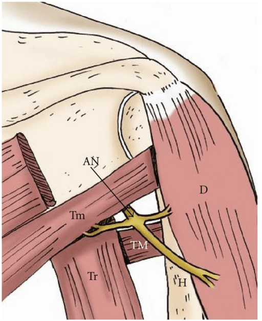
Why compression happens. The space is dynamic. In abduction and external rotation, the cocking position of the throw, the borders crowd together and the cross-sectional area of the space falls. In repetitive overhead athletes, hypertrophy of the surrounding muscles and fibrous bands, most often between the teres muscles and the long head of triceps, narrow the space further and tether the neurovascular bundle.
The nerve. Chronic compression or traction of the axillary nerve produces a focal neuropathy. Because the teres minor branch is often involved, isolated teres minor denervation and fatty atrophy is the classic, though non-specific, imaging hallmark. More severe compression of the main axillary nerve also denervates the deltoid.
The artery. Repetitive compression of the PCHA against fibrous bands or the humerus causes intimal injury, which can lead to thrombosis, aneurysm formation and distal embolisation. The clinical result may be digital ischaemia, splinter haemorrhages or cold intolerance in the dominant hand of a young athlete, a presentation that is easy to miss if QSS is not considered.
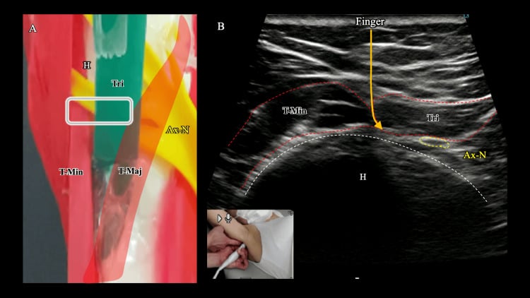
For the borders in more detail, and the related triangular space and triangular interval, see the dedicated Quadrangular and Triangular Spaces anatomy topic and Axillary Nerve Anatomy.
Paralabral Cyst QSS: The Space-Occupying Cause You Must Not Miss
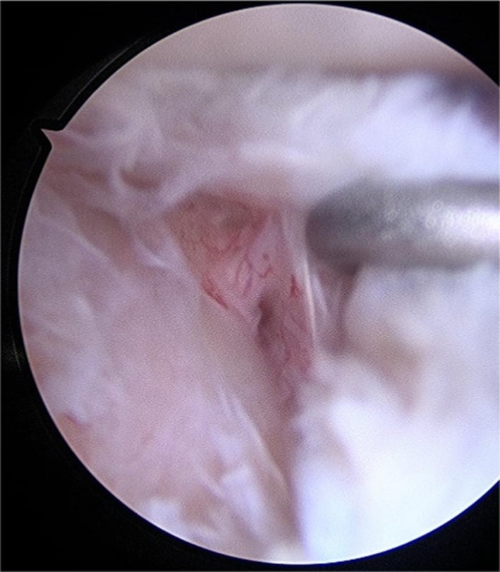
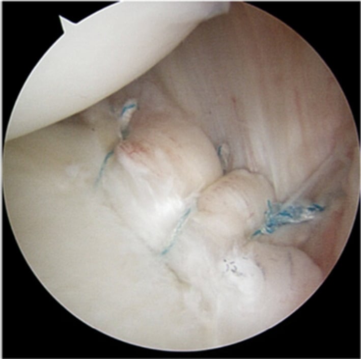
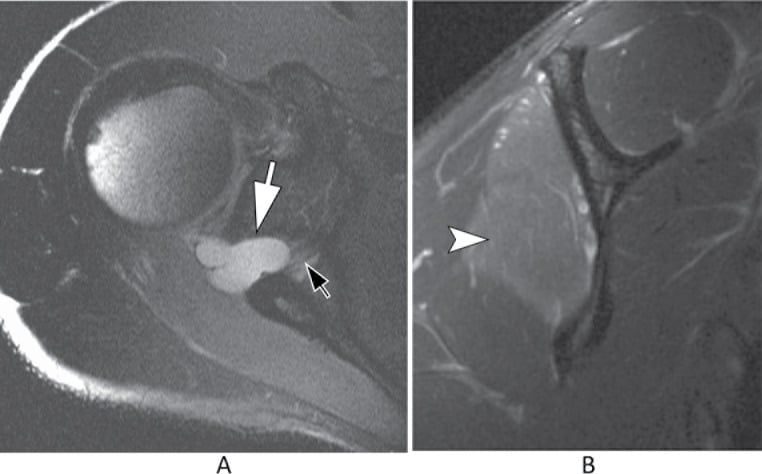
The mechanism. A posteroinferior labral tear acts as a one-way valve. Joint fluid is pumped out through the tear and loculates into a paralabral (ganglion) cyst that tracks posteroinferiorly into the quadrilateral space, where it compresses the axillary nerve.
The analogue. This is the direct counterpart of the spinoglenoid-notch cyst that compresses the suprascapular nerve, which arises from a superior (SLAP-type) labral tear. The valve principle is the same; the tear location and the nerve differ.
Why it changes the plan. Unlike fibrous-band QSS, this is a structural lesion with a treatable source, and MRI shows both the cyst in the quadrilateral space and the underlying labral tear. It is one of the QSS presentations where surgery is clearly indicated rather than a six-month rehabilitation problem.
Treat the tear, not just the cyst. Aspirating or decompressing the cyst alone risks recurrence, because the labral tear keeps refilling it. Definitive treatment addresses the tear, with arthroscopic labral repair to close the one-way valve, with or without cyst decompression.
Classification
QSS is most usefully classified by the predominant compressed structure, because this drives both the presentation and the management.
- Neurogenic QSS - axillary nerve compression; the more common subtype
- Vascular QSS - PCHA compression; rarer, but potentially limb-threatening
By cause. The underlying cause is useful for surgical planning. Fibrous bands are the most commonly implicated; true space-occupying lesions are less common but important to find.
- Fibrous bands - most common; released at decompression
- Muscle hypertrophy - overhead athletes (pitchers, swimmers)
- Space-occupying lesion - paralabral cyst (look for an inferior labral tear), osteochondroma, soft-tissue tumour, PCHA aneurysm
- Post-traumatic - scarring after proximal humerus fracture or dislocation, or iatrogenic after shoulder surgery
Clinical Assessment
History. Ask about the sport and the dominant arm. The typical patient is a young overhead athlete with insidious, poorly localised posterolateral shoulder and lateral arm pain that is worse with the arm abducted and externally rotated. They may describe paraesthesia down the lateral arm that does not follow a dermatome, and fatigue and weakness with overhead activity.
The vascular history. Ask specifically about cold hands, colour change and finger pain. Vascular QSS presents with cold intolerance, digital ischaemia, hand claudication or pain in the dominant arm of a young athlete.
Examination. Inspect, palpate and provoke, then test the nerve and the circulation to the hand.
- Inspection - deltoid or teres minor wasting in chronic cases, often subtle
- Palpation - point tenderness over the quadrilateral space, felt just lateral to the lateral scapular border; a useful, if non-specific, sign
- Provocation - holding the arm in abduction and external rotation for about one minute reproduces the pain and paraesthesia
- Neurology - deltoid and teres minor power, and the regimental-badge sensory patch over the lateral deltoid
- Vascular - distal pulses and capillary refill, and the fingertips for emboli or splinter haemorrhages; distal perfusion may be diminished and a bruit may be present
The differentials. There is no gold-standard test for QSS. Rotator cuff disease, suprascapular neuropathy and cervical radiculopathy are far more common and must be actively excluded first; QSS is what remains when they have been. Wasting separates the suprascapular nerve: suprascapular neuropathy takes infraspinatus, with or without supraspinatus, not teres minor.
- Distinguishing Feature
- Posterolateral pain worse in abduction-ER; teres minor atrophy
- Confirming Test
- MRI + diagnostic block; angiography if vascular
- Distinguishing Feature
- Infraspinatus (+/- supraspinatus) wasting, NOT teres minor
- Confirming Test
- MRI for a spinoglenoid, suprascapular notch or paralabral cyst; EMG suprascapular nerve
- Distinguishing Feature
- Impingement signs, painful arc, cuff weakness, night pain
- Confirming Test
- Ultrasound / MRI cuff
- Distinguishing Feature
- Neck pain, dermatomal symptoms, positive Spurling
- Confirming Test
- MRI cervical spine; EMG
- Distinguishing Feature
- Acute severe pain then patchy weakness/atrophy
- Confirming Test
- EMG (multifocal), clinical course
Investigations
There is no single diagnostic test for QSS. The work-up is built to exclude mimics and to support the clinical impression.
Plain radiographs. Usually normal. They are obtained to exclude bony lesions such as osteochondroma, an old fracture or calcification.
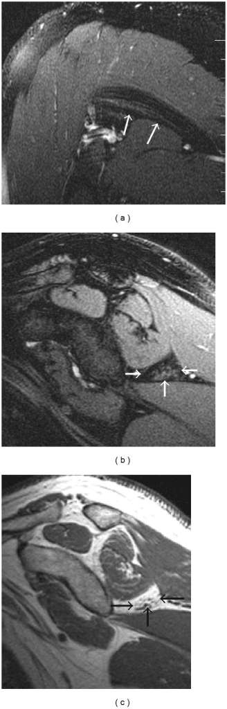
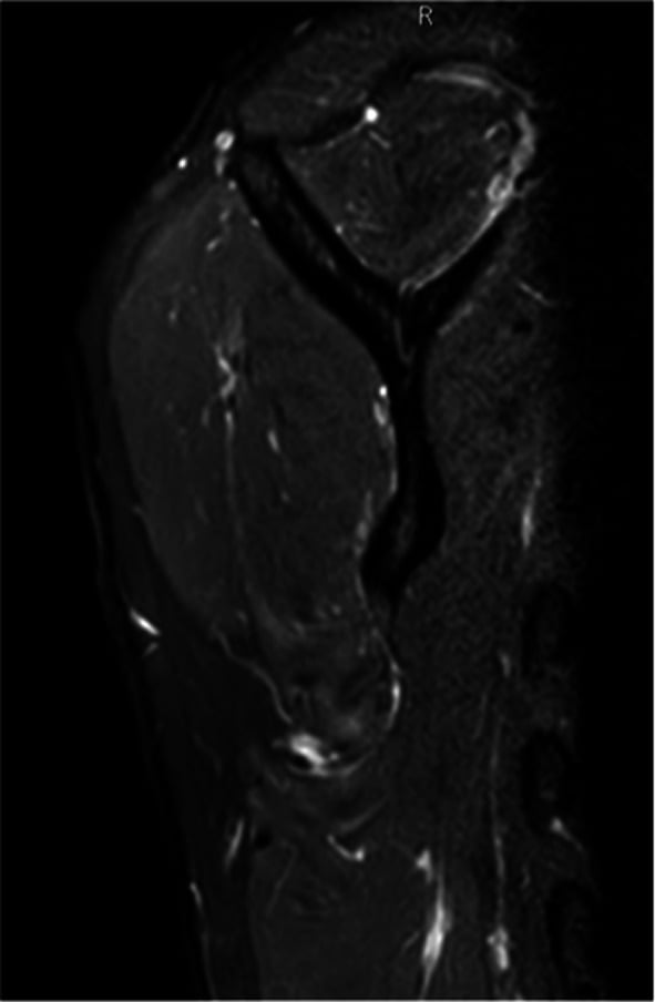
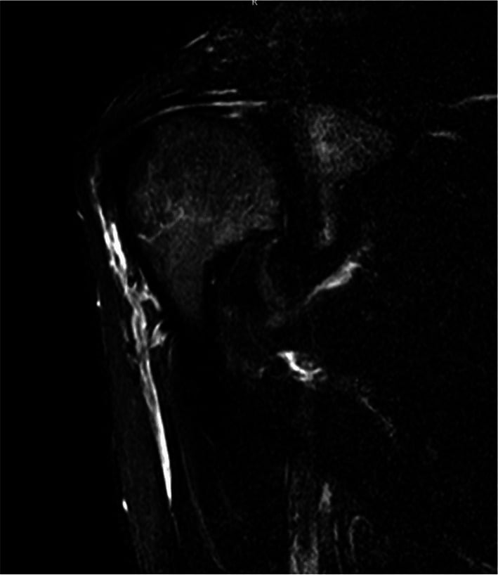
MRI. The most useful single test. It looks for three things:
- Teres minor denervation and fatty atrophy (sensitive but not specific)
- Deltoid denervation, in more advanced axillary nerve involvement
- Space-occupying lesions - paralabral cyst, mass or PCHA aneurysm
Isolated teres minor atrophy on MRI is sensitive but not specific. True compressive QSS is rare, and large MRI series show that isolated teres minor atrophy is far more often explained by rotator cuff disease or idiopathic traction than by genuine compression.
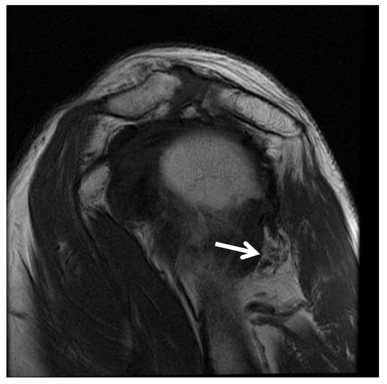
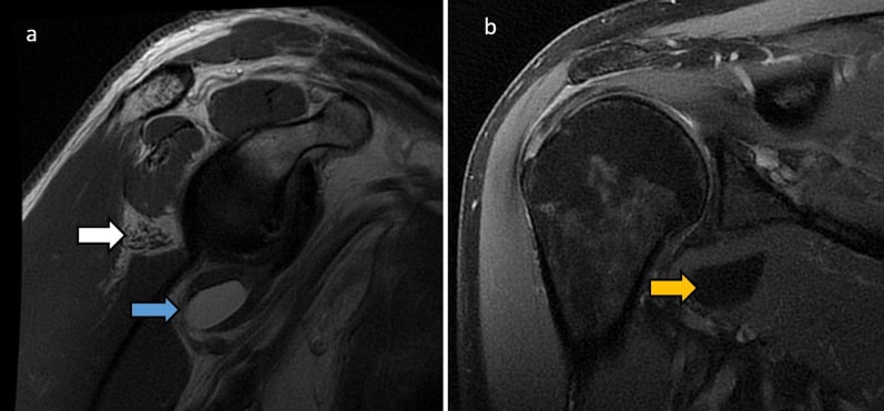
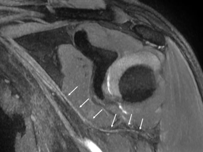
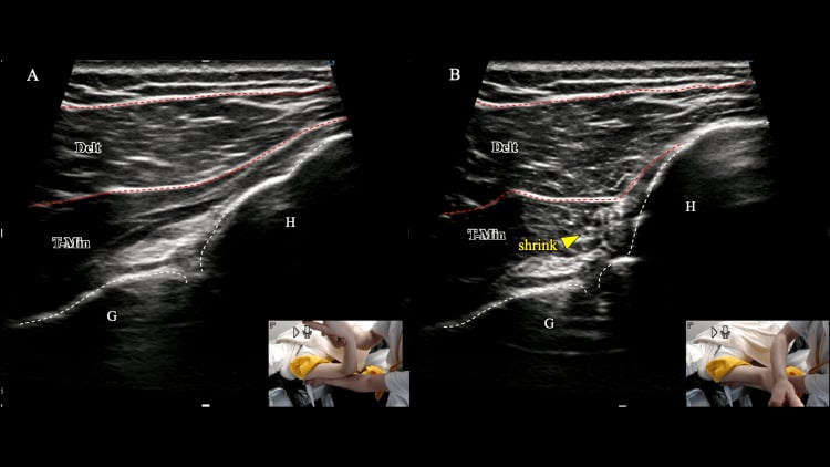
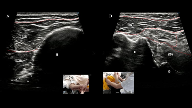
Ultrasound. Increasingly used. With colour Doppler it can assess the PCHA dynamically during abduction-external rotation, and it can guide a diagnostic block.
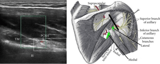
Electromyography and nerve conduction. These may show axillary neuropathy and help exclude suprascapular and cervical causes, but can be normal in early or purely vascular disease.
Vascular imaging. Arterial Doppler ultrasound comes first when the vascular subtype is suspected. CT, MR or catheter angiography is reserved for suspected vascular QSS, and dynamic imaging in abduction-external rotation may be required to demonstrate PCHA occlusion or aneurysm that is not seen in the neutral position.
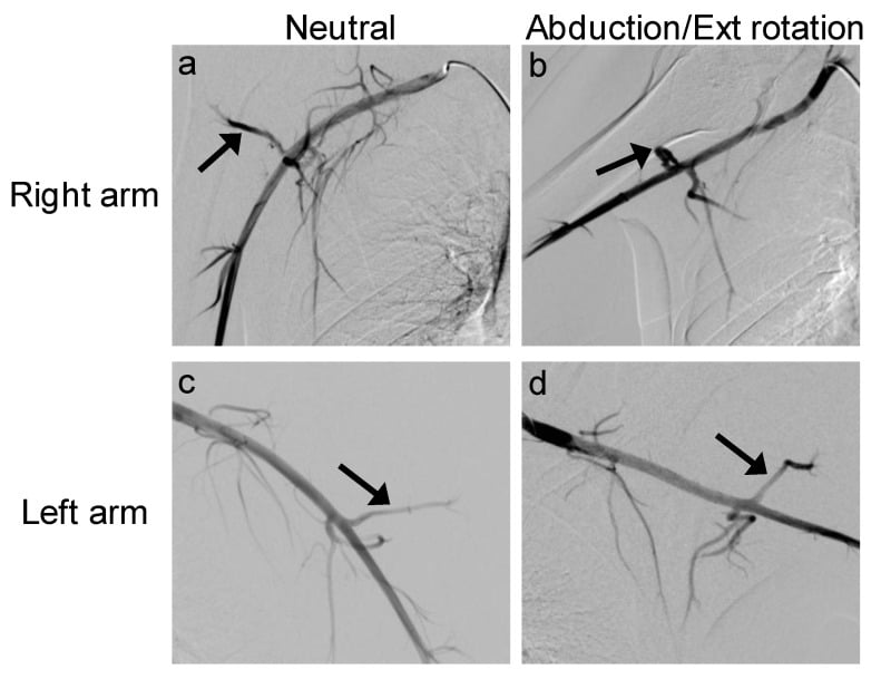
A young dominant-arm athlete presenting with digital ischaemia, cold intolerance, or distal emboli may have vascular QSS. This warrants urgent vascular imaging (Doppler then angiography) and vascular surgical involvement - acute ischaemia can require emergent thrombolysis or thrombectomy.
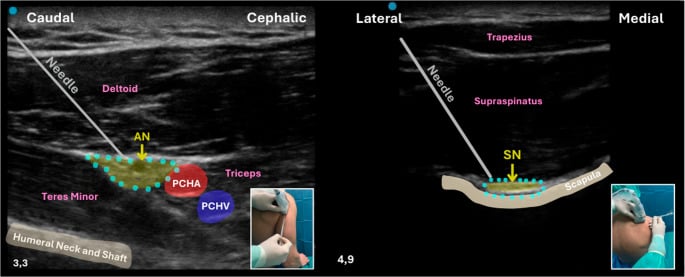
The Diagnostic Quadrilateral-Space Block
The technique. An image-guided (ultrasound or fluoroscopic) injection of local anaesthetic, with or without corticosteroid, is placed into the quadrilateral space around the axillary nerve.
A positive result. Meaningful, temporary relief of the patient's characteristic posterolateral pain after the block, in the right clinical context, supports the quadrilateral space as the pain source and predicts a better response to decompression. Added corticosteroid can also be therapeutic.
Why it carries weight. QSS is a diagnosis of exclusion and MRI teres minor atrophy is non-specific, so a positive block is one of the few findings that localises the problem to the space. That makes it one of the most useful confirmatory findings in the work-up.
Its limits. The block is supportive, not definitive. A placebo response is possible and a poorly targeted injection can give a false negative. A positive block does not distinguish neurogenic from vascular disease, so it never replaces excluding the mimics or screening for the vascular subtype.
Management Algorithm
Management is stepwise and depends on subtype. The default for neurogenic QSS is a prolonged non-operative trial; the threshold for early surgery is much lower in vascular disease or when a structural lesion is found.
Non-operative care is first-line. A trial of at least 6 months is recommended before considering surgery in neurogenic QSS, because many athletes improve with load management and rehabilitation. The figure is a convention drawn from Level 4 case series rather than a studied duration (see the Evidence Base).
- Activity and training modification - reduce provocative overhead loading; correct throwing mechanics
- Physiotherapy - posterior capsule and teres muscle stretching, scapular stabilisation, rotator cuff and periscapular strengthening
- NSAIDs for symptom control
- Image-guided injection - a quadrilateral space corticosteroid/local anaesthetic injection can be both diagnostic and therapeutic
Surgical Technique
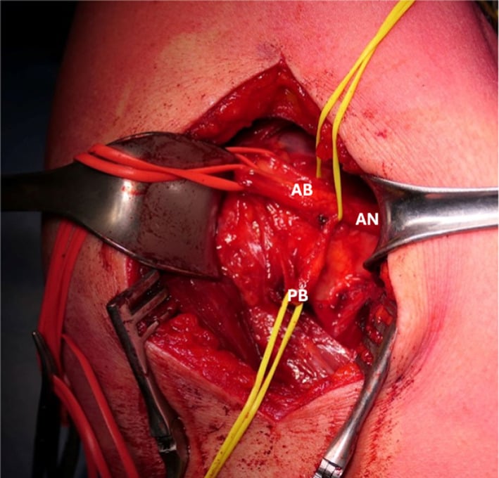
Open posterior quadrilateral space decompression. The nerve to teres minor is the key landmark: follow it to the main axillary nerve.
- Positioning - lateral decubitus or prone/semi-prone, with the affected arm draped free
- Incision - centred over the posterior border of the deltoid, lateral to the posterior axillary fold
- Interval - retract the posterior border of deltoid to reveal the interval between teres minor and teres major
- Identify and protect - find the nerve to teres minor, trace it deep to the axillary nerve, and protect the posterior circumflex humeral vessels throughout; uncontrolled bleeding obscures the field and risks vascular injury
- Decompress - divide the compressive fibrous bands around the nerve; free gliding of the nerve confirms adequate release, and should be checked before closing
- Lesion - excise any paralabral cyst or mass, or address an aneurysm as indicated; for a paralabral cyst, address the underlying labral tear as well
Complications
Of the condition. Untreated or progressive QSS leaves persistent posterolateral shoulder pain and loss of overhead performance, and a progressive axillary neuropathy with teres minor and deltoid weakness and atrophy. In the vascular subtype, PCHA thrombosis, aneurysm and distal embolisation causing digital ischaemia are potentially limb-threatening.
Of surgical decompression. The operation carries its own risks.
- Iatrogenic axillary nerve injury, the very structure being protected
- Injury to the posterior circumflex humeral vessels, with bleeding or haematoma
- Incomplete decompression with persistent symptoms
- Recurrence, for example of a paralabral cyst if the labral tear is not addressed
- Standard wound complications - infection, stiffness
The overtreatment complication. Isolated teres minor atrophy is common and usually not due to compressive QSS, so the most clinically relevant pitfall is operating on a patient who does not have true QSS. Reserve surgery for refractory cases with a coherent clinical picture or a demonstrable structural lesion. The other pitfall is failing to recognise and manage the vascular subtype.
Postoperative Rehabilitation
Rehabilitation after decompression is staged and prioritises early protected motion with progressive return to overhead loading.
Sling for comfort, wound care, gentle pendulum and passive range of motion. Protect the axillary nerve repair/neurolysis.
Progress to active-assisted then active range of motion; begin scapular setting and periscapular activation. Avoid provocative abduction-external rotation loading.
Rotator cuff and deltoid strengthening, scapular stabilisation, and posterior capsule flexibility work.
Sport-specific and throwing progression once strength and painless overhead motion are restored; correct underlying mechanics. Return to sport timelines vary and are guided by symptom resolution and strength recovery.
Outcomes and Prognosis
The evidence base is limited to small case series and case reports (Level 4), so prognostic statements must be cautious.
After decompression. Small series report good symptom relief and return to sport in appropriately selected refractory neurogenic cases. In published case material, athletes who undergo decompression for refractory neurogenic QSS can regain deltoid bulk and strength and return to full sport, with durable relief reported over months to a couple of years of follow-up.
Vascular QSS. With timely diagnosis and combined vascular and decompressive surgery, distal ischaemia can be salvaged and baseline function restored. Delay risks permanent tissue loss.
Selection decides the result. Outcomes are best when the diagnosis is genuine. Operating on non-specific teres minor atrophy without a coherent clinical picture predicts a poor result.
Key Points and Practice
Q: Which two structures pass through the quadrilateral space and are compressed in QSS?
A: The axillary nerve and the posterior circumflex humeral artery (PCHA).
Q: What is the most useful single imaging test in suspected QSS, and what is its classic (if non-specific) finding?
A: MRI, looking for teres minor (+/- deltoid) denervation / fatty atrophy - sensitive but not specific.
Q: A young dominant-arm athlete presents with digital ischaemia and a dusky fingertip. Which QSS subtype is this and what is the priority?
A: Vascular QSS (PCHA thrombosis/aneurysm/emboli) - urgent vascular imaging and surgery; it can be limb-threatening.
Q: How long is non-operative treatment generally trialled before considering surgery in neurogenic QSS?
A: At least 6 months.
Guidelines, Registries and Global Practice
There are no formal society guidelines or registries specific to quadrilateral space syndrome - it is too rare, and the literature is Level 4 (case series and reports). Practice is therefore consensus-based and consistent across regions.
- QSS is a diagnosis of exclusion; rule out the common shoulder mimics first (rotator cuff disease, suprascapular neuropathy, cervical radiculopathy).
- MRI is the primary imaging test worldwide; dynamic ultrasound is a useful, inexpensive, widely available adjunct for the neurovascular bundle and for guided blocks.
- A positive image-guided quadrilateral space block is a widely accepted supportive test where the diagnosis is uncertain.
- Non-operative care for at least 6 months is the accepted first-line for neurogenic disease; surgical decompression is reserved for refractory or structural disease.
- The vascular subtype is managed jointly with vascular surgery, with endovascular therapy for acute ischaemia - a pathway that depends on local interventional radiology and vascular surgery availability.
access to dynamic angiography, musculoskeletal ultrasound, and catheter-directed endovascular therapy differs by resource setting, which influences how quickly the vascular subtype is diagnosed and treated.
Related pages: Axillary Nerve Anatomy for the nerve's course, its teres minor branch and the reason isolated teres minor atrophy points here rather than to the cuff; Teres Minor Anatomy and Teres Major Anatomy for the two muscles that form the upper and lower borders of the space and whose hypertrophy in an overhead athlete narrows it; Thoracic Outlet Syndrome for the other compressive neurovascular syndrome of the throwing shoulder, which shares the vascular presentation and must be excluded alongside it; Latissimus Dorsi and Teres Major Rupture for the injury that produces posterior shoulder pain in the same athlete through a mechanical rather than a neurological route; and Suprascapular Nerve Entrapment for the mimic that must be ruled out before this diagnosis of exclusion can be made, and which shares the paralabral-cyst mechanism described above.
Exam Viva Scenarios
Practise clinical reasoning and management decisions out loud
“A 24-year-old baseball pitcher presents with insidious posterolateral pain in his dominant shoulder and lateral arm tingling, worse when his arm is cocked back to throw. Cuff strength is preserved and his neck examination is normal. How would you approach this?”
“A 22-year-old elite volleyball player reports cold intolerance and painful, dusky fingertips in her dominant hand, with one fingertip showing a small area of discolouration. Shoulder movement is full but spiking provokes arm symptoms. What is your concern and how do you proceed?”
“A radiologist reports isolated fatty atrophy of the teres minor on a shoulder MRI ordered for impingement-type pain in a 58-year-old labourer. The referring team asks whether this means he has quadrilateral space syndrome. How do you advise them?”
Anatomy
- Superior: teres minor
- Inferior: teres major
- Medial: long head of triceps
- Lateral: surgical neck of humerus
- Contents: axillary nerve + PCHA
Two Subtypes
- Neurogenic: axillary nerve - pain, paraesthesia, teres minor atrophy
- Vascular: PCHA - thrombosis, aneurysm, distal emboli
- Vascular can be limb-threatening
Clinical
- Overhead athlete, dominant arm
- Posterolateral pain worse in abduction-ER
- Point tenderness over the space
- Diagnosis of exclusion
Investigations
- MRI: teres minor / deltoid denervation
- Dynamic ultrasound + Doppler for PCHA
- Angiography (abduction-ER) if vascular
- Quadrilateral space block to confirm
Management
- Neurogenic: non-op 6+ months first
- Decompress bands / neurolysis if refractory
- Vascular: urgent thrombolysis/thrombectomy
- Aneurysm resection + decompression
Exam Traps
- Isolated teres minor atrophy = sensitive NOT specific
- Don't miss the vascular subtype
- Suprascapular nerve = infraspinatus, NOT teres minor
- Don't operate on incidental atrophy
Evidence Base
The following sources inform current understanding of quadrilateral space syndrome.
Quadrilateral Space Syndrome: A Review
- QSS is a relatively rare compression of the axillary nerve and PCHA in the quadrilateral space
- Fibrous bands are most commonly implicated; true space-occupying lesions are less common
- Symptoms are non-dermatomal posterolateral pain aggravated by abduction-external rotation
- Lidocaine block within the space giving pain relief is a useful diagnostic finding
- At least 6 months of non-operative treatment is recommended before surgery
Quadrilateral Space Syndrome (vascular and interventional perspective)
- Both the axillary nerve and PCHA are susceptible to compression in the quadrilateral space
- Overhead athletes are at greater risk; presentation may be neurogenic or vascular
- MRI may show teres minor or deltoid atrophy; angiography may show PCHA aneurysm or occlusion
- Emboli from the PCHA can cause acute upper-limb ischaemia requiring catheter-directed thrombolysis or thrombectomy
Exploring the Quadrilateral Space: Clinical Anatomy, Pathology and Imaging Insights
- MRI is the gold-standard imaging modality for QSS
- Ultrasound complements MRI by dynamically assessing neurovascular structures with Doppler
- CT and radiographs help identify bony lesions such as osteochondroma or tumour
- Conservative care (physiotherapy, ultrasound-guided steroid injection) is first-line; surgery is reserved for refractory or structural/vascular disease
Axillary Nerve Decompression for Neurogenic QSS in a Volleyball Player
- 21-year-old competitive volleyball player with 5-year progressive arm weakness and deltoid atrophy
- EMG showed deltoid denervation; MRI showed axillary nerve compression in the quadrangular space
- Posterior microsurgical decompression divided a compressive fibrotic band after failed conservative care
- Restored deltoid mass and strength by 6 months with durable relief and full return to sport at 2 years