Two Pathways of Bone Formation | Membranous vs Cartilage Template | Development
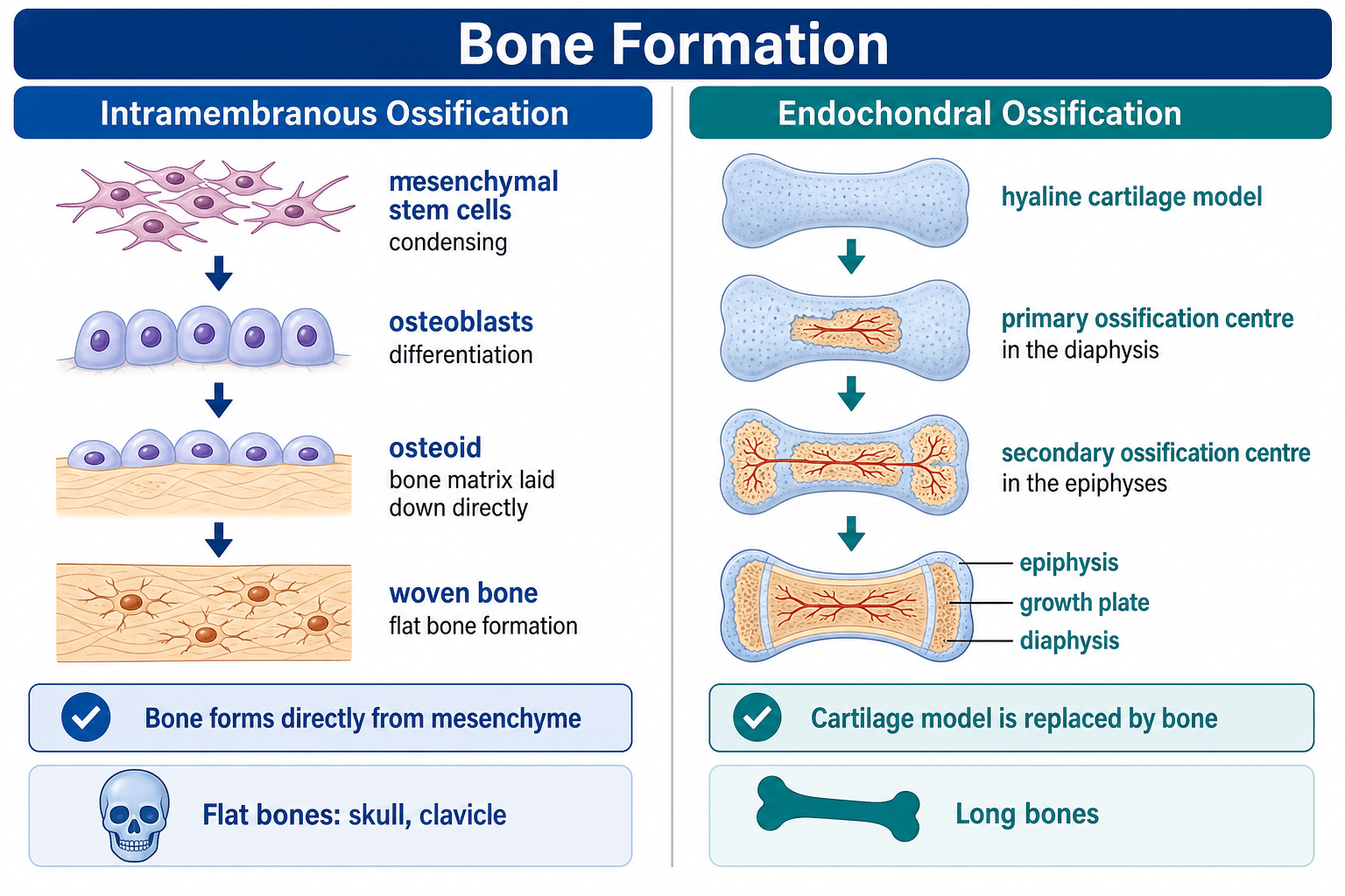
- Intramembranous: bone forms directly from mesenchymal condensation (no cartilage intermediate)
- Endochondral: bone forms by replacing cartilage template through coordinated chondrocyte maturation
- Flat bones (skull, clavicle, mandible) form via intramembranous ossification
- Long bones form via endochondral ossification with growth plates for longitudinal growth
- Both pathways produce lamellar bone; woven bone is emergency/pathologic
- “Clavicle is unique: intramembranous ossification but medial physis for growth
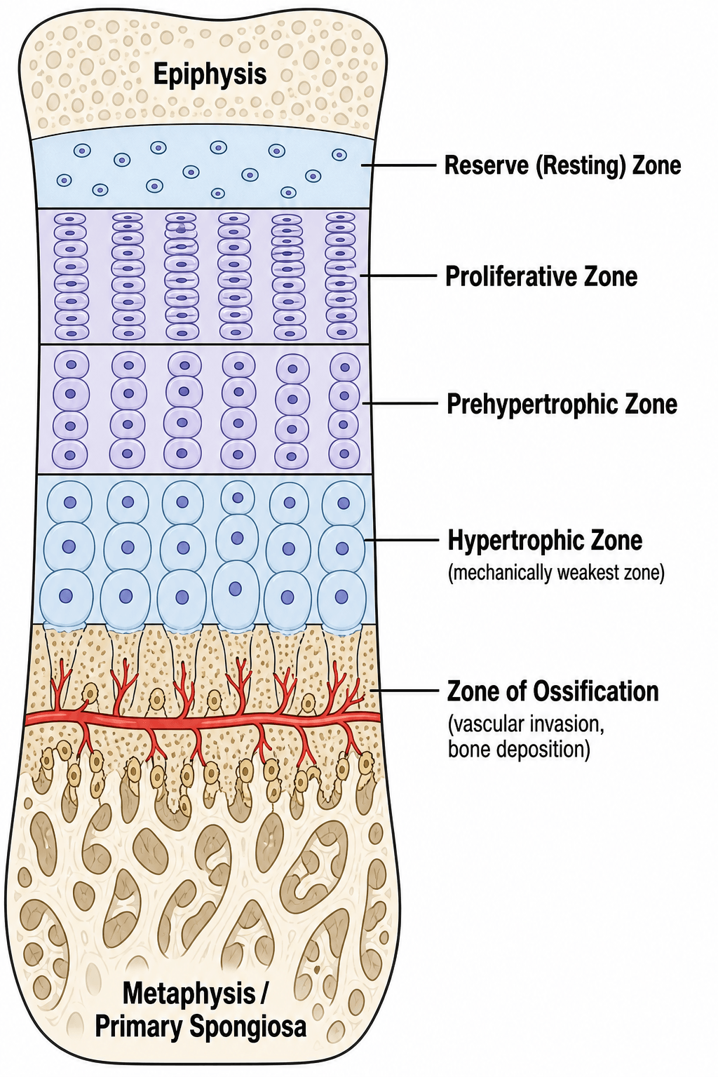
- “Growth plate zones: Reserve, Proliferative, Hypertrophic, Ossification
- “Salter-Harris fractures exploit growth plate zone weakness
- “Distraction osteogenesis uses intramembranous ossification
Fundamentally different. Intramembranous = direct mesenchyme to bone. Endochondral = mesenchyme to cartilage to bone. Same end product (lamellar bone).
Location determines pathway. Flat bones (skull, clavicle) = intramembranous. Long bones, vertebrae, pelvis = endochondral. Rib cage mixed.
RZPHO sequence. Reserve → Proliferative → Hypertrophic (maturation, calcification) → Ossification. Injury pattern basis for Salter-Harris classification.
Fracture healing uses both. Primary (intramembranous) direct bone formation. Secondary (endochondral) via cartilage callus. Distraction osteogenesis is intramembranous.
Overview and Development
Ossification is the process of bone formation. There are two distinct pathways by which bone develops and heals:
- Bone forms directly from mesenchymal stem cells without a cartilage intermediate
- Occurs in flat bones: skull, facial bones, mandible, clavicle
- Also used in primary fracture healing and distraction osteogenesis
- Bone forms by replacing a cartilage template
- Occurs in long bones, vertebrae, pelvis, and most of the skeleton
- Growth plates are persistent endochondral ossification zones
- Also used in secondary fracture healing
Despite fundamentally different mechanisms, both intramembranous and endochondral ossification produce identical lamellar bone. The pathway is determined by anatomical location and mechanical environment, not by final bone type.
Mechanisms: Intramembranous
Intramembranous ossification forms bone directly from mesenchymal tissue WITHOUT a cartilage intermediate. This is the embryonic pathway for flat bones (skull vault, facial bones, mandible, clavicle) and also the mechanism of fracture healing via primary union and distraction osteogenesis.
Intramembranous Ossification Steps
Mesenchymal stem cells aggregate at ossification center, forming condensed tissue with increased cell density and vascularity.
MSCs differentiate into osteoblasts under influence of Runx2 and Osterix transcription factors. Cells secrete osteoid (unmineralized matrix).
Osteoblasts deposit collagen type I and non-collagenous proteins forming osteoid matrix. This occurs along radiating spicules from ossification center.
Hydroxyapatite deposition within osteoid after 10-day lag phase. Some osteoblasts become embedded as osteocytes. Trabeculae form.
Initial woven bone (disorganized collagen) is gradually replaced by organized lamellar bone through remodeling.
Periosteum forms at surface. Appositional growth creates cortical bone. Trabecular spaces become marrow cavities.
- Skull: Frontal, parietal, occipital, temporal (flat parts)
- Face: Maxilla, zygomatic, nasal
- Jaw: Mandible
- Shoulder: Clavicle (unique: has medial physis!)
- No cartilage intermediate
- Direct vascular invasion
- Multiple ossification centers coalesce
- Rapid bone formation
- Used in distraction osteogenesis
Endochondral Ossification
Endochondral ossification forms bone by REPLACING a cartilage template. This is the pathway for most of the skeleton (long bones, vertebrae, pelvis, ribs) and the mechanism of secondary fracture healing via cartilaginous callus. The growth plate is an endochondral ossification zone that persists until skeletal maturity.
Endochondral Ossification Steps
Mesenchymal cells condense and differentiate into chondrocytes, forming hyaline cartilage model of future bone. Shape matches final bone.
Central chondrocytes enlarge (hypertrophy), matrix calcifies. Hypertrophic cells secrete VEGF, attracting blood vessels.
Vascular invasion at mid-diaphysis. Chondrocytes undergo apoptosis. Osteoblasts arrive and deposit bone on calcified cartilage scaffold. Occurs week 8 fetal development.
Intramembranous bone forms around mid-shaft via periosteum, providing structural support during cartilage replacement.
Epiphyseal ossification centers develop (birth to adolescence). Same process: vascular invasion, chondrocyte apoptosis, bone deposition.
Cartilage persists between diaphysis and epiphysis as growth plate. Endochondral ossification continues here until skeletal maturity, driving longitudinal growth.
Estrogen-mediated closure at skeletal maturity. Cartilage fully replaced by bone, forming epiphyseal line scar.
Growth Plate Structure and Function
Five Functional Zones
- Cell Characteristics
- Sparse, small chondrocytes
- Matrix
- High proteoglycan
- Function
- Stem cell niche
- Cell Characteristics
- Columnar stacks, flat cells
- Matrix
- Type II collagen
- Function
- Rapid cell division
- Cell Characteristics
- Cells begin enlarging
- Matrix
- Transition matrix
- Function
- Maturation initiation
- Cell Characteristics
- Large cells (10x volume)
- Matrix
- Type X collagen, calcified
- Function
- Matrix mineralization
- Cell Characteristics
- Chondrocyte apoptosis
- Matrix
- Calcified cartilage scaffold
- Function
- Vascular invasion, bone deposition
The hypertrophic zone is the weakest point in the growth plate due to large cells with minimal matrix. This is where Salter-Harris fractures propagate. Zone of Ranvier (peripheral fibrous ring) provides lateral support and circumferential growth.
Comparison of Ossification Pathways
- Intramembranous
- None (direct)
- Endochondral
- Cartilage template
- Intramembranous
- Flat bones, clavicle
- Endochondral
- Long bones, axial skeleton
- Intramembranous
- Early, throughout
- Endochondral
- Late, after hypertrophy
- Intramembranous
- Week 8 fetal life
- Endochondral
- Week 8 (primary center)
- Intramembranous
- Appositional only
- Endochondral
- Interstitial (physis) + appositional
- Intramembranous
- Primary union, distraction
- Endochondral
- Secondary union (callus)
Despite different pathways, both intramembranous and endochondral ossification produce identical lamellar bone. The pathway is determined by anatomical location and mechanical environment, not by final bone type. Woven bone (disorganized collagen) is an immature or pathological form seen in rapid ossification, always replaced by lamellar bone.
Differential of Impaired Ossification (Exam Discriminators)
- Molecular defect
- FGFR3 gain-of-function (G380R)
- Pathway predominantly affected
- Endochondral (proliferative zone)
- Discriminating clinical clue
- Rhizomelic short limbs, macrocephaly, normal trunk and intellect
- Molecular defect
- RUNX2 haploinsufficiency
- Pathway predominantly affected
- Both (osteoblast maturation) — clavicle/skull
- Discriminating clinical clue
- Absent/hypoplastic clavicles, wide fontanelles, dental anomalies
- Molecular defect
- Type I collagen (COL1A1/2)
- Pathway predominantly affected
- Both (matrix quality)
- Discriminating clinical clue
- Fragile bones, blue sclerae, dentinogenesis imperfecta
- Molecular defect
- Vitamin D / phosphate handling
- Pathway predominantly affected
- Endochondral (defective mineralisation of hypertrophic zone)
- Discriminating clinical clue
- Widened physis, metaphyseal cupping/fraying, bowing
- Molecular defect
- Cathepsin K loss (pyknodysostosis)
- Pathway predominantly affected
- Remodelling (osteoclast)
- Discriminating clinical clue
- Dense brittle bone + acro-osteolysis distinguishes pyknodysostosis
Clinical Applications
Ossification in Fracture Repair
Primary (Direct) Healing:
- Absolute stability (compression plating)
- Uses intramembranous ossification
- Cutting cone crosses fracture
- No visible callus
Secondary (Indirect) Healing:
- Relative stability (IM nail, cast)
- Uses endochondral ossification
- Cartilaginous callus replaced by bone
- Visible external callus
At fracture site with movement, low oxygen tension favors cartilage formation over direct bone. Cartilage is more tolerant of motion and hypoxia. As vascularity improves and stability increases, cartilage is replaced by bone via endochondral ossification - recapitulating embryonic development.
Guidelines, Registries & Global Practice
Global Epidemiology of Ossification Disorders
- Global frequency
- ~1 in 15,000–30,000 live births
- Pathway affected
- Endochondral (proliferative zone)
- Global frequency
- ~1 in 15,000–20,000
- Pathway affected
- Both (matrix quality)
- Global frequency
- ~1 in 1,000,000
- Pathway affected
- Both (osteoblast maturation) — clavicle/skull
- Global frequency
- ~1–10 per 100,000 adolescents (rising with obesity)
- Pathway affected
- Endochondral (hypertrophic zone shear)
Side-by-Side Society Guidance
- Relevant position
- Fracture-healing biology — absolute stability favours direct (intramembranous/contact) healing; relative stability favours endochondral callus
- Relevant position
- Paediatric physeal injury management; vitamin D / metabolic bone optimisation in healing
- Relevant position
- Standards for paediatric fracture and limb-reconstruction care, including physeal-sparing principles
- Relevant position
- Limb-lengthening and bone-transport (distraction osteogenesis) standards and complication reporting
- Relevant position
- Multidisciplinary care; vosoritide as the first disease-modifying option in eligible children
Registry and Outcome Notes
- Distraction osteogenesis / limb reconstruction registries (e.g. ASAMI/ILLRS-affiliated databases) track external-fixator vs magnetic intramedullary lengthening — both rely on tension-stress intramembranous regenerate.
- Magnetic motorised nails (e.g. lengthening intramedullary devices) have shifted practice from external frames in high-resource settings; circular frames remain the workhorse where implant cost is prohibitive.
High- vs Limited-Resource Practice Variation
- High-resource: magnetic lengthening nails, vosoritide for eligible achondroplasia, advanced 3D deformity planning.
- Limited-resource: Ilizarov/circular external fixation remains the global standard for lengthening and bone transport — low implant cost, no power source, supports infected/segmental defect reconstruction.
- SUFE and physeal injuries present later in settings with limited access to imaging, increasing avascular necrosis and growth-arrest rates.
Controversies and Areas of Uncertainty
- Optimal distraction rate/rhythm: ~1 mm/day in divided steps is the Ilizarov standard, but younger patients and accordion/dynamisation protocols may tolerate variation; over-rapid distraction risks poor regenerate, too slow risks premature consolidation.
- Definition of "primary" healing: truly gap-free contact (Haversian/cutting-cone) healing is rare clinically; most "absolute stability" constructs heal through a mix of contact and small-gap intramembranous bone.
- Disease-modifying dysplasia therapy: vosoritide improves growth velocity (a surrogate), but durable effects on final height, body proportion, foramen magnum stenosis and quality of life are still being established.
- Physeal-bar prevention: no intervention reliably prevents bar formation after high-grade Salter-Harris injuries; interposition grafting outcomes remain variable.
The Master Switch: Why the Same Mesenchyme Takes Two Roads
The whole topic rests on two pathways arising from the same osteochondroprogenitor in a mesenchymal condensation — but it never says what decides the route. The answer is the balance of a few master regulators, which is exactly what an examiner probes when they ask "why does this bone form one way and that bone the other?"
- Sox9 — the master chondrogenic factor. Sox9 drives the progenitor to become a chondrocyte and lay down the hyaline cartilage template, committing it to the endochondral route. It is required for the condensation and for chondrocyte differentiation; SOX9 loss-of-function causes campomelic dysplasia.
- Runx2 (Cbfa1) then Osterix (Sp7) — the master osteogenic factors. These drive the progenitor to become an osteoblast. In the intramembranous route the cell goes straight to Runx2/Osterix and secretes osteoid with no Sox9 cartilage phase. Crucially, Runx2 is essential for the osteoblasts of both pathways — the Komori Runx2-null mouse made no bone at all by either route (see evidence cards) — so the difference is not a different end-cell but whether a cartilage intermediate is made first.
- Wnt/beta-catenin — the tipping switch. Canonical Wnt signalling tips a bipotential osteochondroprogenitor toward osteoblast (high Wnt → Runx2/Osterix) versus chondrocyte (low Wnt permits Sox9). High Wnt also drives the periosteal bone collar and the secondary ossification centres that flank the endochondral template.
So intramembranous versus endochondral is set upstream, by the Sox9-versus-Runx2 balance under Wnt control, not by the final bone (both yield lamellar bone). In endochondral bone the Sox9 chondrocytes build and then hypertrophy on the Ihh-PTHrP clock described above, after which Runx2/Osterix osteoblasts invade and replace them.
Q: Both pathways start from the same mesenchymal progenitor — what determines intramembranous vs endochondral? A: The Sox9 (chondrocyte) vs Runx2/Osterix (osteoblast) balance, tipped by Wnt/beta-catenin. High Wnt → Runx2/Osterix → direct osteoblast (intramembranous); low Wnt → Sox9 → cartilage template first (endochondral). Runx2 is required for both (its loss blocks all ossification), which is why RUNX2 haploinsufficiency hits the most membranous bones — clavicle and skull vault — hardest. Detailed dysplasia phenotypes are routed to the skeletal-dysplasias and cleidocranial-dysostosis topics.
The Clavicle: The Exception That Uses Both Pathways
The clavicle is flagged as "unique" throughout this topic — here is the biology behind the label, and why it matters clinically.
- It is the first bone in the body to begin ossifying (around week 5 of fetal life), and its central shaft forms by intramembranous ossification from two primary centres that fuse.
- Yet it also has two cartilaginous growth zones that ossify endochondrally: a medial (sternal) physis that contributes the large majority of the bone's longitudinal growth, and a smaller lateral (acromial) end. So the clavicle is genuinely a hybrid — intramembranous body, endochondral ends.
- The medial clavicular physis is the last physis in the body to appear and to fuse — the medial epiphysis ossifies only in the late teens and fuses at roughly 22 to 25 years.
Why this matters clinically:
- In adolescents and young adults a presumed sternoclavicular dislocation is usually a medial clavicular physeal (Salter-Harris) injury — the strong sternoclavicular ligaments hold the epiphysis while the metaphysis displaces. This changes management, and a posterior displacement can threaten mediastinal structures.
- The lateral end's thick periosteal sleeve means paediatric "AC" injuries are often periosteal-sleeve avulsions that remodel, unlike adult acromioclavicular dislocations.
- Congenital pseudarthrosis of the clavicle (classically right-sided) is a failure of the two intramembranous primary centres to unite — a defect of this membranous fusion.
The detailed fracture and sternoclavicular-joint management belongs to the clavicle-fracture and sternoclavicular topics; the point here is that the clavicle is the single bone that demonstrates both ossification pathways at once.
MCQ Practice Points
Q: Which bones form via intramembranous ossification? A: Flat bones of skull, facial bones, mandible, and clavicle. All other bones use endochondral ossification.
Q: Which growth plate zone is the weakest and site of Salter-Harris fracture propagation? A: Hypertrophic zone - large cells with minimal surrounding matrix make this the mechanically weakest area.
Q: What type of ossification occurs in distraction osteogenesis? A: Intramembranous ossification - direct bone formation along axis of tension stress without cartilage intermediate.
Q: Why does secondary fracture healing use endochondral ossification? A: Low oxygen tension and movement at fracture site favors cartilage formation. Cartilage is more tolerant of hypoxia and motion. As vascularity improves, cartilage is replaced by bone via endochondral pathway.
Q: What is unique about clavicle ossification? A: Only long bone formed by intramembranous ossification but has a medial growth plate (physis) for longitudinal growth. First bone to ossify (week 5-6 fetal life).
At a Glance
Bone formation occurs through two fundamentally different pathways that produce the same final product—lamellar bone. Intramembranous ossification involves direct transformation of mesenchymal condensations into bone without a cartilage intermediate, forming flat bones (skull vault, clavicle, facial bones, mandible). Endochondral ossification proceeds through a hyaline cartilage template that is progressively replaced by bone, forming long bones and axial skeleton with growth plates enabling longitudinal growth. The growth plate demonstrates organized zones: Reserve → Proliferative → Hypertrophic → Ossification (RZPHO), with the hypertrophic zone's weakness explaining Salter-Harris fracture patterns. The clavicle is unique: it forms via intramembranous ossification yet possesses a medial physis for growth. Clinically, distraction osteogenesis utilizes intramembranous ossification, while secondary fracture healing proceeds through an endochondral callus.
RZPHOGrowth Plate Zones (Endochondral)
Hook:RZPHO: Real Zebras Produce Huge Offspring - the sequential zones of endochondral bone growth!
SCFMIntramembranous Bones
Hook:SCFM: Skull, Clavicle, Face, Mandible form directly without cartilage!
Exam Viva Scenarios
Practise clinical reasoning and management decisions out loud
“Compare and contrast intramembranous and endochondral ossification.”
“Describe the zones of the growth plate and explain the clinical relevance to Salter-Harris fractures.”
“A child presents with disproportionate short stature affecting the limbs more than the trunk, with normal intelligence and macrocephaly. How does ossification biology explain the phenotype, and how does this contrast with a child who has clavicular and skull-vault defects?”
Intramembranous
- Direct mesenchyme → bone (no cartilage)
- Flat bones: skull, face, mandible, clavicle
- MSC → osteoblast → osteoid → mineralization
- Used in: primary fracture healing, distraction osteogenesis
Endochondral
- Mesenchyme → cartilage → bone (cartilage template)
- Long bones, axial skeleton, pelvis
- Cartilage model → hypertrophy → vascular invasion → ossification
- Growth plate = persistent endochondral zone until closure
Growth Plate Zones (RZPHO)
- Reserve: sparse cells, stem cell niche
- Proliferative: columnar stacks, rapid division
- Hypertrophic: large cells, calcified matrix (WEAKEST)
- Ossification: apoptosis, vascular invasion, bone deposition
Growth Plate Regulation
- Ihh-PTHrP loop maintains proliferative zone
- Growth hormone → IGF-1 → promotes growth
- Estrogen (high dose) → physeal closure
- Zone of Ranvier = peripheral fibrous support
Clinical Applications
- Primary fracture healing = intramembranous
- Secondary fracture healing = endochondral (cartilage callus)
- Salter-Harris fractures through hypertrophic zone
- SUFE = shear through hypertrophic zone of proximal femur
Key Differences
- Intramembranous: early vascular, direct bone, appositional growth
- Endochondral: late vascular, cartilage first, interstitial + appositional
- Both produce lamellar bone (same end product)
- Woven bone = immature form, always replaced
Evidence and References
Developmental Regulation of the Growth Plate
- Landmark review synthesising the signalling networks controlling endochondral bone formation
- Detailed the Ihh-PTHrP negative feedback loop: Ihh from prehypertrophic/hypertrophic chondrocytes drives PTHrP, which delays hypertrophy and maintains the proliferative pool
- Integrated FGF, BMP and Wnt signalling with Runx2/Sox9 transcriptional control
- Disruption of these pathways underlies human skeletal dysplasias
The Amazing Osteocyte
- Osteocytes comprise 90 to 95 percent of all bone cells and are the longest-lived bone cell
- Act as the principal mechanosensors orchestrating both osteoblast and osteoclast activity
- Function as endocrine cells regulating phosphate metabolism (via FGF23) and calcium availability
- Glucocorticoids and inflammatory cytokines induce osteocyte death, impairing remodelling
Cbfa1/Runx2 Is Essential for Both Ossification Pathways
- Cbfa1 (Runx2) knockout mice showed a complete lack of ossification and died at birth from respiratory failure
- Both intramembranous and endochondral ossification were blocked, owing to maturational arrest of osteoblasts
- Established Runx2 as the master transcription factor for osteoblast differentiation
- Cartilage anlage formed normally but no mineralisation occurred
The Tension-Stress Effect on the Genesis and Growth of Tissues (Part I)
- Canine tibial study establishing that gradual tension stress on living tissue stimulates regeneration
- Increased fixator stability and preservation of periosteum, marrow and medullary blood supply enhanced bone formation
- New bone formed parallel to the tension vector, even when applied perpendicular to the mechanical axis
- Bone marrow preservation during osteotomy was critical for osteogenesis
FGFR3 Mutations Cause Achondroplasia
- Identified a recurrent G380R missense mutation in the transmembrane domain of FGFR3 in 23 of 23 achondroplasia cases
- Confirmed achondroplasia as a fully penetrant autosomal dominant trait (incidence approximately 1 in 15,000)
- Mutation is gain-of-function: constitutively active FGFR3 inhibits chondrocyte proliferation in the proliferative zone
- Most cases sporadic with paternal-origin de novo mutation
Vosoritide in Children with Achondroplasia (Phase 3 Extension)
- C-type natriuretic peptide analogue antagonising FGFR3 downstream signalling to restore endochondral growth
- Annualised growth velocity rose from 4.26 to 5.52 cm/year at 104 weeks in the treated cohort
- Placebo crossover children gained a comparable increase after starting vosoritide
- No new safety signals over 2 years of daily treatment