Marble Bone Disease | Osteoclast Dysfunction | Dense But Brittle Bones
Types of Osteopetrosis
Critical Must-Knows
- Osteoclast Dysfunction: Failure of bone resorption, NOT increased bone formation
- Malignant Infantile (ARO): Life-threatening, pancytopenia, cranial nerve compression, death by age 10 without BMT
- Benign Adult (ADO): Often incidental, may present with fractures, Type II more common than Type I
- Radiographic Triad: Sandwich vertebrae, Erlenmeyer flask deformity, bone-in-bone appearance
- Surgical Challenge: Dense but brittle bone, difficult drilling, high implant failure, delayed/nonunion
Clinical Pearls
- "Osteoclasts present but dysfunctional (carbonic anhydrase II or chloride channel defects)
- "Dense on X-ray but WEAK in reality - paradoxically fragile
- "Bone marrow failure from obliterated medullary canal causes pancytopenia
- "Foraminal narrowing causes optic nerve and facial nerve palsies
Clinical Imaging
Imaging Gallery


Critical Osteopetrosis Exam Points
Dense But Brittle Paradox
Bone is radiodense but mechanically weak. Osteopetrotic bone lacks normal architecture - unmineralized cartilage cores persist within bone, making it brittle like chalk. Despite appearing strong on X-ray, these patients fracture easily.
Malignant Form = Emergency
ARO without BMT is fatal by age 10. Pancytopenia from medullary obliteration, hepatosplenomegaly (extramedullary hematopoiesis), blindness from optic canal stenosis, recurrent infections. BMT is curative if done early.
Surgical Nightmares
Technical challenges: Drill bits break, screws strip, bone cuts difficult, tourniquet time prolonged. High failure rates: Delayed union, nonunion, and implant failure are all markedly more frequent than in normal bone. Use large drill bits, sharp instruments, patience.
Classic Radiographic Findings
Sandwich vertebrae: Sclerotic endplates with lucent center. Erlenmeyer flask: Flared metaphyses (distal femur). Bone-in-bone: Endobone appearance. Diffuse sclerosis throughout skeleton.
Comparison of Osteopetrosis Types
| Feature | ARO (Malignant Infantile) | ADO Type II (Benign Adult) |
|---|---|---|
| Autosomal recessive | Autosomal dominant | |
| 1:250,000 | 1:20,000 (most common form) | |
| TCIRG1 (50%), CLCN7, OSTM1 | CLCN7 (70%) | |
| Infancy (first year) | Adolescence/adulthood | |
| Fatal by age 10 without BMT | Normal | |
| Obliterated - pancytopenia | Preserved - normal counts | |
| Blindness, deafness, facial palsy common | Rare cranial nerve issues | |
| Marked (extramedullary hematopoiesis) | None | |
| Very high | Moderate (lower limbs) | |
| Bone marrow transplant (curative) | Supportive, fracture management |
MARBLEOsteopetrosis Features
| M | Medullary obliteration Bone marrow cavity replaced by bone - pancytopenia |
| A | Absent resorption Osteoclast dysfunction - no bone remodeling |
| R | Radiodense skeleton Diffuse sclerosis on X-ray throughout |
| B | Brittle bones Paradoxically fragile despite density |
| L | Liver/spleen enlargement Extramedullary hematopoiesis in severe forms |
| E | Eye problems Optic nerve compression from foraminal stenosis |
| M | Medullary obliteration Bone marrow cavity replaced by bone - pancytopenia | R | Radiodense skeleton Diffuse sclerosis on X-ray throughout | L | Liver/spleen enlargement Extramedullary hematopoiesis in severe forms |
| A | Absent resorption Osteoclast dysfunction - no bone remodeling | B | Brittle bones Paradoxically fragile despite density | E | Eye problems Optic nerve compression from foraminal stenosis |
Hook:MARBLE bone disease - dense like marble but breaks like chalk!
SEBERadiographic Features
| S | Sandwich vertebrae Sclerotic endplates with lucent center - rugger jersey spine |
| E | Erlenmeyer flask Flared metaphyses, loss of normal modeling (distal femur) |
| B | Bone-in-bone Endobone appearance - ghost of previous bone within |
| E | Everywhere sclerotic Diffuse increased density throughout skeleton |
| S | Sandwich vertebrae Sclerotic endplates with lucent center - rugger jersey spine | B | Bone-in-bone Endobone appearance - ghost of previous bone within |
| E | Erlenmeyer flask Flared metaphyses, loss of normal modeling (distal femur) | E | Everywhere sclerotic Diffuse increased density throughout skeleton |
Hook:SEBE - See Every Bone Enhanced on X-ray!
HARDSurgical Challenges
| H | Hardware failure Screws strip, plates loosen in abnormal bone |
| A | Access difficult Drilling extremely hard, bits break, prolonged surgery |
| R | Resorption absent No bone remodeling means delayed/nonunion common |
| D | Delayed healing Markedly elevated delayed-union and nonunion risk, poor fracture consolidation |
| H | Hardware failure Screws strip, plates loosen in abnormal bone | R | Resorption absent No bone remodeling means delayed/nonunion common |
| A | Access difficult Drilling extremely hard, bits break, prolonged surgery | D | Delayed healing Markedly elevated delayed-union and nonunion risk, poor fracture consolidation |
Hook:Operating on osteopetrosis is HARD - be prepared for technical challenges!
Overview and Epidemiology
Definition
Osteopetrosis (marble bone disease, Albers-Schonberg disease) is a group of rare inherited skeletal disorders characterized by defective osteoclast-mediated bone resorption. This leads to accumulation of primary spongiosa and calcified cartilage, resulting in generalized skeletal sclerosis. Despite increased radiographic density, the bone is structurally abnormal and paradoxically brittle.
Epidemiology
Incidence by Type:
- Autosomal Recessive Osteopetrosis (ARO): 1 in 250,000 births - severe, infantile onset
- Autosomal Dominant Osteopetrosis (ADO): 1 in 20,000 - mild, adult onset
- Intermediate Autosomal Recessive: Variable, childhood onset
Demographics:
- Equal male:female ratio
- Higher incidence in consanguineous populations (ARO)
- ADO Type II is the most common form overall
Genetics
Autosomal Recessive (Malignant/Infantile):
- TCIRG1 (50%): Encodes a3 subunit of vacuolar H+-ATPase (proton pump)
- CLCN7 (15%): Chloride channel 7
- OSTM1: Osteopetrosis-associated transmembrane protein
- TNFSF11 (RANKL): Osteoclast-poor form
- TNFRSF11A (RANK): Osteoclast-poor form
Autosomal Dominant (Benign/Adult):
- CLCN7 (70%): Most common cause of ADO Type II
- LRP5: Associated with ADO Type I
Genetics Pearl
CLCN7 mutations can cause both ARO and ADO - the severity depends on whether mutations are biallelic (recessive, severe) or monoallelic (dominant, mild). This explains phenotypic variability.
Pathophysiology
The Core Defect: Osteoclast Failure
Osteopetrosis is fundamentally a disease of osteoclast dysfunction - the osteoclasts are present (in most forms) but cannot resorb bone effectively.
Normal Osteoclast Function:
- Osteoclast attaches to bone surface (sealing zone)
- Proton pump (H+-ATPase) acidifies resorption lacuna
- Chloride channel (CLCN7) provides charge balance
- Acid dissolves hydroxyapatite mineral
- Cathepsin K digests collagen matrix
- Resorption products endocytosed
Pathologic Mechanisms:
| Gene | Protein | Function | Result of Mutation |
|---|---|---|---|
| TCIRG1 | Proton pump a3 subunit | Acid secretion | Cannot acidify lacuna |
| CLCN7 | Chloride channel 7 | Charge balance | Cannot maintain acid pH |
| CA2 | Carbonic anhydrase II | H+ generation | No protons for pump |
| OSTM1 | CLCN7 partner | Channel stability | CLCN7 degraded |
| RANKL/RANK | Osteoclast formation | Differentiation | Osteoclast-poor form |
Consequence: No bone resorption despite normal osteoblast function leads to:
- Accumulation of calcified cartilage cores
- Primary spongiosa not converted to mature bone
- Generalized skeletal sclerosis with abnormal architecture
Clinical Presentation
Autosomal Recessive Osteopetrosis

Presentation: First year of life, often within months of birth.
Classic Features:
Hematologic:
- Severe pancytopenia - pallor, fatigue, failure to thrive
- Recurrent infections (pneumonia, sepsis)
- Bleeding/bruising (thrombocytopenia)
- Hepatosplenomegaly - massive, from extramedullary hematopoiesis
Neurological:
- Progressive blindness - optic nerve compression (50-80%)
- Deafness - auditory nerve compression
- Facial palsy - facial nerve compression
- Hydrocephalus (foramen magnum stenosis)
- Developmental delay
Skeletal:
- Macrocephaly - skull thickening
- Frontal bossing
- Fractures (even birth trauma)
- Failure to thrive
Dental:
- Delayed tooth eruption
- Dental abscesses
- Osteomyelitis of mandible (major morbidity)
Natural History:
- Fatal by age 10 without bone marrow transplant
- Death from infection, bleeding, or anemia
- Progressive blindness and neurological deterioration
ARO is a Medical Emergency
Early diagnosis is critical. Bone marrow transplant in infancy offers the best chance of cure. In the largest international registry, 5-year survival was about 62% with an HLA-matched sibling versus about 42% with alternative donors. Delayed transplant has worse outcomes because of established, often irreversible, neurological damage.
Investigations
Blood Tests
Hematology:
- Full blood count: Pancytopenia in ARO, usually normal in ADO
- Blood film: Nucleated red cells, immature white cells (extramedullary hematopoiesis)
- Reticulocyte count: May be elevated (hemolysis)
Biochemistry:
- Calcium: Normal, low, or high (variable)
- Phosphate: Usually normal
- Alkaline phosphatase: May be elevated (osteoblast activity)
- Acid phosphatase (TRAP): Elevated (osteoclast marker)
- Creatine kinase BB isoenzyme: Elevated (osteoclast marker in ARO)
- PTH: May be elevated (secondary hyperparathyroidism)
Genetic Testing:
- TCIRG1, CLCN7, OSTM1, TNFSF11, TNFRSF11A
- Important for prognosis and family counseling
- Determines eligibility for BMT
Bone Marrow:
- Difficult aspiration (sclerotic bone)
- Trephine biopsy may show abnormal architecture
- Reduced or absent hematopoietic tissue
Management
Treatment by Disease Type
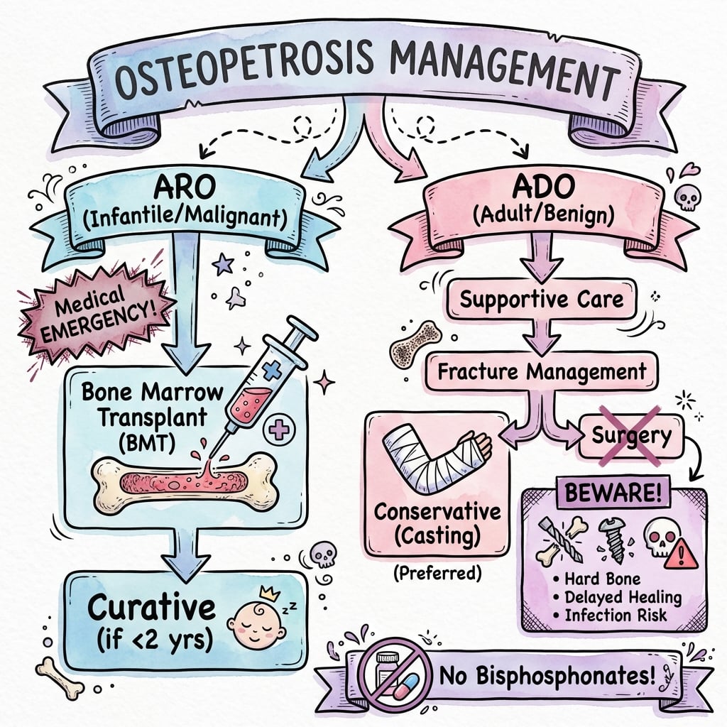
Autosomal Recessive Osteopetrosis (ARO):
Bone Marrow Transplant (BMT) / Hematopoietic Stem Cell Transplant:
- ONLY curative treatment for most forms of ARO
- Donor osteoclast precursors can form functional osteoclasts
- Best outcomes if performed early in infancy (before irreversible neurological damage)
- International registry 5-year survival: approximately 62% with an HLA-matched sibling donor
- Approximately 42% with alternative (mismatched related or unrelated) donors
- Graft failure is the leading cause of post-transplant death
Indications for BMT:
- Confirmed ARO with functional osteoclast defects
- Severe hematologic involvement
- Progressive disease
Contraindications:
- RANKL/RANK mutations (osteoclast-poor form - donor cells cannot form osteoclasts)
- Established severe neurological damage (may proceed if other organs threatened)
Supportive Care (ARO):
- Transfusions for anemia/thrombocytopenia
- Antibiotics for infections
- Vitamin D and calcium supplementation (if hypocalcemic)
- Interferon-gamma: May enhance osteoclast function
- Corticosteroids: Short-term for pancytopenia (limited role)
Autosomal Dominant Osteopetrosis (ADO):
- No specific medical treatment - supportive care only
- Manage fractures and complications
- Optimize bone health (vitamin D, calcium)
- Avoid bisphosphonates (further impair resorption)
- Dental hygiene to prevent osteomyelitis
No Bisphosphonates
Do NOT give bisphosphonates in osteopetrosis. These drugs inhibit osteoclast function - the exact problem in osteopetrosis. They will worsen the disease.
Surgical Management
Orthopaedic Challenges
Technical Difficulties:
Drilling and Cutting
- Extremely hard cortical bone
- Drill bits break frequently (use fresh, sharp bits)
- Prolonged drilling time generates heat
- Saw blades dull rapidly
- Use larger diameter drill bits (less likely to break)
- Intermittent drilling with irrigation to prevent thermal necrosis
Fixation Problems
- Screws strip easily in abnormal bone
- Plates may not seat properly on irregular surface
- Consider locked plates (angle-stable, less reliance on bone quality)
- IM nails difficult to insert (obliterated canal)
- May need to ream canal if attempting nailing
Fracture Healing:
- Delayed union very common (no bone remodeling)
- Nonunion markedly more common than in normal bone
- Callus forms but may not mature
- Fractures may heal to some degree if aligned and immobilized
Treatment Options:
Conservative:
- Strongly consider for non-displaced fractures
- Cast immobilization for extended periods (3-6 months)
- May achieve union with patience
- Avoids surgical complications
Surgical Indications:
- Displaced fractures
- Failure of conservative treatment
- Femoral neck fractures (high nonunion risk)
- Open fractures
Surgical Technique Tips:
- Use new, sharp instruments (change drill bits frequently)
- Large diameter drills (3.2mm or larger if possible)
- Intermittent drilling with saline irrigation
- Longer screws with larger diameter if bone allows
- Locked plating preferred (less torque on screws)
- Consider external fixation (avoids screw placement in dense bone)
- Bone grafting may help (autograft preferred)
- Prolonged immobilization postoperatively
Complications
Disease-Related Complications
Complications by System
| System | ARO Complications | ADO Complications |
|---|---|---|
| Severe pancytopenia, need for transfusions, infections | Usually none or mild anemia | |
| Blindness (50-80%), deafness, facial palsy, hydrocephalus | Rare cranial nerve involvement | |
| Fractures, growth retardation, skeletal fragility | Fractures (main problem), delayed healing | |
| Delayed eruption, abscesses, mandibular osteomyelitis | Osteomyelitis after dental work | |
| Hepatosplenomegaly, extramedullary hematopoiesis | None |
Surgical Complications
Early Complications:
- Intraoperative fracture: Brittle bone fractures during manipulation
- Drill bit breakage: Bits left in bone (usually left in situ)
- Prolonged operative time: Increased infection risk
- Bleeding: May be significant from abnormal bone
- Thermal necrosis: From prolonged drilling
Late Complications:
- Delayed union (very common - expect as norm)
- Nonunion (markedly more common than in normal bone)
- Implant failure: Screw loosening, plate breakage
- Refracture: After implant removal or adjacent to hardware
- Infection: Increased susceptibility, difficult to eradicate
BMT Complications
- Graft-versus-host disease (GVHD)
- Graft failure
- Infection (during immunocompromised period)
- Veno-occlusive disease
- Late effects of conditioning chemotherapy
Evidence Base
- Authoritative review of osteopetrosis pathophysiology and classification
- Osteoclast development vs function defects distinguished as the unifying mechanism
- Proton pump (TCIRG1), chloride channel (CLCN7) and RANK/RANKL pathways mapped to phenotypes
- Haematopoietic stem cell transplantation framed as the only cure for osteoclast-rich infantile disease
- ARO incidence 1 in 250,000 births; ADO incidence 1 in 20,000 births
- At least 10 causative genes account for roughly 70% of all cases
- Severe infantile forms cause death in the first decade if untreated; adult-onset forms have normal life expectancy
- Diagnosis is clinical and radiographic, confirmed by gene testing where applicable
Viva Scenarios
Use these scenarios to practise clinical reasoning and management decisions
Infant with Osteopetrosis
"You are asked to see a 6-month-old infant diagnosed with autosomal recessive osteopetrosis. The child has pancytopenia, hepatosplenomegaly, and progressive vision loss. How would you manage this patient?"
This infant has **malignant infantile osteopetrosis (ARO)**, a life-threatening condition. My priorities are:
**Immediate Management:**
- Stabilize hematologically - transfusions for anemia, platelets if bleeding
- Treat/prevent infections aggressively - these children are immunocompromised
- Urgent ophthalmology assessment - document vision status
**Definitive Treatment:**
- **Bone marrow transplant is the only curative option** and should be pursued urgently
- HLA typing of patient and family for donor matching
- Best outcomes when transplanted early in infancy, before established neurological damage
- Confirm genetic mutation to ensure BMT eligibility (RANKL mutations won't respond)
**Supportive Care:**
- Consider interferon-gamma as bridge to BMT
- Vitamin D and calcium if hypocalcemic
- Refer to specialist center with BMT experience
Without BMT, prognosis is poor - most die by age 10 from infections, bleeding, or anemia.
Adult Femoral Fracture
"A 35-year-old man with known benign adult osteopetrosis (ADO Type II) presents with a displaced mid-shaft femoral fracture after a fall. How would you manage this fracture?"
This patient has a displaced femoral shaft fracture in the setting of **benign adult osteopetrosis**. This presents significant surgical challenges.
**Initial Assessment:**
- Standard trauma assessment - rule out other injuries
- Neurovascular exam of affected limb
- Blood tests including FBC (usually normal in ADO)
- Full-length femur X-rays
**Treatment Considerations:**
- Displaced femoral shaft fracture requires surgical fixation
- **IM nailing**: Preferred in normal patients but challenging here - medullary canal may be obliterated
- **Plate fixation**: More reliable in osteopetrosis - use locked plates, large diameter screws
- **External fixation**: Consider if internal fixation fails or as definitive treatment
**Surgical Technique:**
- Use fresh, sharp drill bits - change frequently (they will break)
- Use larger diameter drills (less likely to break)
- Intermittent drilling with irrigation (prevent thermal necrosis)
- Locked plating to maximize fixation
- Have external fixator available as backup
**Postoperative:**
- Expect **delayed union** - may take 4-6 months
- Prolonged protected weight-bearing
- Serial X-rays to monitor healing
- Counsel patient about the substantially increased delayed-union and nonunion risk
Radiographic Diagnosis
"You are shown X-rays of the lumbar spine and distal femur showing diffuse sclerosis, 'sandwich vertebrae', and flaring of the distal femoral metaphysis. What is the diagnosis and what are the key radiographic features of this condition?"
These radiographic findings are pathognomonic for **osteopetrosis** (marble bone disease).
**Classic Radiographic Features:**
- **Sandwich vertebrae** (rugger jersey spine): Sclerotic endplates with relatively lucent center. The alternating bands of sclerosis and lucency resemble a rugby jersey stripe pattern.
- **Erlenmeyer flask deformity**: Flared metaphyses, especially at the distal femur. Due to failure of normal bone modeling/tubulation (requires osteoclast resorption).
- **Bone-in-bone appearance**: Endobone phenomenon where a ghost outline of previous bone is visible within current bone structure. Seen in vertebrae, phalanges, iliac wings.
- **Diffuse osteosclerosis**: Generalized increased bone density throughout the skeleton.
**Other Features:**
- Skull: Thickened base, obliterated sinuses
- Long bones: Loss of corticomedullary distinction, transverse fractures
- Pelvis: Dense iliac wings with bone-in-bone
**Important Correlation:**
Despite the dense appearance, the bone is **structurally weak**. The retained calcified cartilage and abnormal architecture make it paradoxically fragile - it breaks like chalk despite looking strong on X-ray.
Guidelines, Registries & Global Practice
Global Epidemiology
| Parameter | Figure | Source population |
|---|---|---|
| ARO (malignant infantile) | approximately 1 in 250,000 births | Worldwide; higher in consanguineous populations |
| ADO (benign adult) | approximately 1 in 20,000 births | Most common form overall |
| Genetic yield | approximately 70% of cases explained by 10+ genes | International cohorts |
| Regional clusters | Costa Rica, Middle East, parts of Northern Europe | Founder and consanguinity effects |
There is no high-level (RCT) guidance for this rare disease; practice rests on registry data and expert consensus.
Consensus Positions Across Societies
| Issue | Working consensus | Comment |
|---|---|---|
| Definitive cure for osteoclast-rich ARO | Allogeneic HSCT | Best survival with HLA-matched sibling; refer in infancy |
| Genotype before transplant | Mandatory | RANKL/TNFSF11 (osteoclast-poor) forms are NOT cured by HSCT |
| Bisphosphonates and denosumab as therapy | Contraindicated as disease treatment | They further suppress osteoclasts; denosumab studied only for post-HSCT hypercalcaemia |
| Fracture fixation | Anticipate hard, brittle bone | Locked plating favoured; external fixation as backup |
| Surveillance | Vision, hearing, FBC, dental | Optic-canal decompression considered before established atrophy |
Registries and Networks
HSCT outcomes are pooled through international transplant registries (CIBMTR in North America, EBMT in Europe), which together generated the largest osteopetrosis transplant cohort to date. Unrelated-donor matching relies on national and international marrow donor registries. Rare-bone-disease reference networks (for example ERN-BOND in Europe) coordinate diagnosis, genetic confirmation and multidisciplinary care.
High- vs Limited-Resource Practice Variation
- Well-resourced settings: next-generation sequencing gene panels, early HSCT in infancy, skull-base and ophthalmology services, and locked-plate plus external-fixation inventories for fracture surgery.
- Limited-resource settings: diagnosis is often clinical and radiographic; transplant access may be delayed or unavailable, shifting care toward transfusion support, infection control and conservative fracture management. Consanguinity raises ARO incidence, so genetic counselling and antenatal diagnosis carry high value where available.
OSTEOPETROSIS
Clinical summary
PATHOPHYSIOLOGY
- •Osteoclast DYSFUNCTION (not absent)
- •Failed bone RESORPTION
- •Dense but BRITTLE bone
- •Medullary canal OBLITERATED
TYPES
- •ARO: Autosomal recessive, infantile, FATAL without BMT
- •ADO Type II: Most common, benign, adult, CLCN7
- •ADO Type I: Rare, cranial vault involvement
- •Intermediate: Variable, childhood onset
RADIOGRAPHIC FEATURES
- •Sandwich vertebrae (rugger jersey)
- •Erlenmeyer flask (flared metaphyses)
- •Bone-in-bone (endobone)
- •Diffuse osteosclerosis
ARO COMPLICATIONS
- •Pancytopenia (marrow obliteration)
- •Blindness (optic canal stenosis)
- •Hepatosplenomegaly (extramedullary hematopoiesis)
- •Death by age 10 without BMT
GENES
- •TCIRG1: 50% of ARO (proton pump)
- •CLCN7: ARO and ADO (chloride channel)
- •RANKL: Osteoclast-poor (no BMT benefit)
- •CA2: With renal tubular acidosis
SURGICAL CHALLENGES
- •Drill bits BREAK - use large diameter, change often
- •Screws STRIP - use locked plates
- •Healing DELAYED - high delayed-union and nonunion risk
- •Consider EXTERNAL FIXATION as alternative
TREATMENT
- •ARO: BMT curative (early, before age 2)
- •ADO: Supportive, fracture management
- •NO bisphosphonates (worsen disease)
- •Interferon-gamma (bridge therapy)
Differential Diagnosis
Sclerosing Bone Disorders
| Condition | Key Differentiator | Genetics |
|---|---|---|
| Osteopetrosis | Osteoclast dysfunction, Erlenmeyer flask, sandwich vertebrae | TCIRG1, CLCN7 |
| Pyknodysostosis | Short stature, open fontanelles, acro-osteolysis, mandible hypoplasia | CTSK (cathepsin K) |
| Engelmann Disease | Diaphyseal involvement, limb pain, symmetric long bone sclerosis | TGFB1 |
| Melorheostosis | Dripping candle wax appearance, dermatomal distribution | LEMD3 (mosaic) |
| Osteopoikilosis | Spotted bones, asymptomatic, bone islands | LEMD3 |
| Sclerotic Metastases | Prostate/breast cancer history, focal lesions | Acquired |
Key Distinguishing Points:
- Pyknodysostosis: Toulouse-Lautrec had this; characterized by short stature, fragile bones, but with ACRO-OSTEOLYSIS (absent in osteopetrosis) and open fontanelles
- Engelmann Disease: Affects diaphyses primarily, causes pain and weakness, autosomal dominant
- Melorheostosis: Unilateral, follows sclerotome/dermatomal pattern, looks like dripping candle wax
Controversies & Areas of Uncertainty
The rarity of osteopetrosis means most management rests on registries and expert opinion rather than randomised trials. Several genuine areas of debate persist:
-
Optimal timing and conditioning for HSCT. Earlier transplantation in infancy is associated with better outcomes, but the ideal age threshold, conditioning regimen, and donor hierarchy beyond matched siblings remain unsettled. Graft failure and hepatic/pulmonary toxicity are the dominant limitations rather than the transplant decision itself.
-
Reversibility of neurosensory deficits. Whether established optic atrophy or hearing loss can be salvaged by transplant or surgical decompression is uncertain; most evidence suggests vision rarely improves once atrophy is fixed, which is why decompression is considered only before established damage.
-
Role of optic-canal decompression. Indications and benefit are inconsistent across centres, and it is reserved for selected, progressive cases rather than offered routinely.
-
Pharmacological alternatives to transplant. Interferon-gamma-1b offers only modest, mainly supportive benefit; recombinant RANKL for osteoclast-poor (TNFSF11) disease and gene therapy for TCIRG1/CLCN7 remain investigational, with limited human data.
-
Denosumab in osteopetrosis. As a disease therapy it is contraindicated (it suppresses osteoclasts), yet it has been explored narrowly for transplant-related hypercalcaemia - a nuance that is easy to misstate.
-
Fracture fixation strategy. There is no consensus trial comparing plating, intramedullary fixation, and external fixation; choice is individualised, and the often-quoted precise nonunion percentages are extrapolated from small series rather than robust cohort data.
Self-Assessment Questions
Q1: Pathophysiology
What is the fundamental defect in osteopetrosis?
A: Osteoclast dysfunction leading to failure of bone resorption. The osteoclasts are present (in most forms) but cannot resorb bone effectively due to defects in proton pump (TCIRG1), chloride channel (CLCN7), or carbonic anhydrase (CA2). This leads to accumulation of calcified cartilage and primary spongiosa.
Q2: Genetics
Which gene is most commonly mutated in autosomal recessive osteopetrosis?
A: TCIRG1 (50% of ARO cases). This gene encodes the a3 subunit of the vacuolar H+-ATPase (proton pump) essential for acidification of the resorption lacuna.
Q3: Radiology
Name three classic radiographic features of osteopetrosis.
A: Sandwich vertebrae (rugger jersey spine - sclerotic endplates with lucent center), Erlenmeyer flask deformity (flared metaphyses from failed tubulation), and bone-in-bone appearance (endobone phenomenon).
Q4: Treatment
Why is bone marrow transplant curative for most forms of ARO?
A: Osteoclasts are derived from hematopoietic stem cells. Donor HSCs can differentiate into functional osteoclasts that restore bone resorption. However, RANKL-deficient forms do NOT respond because osteoclast precursors cannot differentiate without RANKL signal.
Q5: Surgical
Why are delayed union and nonunion common after fractures in osteopetrosis?
A: Fracture healing depends on coordinated bone remodelling, which requires functional osteoclasts. In osteopetrosis the osteoclast defect abolishes normal remodelling, so callus forms but cannot be reorganised into mature lamellar bone. The result is a markedly elevated rate of delayed union and nonunion compared with normal bone, alongside the technical difficulty of fixation in dense, brittle bone.
Q6: Contraindication
Why are bisphosphonates absolutely contraindicated in osteopetrosis?
A: Bisphosphonates inhibit osteoclast function - the exact problem in osteopetrosis. Administration would further impair bone resorption and worsen the disease.