Radiation Safety, Modality Selection & the Growing Skeleton
Ultrasound: DDH (less than 6mo), joint effusion, soft tissue, guided aspiration — NO radiation
Radiograph: Fracture screening, alignment, ossification centre assessment — LOW radiation
MRI: Complex fractures (physeal), infection, tumour, cartilage — NO radiation
CT: Minimise in children — use ONLY for complex fractures, spinal trauma, or tumour characterisation
Fluoroscopy: Intraoperative only — mandatory ALARA (pulse mode, collimation, shielding)
Key: Non-ionising modalities (USS, MRI) first. CT only when absolutely essential and with paediatric protocols.
- Children are 3-5 times more radiosensitive than adults due to rapidly dividing cells and longer remaining lifespan for stochastic effects to manifest.
- ALARA principle is PARAMOUNT in paediatric imaging: use non-ionising modalities (USS, MRI) first whenever possible.
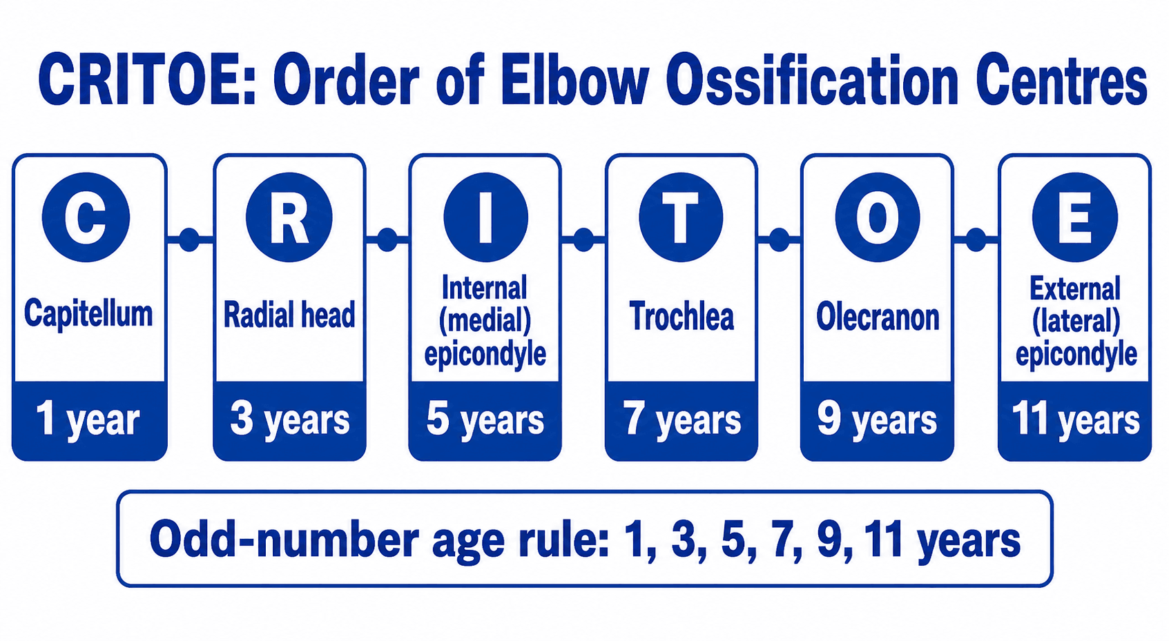
- Ossification centres appear sequentially: CRITOE for the elbow, specific timetables for the hip, knee, and wrist.
- The growing skeleton creates unique imaging challenges: unfused growth plates mimic fractures, ossification centres mimic avulsion fractures, and cartilaginous structures are radiolucent.
- Ultrasound is the preferred first-line investigation for many paediatric conditions: DDH (before 6 months), joint effusion, soft tissue masses, and guided procedures.
- “CRITOE: Capitellum (1yr), Radial head (3yr), Internal (medial) epicondyle (5yr), Trochlea (7yr), Olecranon (9yr), External (lateral) epicondyle (11yr) — the order of elbow ossification centre appearance.
- “The trapped medial epicondyle: following an elbow dislocation, the medial epicondyle avulsion may be trapped within the joint mimicking the trochlea. Compare with the contralateral elbow and check CRITOE sequence — if the trochlea appears BEFORE the medial epicondyle, it is a displaced medial epicondyle.
- “Image Gently campaign principles: reduce dose (lower kVp, mAs), use appropriate collimation (include only what is necessary), and scan once (avoid repeat/unnecessary imaging).
- “Remodelling potential: greatest in younger children, closer to the physis, and in the plane of motion of the adjacent joint. Angular deformity opposite the direction of joint motion has the LEAST remodelling potential.
- “Greenstick and torus (buckle) fractures are unique to children due to the more porous, elastic nature of the paediatric cortex.
Paediatric imaging principles are frequently tested in fellowship examinations. You must know: why children are more radiosensitive (dividing cells, longer lifespan, smaller body = higher organ doses), ALARA implementation, CRITOE ossification centre sequence, the trapped medial epicondyle pitfall, age-appropriate imaging selection (USS for DDH, MRI for complex injuries), and the unique fracture patterns of the paediatric skeleton (greenstick, torus, physeal). Classic traps: ordering CT instead of MRI for paediatric assessment, not knowing CRITOE, and confusing ossification centres with fractures.
CRITOEElbow Ossification Centre Sequence
Hook:CRITOE: 1-3-5-7-9-11 years. If the trochlea appears 'before' the medial epicondyle — the medial epicondyle is TRAPPED in the joint.
CHILDWhy Children Are More Radiosensitive
Hook:CHILD: why we must be extra careful — Children have dividing cells, Higher doses per mAs, Immature DNA repair, Longer lifespan, and Dose accumulation.
PGTBSPaediatric Fracture Patterns
Hook:PGTBS: Plastic deformation, Greenstick, Torus, Bow, Salter-Harris — the spectrum of paediatric fracture patterns.
Overview
Paediatric imaging requires a fundamentally different approach from adult imaging. Three principles underpin all paediatric imaging decisions: (1) radiation safety — children are 3-5 times more radiosensitive than adults and have a longer remaining lifespan for stochastic effects to manifest; (2) the growing skeleton — cartilaginous structures are radiolucent, ossification centres appear sequentially and can mimic pathology, and growth plates create unique injury patterns; (3) clinical context — children cannot reliably describe symptoms, examination findings may be non-specific, and the differential diagnosis for musculoskeletal complaints is different from adults.
The ALARA principle (As Low As Reasonably Achievable) is paramount in paediatric imaging. Practical implementation: (1) Always consider whether imaging is necessary — clinical assessment alone may suffice (Ottawa ankle rules apply from age 6). (2) Use non-ionising modalities FIRST (USS for soft tissue, MRI for complex assessment). (3) If ionising imaging is needed, use PAEDIATRIC PROTOCOLS with reduced kVp and mAs. (4) COLLIMATE tightly — include ONLY the anatomy needed (avoid whole-body scatter). (5) Shield radiosensitive organs (gonads, thyroid) when they are in or near the beam. (6) Scan ONCE — avoid unnecessary repeat imaging. The Image Gently campaign provides protocols for paediatric dose reduction.
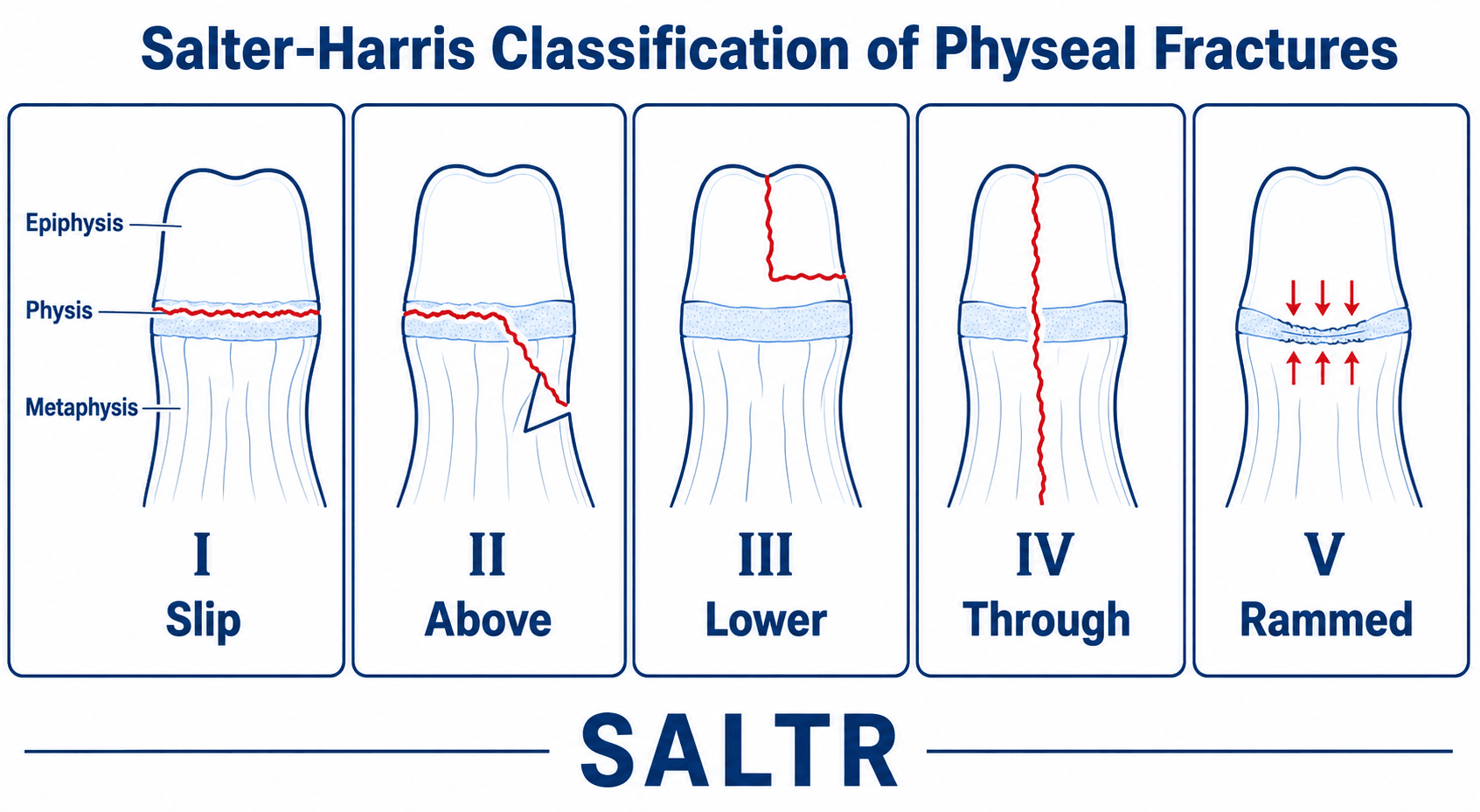
Key imaging challenges: (1) Ossification centres: appear at predictable ages but can be confused with fracture fragments. The most critical example is the TRAPPED MEDIAL EPICONDYLE in elbow dislocation — compare with CRITOE sequence and obtain comparison views of the uninjured side. (2) Growth plates: the physis is the weakest link (weaker than ligaments) — what would be a ligament injury in an adult is a physeal fracture in a child. Salter-Harris Type I may have a NORMAL radiograph. (3) Remodelling: children can correct angular deformity through growth. Greatest potential in younger children, near the physis, in the plane of motion. (4) Non-ossified cartilage: articular cartilage and epiphyseal cartilage are radiolucent — MRI or USS may be needed to evaluate these structures.
Systematic Approach
Paediatric Imaging Modality Selection
- Preferred Imaging
- Ultrasound (hip USS, Graf classification)
- Rationale
- Femoral head not yet ossified. USS shows cartilaginous anatomy. No radiation
- Preferred Imaging
- AP pelvis radiograph (Perkins, Hilgenreiner lines)
- Rationale
- Femoral head ossification centre now visible. Radiograph is standard
- Preferred Imaging
- AP pelvis + frog lateral radiograph. Blood tests (CRP, FBC)
- Rationale
- Differential: irritable hip vs Perthes vs septic arthritis. USS if effusion suspected
- Preferred Imaging
- AP + lateral elbow radiograph. Comparison views if needed
- Rationale
- Assess fat pads, anterior humeral line, CRITOE centres. Compare with contralateral side
- Preferred Imaging
- MRI (if management would change)
- Rationale
- Salter-Harris I may have normal radiographs. MRI shows physeal oedema and confirms the diagnosis
- Preferred Imaging
- Ultrasound (detects effusion + guides aspiration)
- Rationale
- NO radiation. Real-time guided aspiration for diagnostic synovial fluid analysis
- Preferred Imaging
- Skeletal survey (full-body radiograph series)
- Rationale
- Standardised protocol: AP/lateral skull, AP chest, AP abdomen, AP all limbs. Follow-up repeat survey at 2 weeks
- Preferred Imaging
- MRI (NO radiation, excellent soft tissue contrast)
- Rationale
- Avoids CT radiation. Shows cartilaginous structures, physeal involvement, marrow pathology
Differential: Normal Variant versus Pathology
The single most common interpretive error in paediatric imaging is mistaking a normal developmental appearance for pathology (or vice versa). Use this differential whenever a finding looks abnormal in a child.
- Benign developmental explanation
- Physis (growth plate) — smooth, regular, expected location
- True pathology to exclude
- Salter-Harris fracture — irregular widening, metaphyseal/epiphyseal extension
- Discriminator
- Compare with contralateral side; physis is symmetric and smooth
- Benign developmental explanation
- Ossification centre appearing on schedule (CRITOE order)
- True pathology to exclude
- Avulsion fracture — fragment displaced, surrounding soft-tissue swelling
- Discriminator
- Check CRITOE order; an out-of-sequence centre means a fragment, not a centre
- Benign developmental explanation
- Normal variant ossification (e.g. distal femoral, calcaneal apophysis)
- True pathology to exclude
- Osteochondritis/Perthes — sclerosis, collapse, fragmentation with symptoms
- Discriminator
- Bilateral and asymptomatic favours variant; unilateral and painful favours disease
- Benign developmental explanation
- Normal metaphyseal step or developmental cortical irregularity
- True pathology to exclude
- Torus (buckle) fracture — focal buckle with point tenderness and mechanism
- Discriminator
- Clinical tenderness and mechanism; comparison view of the other limb
- Benign developmental explanation
- Physiological bowing of infancy/toddler
- True pathology to exclude
- Plastic deformation fracture or rickets/dysplasia
- Discriminator
- Age, symmetry, metaphyseal cupping/fraying (rickets) vs trauma history
- Benign developmental explanation
- —
- True pathology to exclude
- Non-accidental injury — fractures of varying ages, classic metaphyseal lesions, rib fractures
- Discriminator
- Always consider NAI; correlate with history, perform skeletal survey, safeguard
Controversies & Areas of Uncertainty
- Universal versus selective DDH ultrasound screening. Universal Graf ultrasound (used in parts of Europe) detects more dysplasia but increases over-treatment and cost; selective screening (US/UK) may miss some late-presenting cases. No high-quality trial has resolved which strategy improves long-term hip outcomes, so practice remains region-dependent.
- Magnitude of low-dose CT cancer risk. Cohort data (Pearce, Mathews) show dose-response associations, but reverse causation (scanning sicker children) and the validity of the linear-no-threshold model at low doses are debated. The pragmatic consensus is to optimise and justify rather than to refuse necessary CT.
- Routine comparison radiographs of the uninjured limb. Long taught for the elbow, comparison views add radiation and often little value when a structured CRITOE/line analysis is applied; many paediatric centres now reserve them for genuinely equivocal cases.
- Point-of-care ultrasound for fractures. Evidence for USS in occult elbow and for distal forearm fractures is growing but mostly single-centre and operator-dependent; it is an adjunct, not yet a replacement for radiographs in most guidelines.
Clinical Applications
Ossification Centre Assessment
Understanding the sequence and timing of ossification centre appearance is fundamental to paediatric imaging interpretation. The key clinical relevance is distinguishing NORMAL ossification centres from fracture fragments.
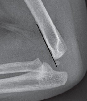
Elbow (CRITOE): The most commonly tested ossification centre sequence. Capitellum (1yr), Radial head (3yr), Internal (medial) epicondyle (5yr), Trochlea (7yr), Olecranon (9yr), External (lateral) epicondyle (11yr). The critical clinical application is the TRAPPED MEDIAL EPICONDYLE: following a paediatric elbow dislocation, the medial epicondyle (which has been avulsed by the ulnar collateral ligament) can become trapped within the joint. On the post-reduction radiograph, it may be misinterpreted as the trochlea. KEY RULE: if the trochlea is 'present' but the medial epicondyle is NOT visible in its normal position, the medial epicondyle is trapped in the joint and requires open surgical removal.
Hip: The femoral head ossification centre appears at 3-6 months. Its absence before this age means that DDH assessment requires ultrasound (radiographs cannot visualise the cartilaginous femoral head). After 6 months, the AP pelvis radiograph using Perkins and Hilgenreiner lines becomes the standard assessment tool.
Wrist: The carpal ossification centres appear in a roughly circular sequence — the capitate (1-3 months) and hamate (2-4 months) appear first. The pisiform is the last carpal bone to ossify (approximately 10-12 years). The distal radial epiphysis ossification centre appears at approximately 1 year.
Comparison views: When uncertain whether an ossification centre is normal or a fracture fragment, obtain comparison views of the contralateral (uninjured) side. Both limbs should show symmetric ossification patterns.
Guidelines, Registries & Global Practice
Paediatric imaging is governed by convergent international frameworks. The unifying principle worldwide is justification plus optimisation (ALARA/ALARP) under the EURATOM Basic Safety Standards and ICRP recommendations, operationalised through diagnostic reference levels (DRLs) and child-sized protocols.
Global epidemiology and burden. Paediatric CT use rose sharply in the 1990s-2000s then plateaued/declined after the dose-risk evidence emerged (Miglioretti 2013; Mathews 2013). Effective dose per paediatric CT varies more than 1000-fold between scans and institutions, so the dominant driver of population radiation risk is unoptimised technique rather than the number of scans. DDH affects roughly 1-3 per 1000 live births for true dislocation, with higher rates for instability/dysplasia, and is more common in girls, breech presentation, and those with a positive family history — driving the global debate over universal versus selective ultrasound screening.
- AAOS / ACR (US)
- ACR Appropriateness Criteria + Image Gently; justify and optimise every study
- BOA-BOAST / NICE (UK)
- IR(ME)R regulations; clinician must justify each exposure; NICE imaging guidance
- AO / EFORT (Europe)
- EURATOM Basic Safety Standards; EuroSafe Imaging; national DRLs
- AAOS / ACR (US)
- AAOS guideline: selective USS for risk factors (breech, family history, clinical instability); no universal USS
- BOA-BOAST / NICE (UK)
- UK NIPE: clinical screening at birth and 6-8 weeks plus selective USS for risk factors
- AO / EFORT (Europe)
- Several European systems (e.g. Germany, Austria) use universal Graf USS screening
- AAOS / ACR (US)
- Radiograph; treat positive fat pad as occult fracture; USS emerging adjunct
- BOA-BOAST / NICE (UK)
- Radiograph plus splint-and-review at 7-14 days; comparison views if needed
- AO / EFORT (Europe)
- Radiograph with CRITOE assessment; USS used in some centres (Burnier)
- AAOS / ACR (US)
- Prefer MRI where it answers the question; reserve CT for cortical/operative planning
- BOA-BOAST / NICE (UK)
- MRI preferred for physeal and soft-tissue questions to avoid dose
- AO / EFORT (Europe)
- MRI-first for cartilage/physis; CT for fracture geometry and 3D planning
Registries and audit. There is no single global paediatric imaging registry, but dose audit is registry-like: national DRL surveys (e.g. UK PHE/HPA, US ACR Dose Index Registry, EU EUCLID project) benchmark paediatric doses by age/weight band, and paediatric DRLs are typically 50-70% lower than adult values for the same examination. For DDH and implant-related paediatric work, arthroplasty registries (NJR, AOANJRR, SHAR) inform downstream practice but do not capture screening imaging.
High- versus limited-resource practice. In well-resourced settings, MRI and high-quality ultrasound are widely available, enabling radiation-free pathways (USS-first for DDH and effusion, MRI for physeal/marrow/tumour questions). In limited-resource settings, MRI access, sedation capacity, and trained paediatric sonographers may be scarce, so radiographs and clinical decision rules carry more weight; here the priorities shift to avoiding unnecessary radiographs, using comparison views judiciously, and reserving CT for clearly decisive indications.
Clinical Decision Scenarios
Practise clinical reasoning and management decisions out loud
“A 7-year-old boy falls off monkey bars and presents with elbow pain and swelling. The lateral elbow radiograph shows a posterior fat pad sign but no visible fracture.”
“An examiner asks you why radiation dose management is particularly important in children and what principles you would apply.”
“A 4-year-old has sustained an elbow dislocation. The post-reduction radiograph appears satisfactory, but you notice that the trochlea ossification centre appears to be present while the medial epicondyle is not visible.”
Radiation Safety (CHILD)
- Children are 3-5x more radiosensitive than adults
- ALARA: justify, use non-ionising modalities first, paediatric protocols, collimate, shield
- USS and MRI preferred over CT whenever possible
- Image Gently: child-size-specific protocols reduce dose by 20-50%
- Pearce et al. 2012: direct evidence linking childhood CT to cancer risk
CRITOE (Elbow Ossification)
- C: Capitellum (1yr), R: Radial head (3yr), I: Internal/medial epicondyle (5yr)
- T: Trochlea (7yr), O: Olecranon (9yr), E: External/lateral epicondyle (11yr)
- CRITICAL: trochlea ALWAYS after medial epicondyle — if trochlea without M.E. = TRAPPED
- Comparison views essential when in doubt about normal vs pathological
- Posterior fat pad sign = occult fracture in trauma
Modality Selection
- DDH: USS before 6 months (Graf), radiograph after 6 months (Perkins/Hilgenreiner)
- Joint effusion/septic arthritis: USS (no radiation, guides aspiration)
- Complex fracture/physeal injury: MRI (no radiation, shows cartilage and physis)
- NAI: skeletal survey (standardised protocol, repeat at 2 weeks)
- CT: ONLY when essential (complex fractures, spinal trauma, tumour)
Paediatric Fracture Patterns
- Plastic deformation: bowed bone, no fracture line (ulna/fibula)
- Torus (buckle): cortical compression failure, subtle bump (distal radius)
- Greenstick: one cortex fractured, opposite bends
- Salter-Harris: I (normal X-ray), II (most common), III-IV (ORIF), V (retrospective)
- Remodelling: best in younger children, near physis, in plane of motion
Evidence Base
Childhood CT and Subsequent Leukaemia and Brain Tumour Risk
- Retrospective cohort of 178,604 patients (leukaemia analysis) and 176,587 (brain tumour analysis) first scanned under age 22 in Great Britain (1985-2002).
- Cumulative red-marrow doses of about 30 mGy or more (mean 51 mGy) gave a relative risk of leukaemia of 3.18 (95% CI 1.46-6.94) versus under 5 mGy.
- Cumulative brain doses of 50-74 mGy (mean 60 mGy) gave a relative risk of brain tumour of 2.82 (95% CI 1.33-6.03).
Pediatric CT Use, Dose and Projected Cancer Risk
- Across seven US health systems, paediatric CT use roughly doubled (under-5s) to tripled (5-14y) between 1996 and 2005 before declining.
- Effective dose per scan varied enormously (0.03 to 69.2 mSv), showing wide, often unjustified, dose variability.
- An estimated 4 million annual paediatric CTs project to about 4,870 future cancers; reducing the highest-quartile doses to the median could prevent roughly 43% of these.
Image Gently Campaign — Optimising Paediatric Dose
- Promotes child-size-specific CT protocols: reduce kVp/mAs for body size, scan only the indicated region, and avoid multiphase scanning.
- Core slogan: image gently — child-size the dose, scan only when needed, and scan only the indicated area once.
- Adopted internationally and complemented by Image Wisely (adults) and the EuroSafe/EURATOM dose-optimisation frameworks.
Cancer Risk After Childhood CT — 11 Million Australians
- Population data-linkage cohort of 10.9 million people; 680,211 exposed to CT under age 20 (mean follow-up 9.5 years; mean dose 4.5 mSv/scan).
- Overall cancer incidence was 24% higher in exposed individuals (IRR 1.24, 95% CI 1.20-1.29), rising by 0.16 per additional CT scan.
- Risk was greater with younger age at exposure, spanning solid cancers, leukaemia and myelodysplasia (absolute excess 9.38 cancers per 100,000 person-years).
Direct cohort evidence (Pearce, Miglioretti, Mathews) plus society campaigns strongly support radiation dose minimisation and protocol optimisation in children.