Seronegative Spondyloarthropathy | DIP Predominance | CASPAR Criteria
- Dactylitis (sausage digit) pathognomonic - tenosynovitis plus joint inflammation
- Nail changes (pitting, onycholysis) correlate with DIP arthritis severity
- Pencil-in-cup deformity on XR is pathognomonic radiographic finding
- Arthrodesis preferred over arthroplasty due to poor bone quality
- Hold biologics 2-4 weeks preoperatively (coordinate with rheumatology)
- “Differentiate from RA: DIP involvement, asymmetry, radiographic periostitis
- “CASPAR criteria: psoriasis + nail + negative RF + dactylitis + juxta-articular bone = 3 or more
- “Synovectomy has limited role compared to RA due to aggressive bone erosion
- “Ray resection most reliable procedure for arthritis mutilans
Overview
Psoriatic arthritis (PsA) is a chronic inflammatory arthropathy affecting 5 to 10 percent of patients with psoriasis. Hand involvement occurs in approximately 70 percent of PsA patients and often determines functional capacity and quality of life. The disease exhibits unique patterns of joint destruction that differ fundamentally from rheumatoid arthritis, requiring distinct surgical strategies.
The hand surgeon must recognize five distinct clinical patterns, understand the impact of disease-modifying medications on surgical outcomes, and coordinate perioperative management with rheumatology. Surgical decision-making balances aggressive disease progression against modern biologic therapy efficacy.
- Prevalence: 0.3-1% general population
- PsA in psoriasis patients: 5-10%
- Peak onset: 30-50 years
- Male:Female ratio 1:1
- Hand involvement: 70%
- Family history positive: 40%
- HLA-B27 association (spondylitis pattern)
- HLA-Cw6 (skin psoriasis)
- TNF-alpha, IL-17, IL-23 pathways
- Enthesitis primary feature
- Osteoproliferation AND erosion
- Synovitis less prominent than RA
- 47% develop erosive disease within 2 years
- Progressive functional decline without treatment
- Arthritis mutilans in 5% (historically 16%)
- Biologics alter natural history
- Nail involvement predicts DIP arthritis
- Dactylitis predicts worse outcomes
Surgical Indications and Principles
Indications for Surgery
Surgical intervention in PsA serves distinct purposes compared to rheumatoid arthritis. The aggressive bone destruction and less prominent synovitis alter the risk-benefit calculation.
- Pain refractory to medical management (6 months optimized therapy)
- Progressive deformity affecting function
- Tendon rupture or impending rupture
- Joint instability limiting ADLs
- Arthritis mutilans salvage
- Neurologic compromise (rare)
- Active skin psoriasis over surgical field
- Uncontrolled systemic disease
- Recent biologic dose (within half-life)
- Poor medical optimization
- Unrealistic patient expectations
Synovectomy
Unlike rheumatoid arthritis where early synovectomy prevents erosions, PsA synovectomy has limited disease-modifying effect. Bone destruction predominates over synovial inflammation.
- Refractory painful synovitis (6 months medical therapy)
- Accessible joints (MCP, PIP, wrist)
- Minimal radiographic erosion
- Failed corticosteroid injection
- Patient unwilling to consider arthrodesis
- Pain relief in 60-70 percent at 2 years
- Disease progression continues in 50 percent
- Inferior results compared to RA
- Consider adjunct to other procedures
The limited efficacy reflects PsA's primary bone pathology rather than synovial inflammation. Set realistic expectations with patients.
Arthrodesis
Joint fusion represents the gold standard for end-stage PsA arthritis in the hand. The aggressive bone destruction makes arthroplasty less reliable.
Distal Interphalangeal Joint Fusion:
Most common surgical procedure in PsA hand. The correlation between nail psoriasis and DIP arthritis makes this joint particularly vulnerable.
- Painful DIP arthritis refractory to medical therapy
- Progressive deformity (swan-neck developing at PIP)
- Pencil-in-cup deformity
- Nail bed compromise from joint destruction
- Post-traumatic arthritis superimposed
- Position: 5-10 degrees flexion (index), progressive flexion ulnarly
- Bone preparation: Remove all sclerotic bone to bleeding surface
- Fixation: K-wire, headless screw, or tension band
- Bone grafting: Consider if significant bone loss
- K-wire: Simple, inexpensive, requires prolonged protection
- Headless compression screw: Higher union rate, early mobilization
- Tension band wire: Good compression, technically demanding
- External fixator: Salvage for severe bone loss
- Union rate: 85-95% with rigid fixation
- Pain relief: 90-95%
- Satisfaction: 85-90%
- Return to function: 6-8 weeks
The procedure reliably addresses pain and prevents proximal deformity progression. Nail bed involvement may require separate management.
Arthroplasty
Joint replacement in PsA carries higher failure rates than in osteoarthritis due to ongoing inflammation and bone quality issues. Patient selection is critical.
PsA Arthroplasty Contraindications: Active joint inflammation, poor bone quality, arthritis mutilans pattern, ongoing high-dose corticosteroids, and skin psoriasis over surgical field are relative/absolute contraindications. Silicone implants show high failure rates. Modern pyrocarbon implants may improve outcomes but long-term data in PsA is limited.
- Silicone MCP arthroplasty: 30-40% failure at 10 years in PsA (vs 15% in RA)
- PIP arthroplasty: Very limited role, consider fusion instead
- CMC arthroplasty: Acceptable outcomes for thumb base arthritis
- Wrist arthroplasty: Contraindicated in PsA due to bone quality
- Quiescent disease on stable medical therapy
- Adequate bone stock
- Intact soft tissue envelope
- No active skin psoriasis
- Realistic expectations
- Compliance with therapy
Pathophysiology
Inflammatory Pathways
Psoriatic arthritis is driven by dysregulated immune pathways involving TNF-alpha, IL-17, and IL-23. Unlike rheumatoid arthritis where synovitis predominates, enthesitis (inflammation at tendon insertions) is the hallmark of PsA. The close anatomical relationship between the nail matrix and DIP joint explains the strong correlation between nail disease and DIP arthritis.
Key Pathophysiologic Features:
- Enthesitis: Primary pathology at tendon insertions
- Osteoproliferation AND erosion: Unique combination vs pure erosion in RA
- HLA associations: HLA-B27 (spondylitis), HLA-Cw6 (skin)
- Cytokine pathways: TNF-alpha, IL-17, IL-23 targeted by biologics
Classification
Psoriatic arthritis is a seronegative spondyloarthropathy affecting 5-10% of psoriasis patients, with hand involvement in 70%. Diagnosis uses CASPAR criteria (91.4% sensitivity, 98.7% specificity): psoriasis history, nail dystrophy, negative RF, dactylitis, and juxta-articular new bone formation. Classic radiographic finding is "pencil-in-cup" deformity at DIPs. Five clinical patterns exist: SADDS - Symmetric polyarticular (RA-like), Asymmetric oligoarticular (most common), DIP predominant, Destructive (arthritis mutilans), Spondylitis. Dactylitis (sausage digit) is pathognomonic, representing tenosynovitis plus joint inflammation. Nail changes correlate with DIP severity. Unlike RA, synovectomy has limited role due to aggressive bone erosion.
Asymmetric Oligoarticular Pattern
The most common presentation involves fewer than five joints in an asymmetric distribution. Ray pattern involvement is characteristic, where multiple joints in one digit are affected while adjacent digits remain normal. This pattern responds well to local corticosteroid injection and targeted DMARDs.
Clinical Features:
- 1-4 joints affected
- Ray distribution common
- Dactylitis frequent (sausage digit)
- Better functional prognosis
- May progress to polyarticular pattern in 25 percent
Symmetric Polyarticular Pattern
Mimics rheumatoid arthritis but includes DIP joints. This pattern presents the greatest diagnostic challenge and accounts for 30 to 40 percent of PsA cases. Functional impairment parallels rheumatoid arthritis severity.
Distinguishing Features from RA:
- DIP joint involvement
- Asymmetric severity despite symmetric distribution
- Absence of rheumatoid nodules
- Radiographic new bone formation
- Nail changes in 80 percent
DIP Predominant Pattern
Classic psoriatic arthritis pattern with isolated DIP involvement. Strong correlation exists between nail psoriasis and underlying DIP arthritis. This pattern often progresses slowly but can develop severe destructive changes.
Surgical Considerations:
- Arthrodesis gold standard
- Arthroplasty contraindicated in active disease
- Nail bed involvement complicates wound healing
- Mucous cyst association
- Consider MCP or PIP involvement before isolated DIP fusion
Arthritis Mutilans
The most destructive pattern features telescoping digits from severe osteolysis. Modern biologic therapy has reduced prevalence from 16 percent to approximately 5 percent. Surgical reconstruction remains challenging.
Arthritis Mutilans Surgical Principles: Ray resection provides better function than attempted reconstruction with bone grafting. Preserve thumb and radial digits when possible. Assess extensor mechanism integrity before planning reconstruction. Consider wrist arthrodesis as foundation for hand function.
Reconstruction Options:
- Ray resection (most reliable)
- Arthrodesis with bone grafting
- Tendon transfers for extension loss
- Wrist fusion for stability
- Consider prosthetic digits for severe cases
Clinical Presentation
CASPAR Criteria
The Classification Criteria for Psoriatic Arthritis (CASPAR) provides standardized diagnosis with high sensitivity and specificity. A score of 3 or more indicates psoriatic arthritis.
- points
- 2
- definition
- Psoriatic skin or scalp lesions present today
- sensitivity
- High specificity for diagnosis
- points
- 1
- definition
- Patient or first/second degree relative history
- sensitivity
- Expands diagnostic capture when skin clear
- points
- 1
- definition
- Onycholysis, pitting, hyperkeratosis
- sensitivity
- Correlates with DIP arthritis
- points
- 1
- definition
- Seronegative spondyloarthropathy
- sensitivity
- Distinguishes from RA
- points
- 1
- definition
- Sausage digit from tenosynovitis plus arthritis
- sensitivity
- Pathognomonic when present
- points
- 1
- definition
- Juxta-articular new bone formation
- sensitivity
- Distinguishes from RA erosive pattern
Investigations
Radiographic Assessment
Plain radiographs demonstrate characteristic features that distinguish PsA from rheumatoid arthritis. The combination of erosive changes AND new bone formation is pathognomonic.
Grading Radiographic Severity:
- Grade 1: Soft tissue swelling, periarticular osteopenia
- Grade 2: Joint space narrowing, small erosions
- Grade 3: Multiple erosions, subluxation, periostitis
- Grade 4: Arthritis mutilans, telescoping, ankylosis
Laboratory Studies
Unlike rheumatoid arthritis, laboratory findings in PsA are relatively nonspecific. The diagnosis relies primarily on clinical and radiographic features.
Key Laboratory Features:
- Rheumatoid factor: Negative (positive in less than 10 percent)
- Anti-CCP antibodies: Negative
- HLA-B27: Positive in 20 percent (50 percent with spondylitis)
- ESR/CRP: Elevated in 50 percent
- Uric acid: Check to exclude gout
- ANA: May be weakly positive
Differential Diagnosis
The DIP-predominant, asymmetric, seronegative pattern with nail change distinguishes PsA from the other common inflammatory and degenerative hand arthropathies. The single highest-yield discriminator versus rheumatoid arthritis is the combination of DIP involvement and juxta-articular new bone formation.
- distribution
- Asymmetric, ray pattern, DIP involvement, dactylitis
- serology
- RF and anti-CCP negative
- radiographic
- Erosions plus new bone (periostitis, pencil-in-cup, ankylosis)
- clue
- Nail dystrophy, skin plaques, enthesitis
- distribution
- Symmetric, MCP and PIP predominant, DIP usually spared
- serology
- RF and/or anti-CCP positive in most
- radiographic
- Marginal erosions, periarticular osteopenia, no new bone
- clue
- Rheumatoid nodules, ulnar drift, MCP subluxation
- distribution
- DIP (Heberden) and PIP (Bouchard), thumb base, symmetric
- serology
- Seronegative
- radiographic
- Osteophytes, central gull-wing erosions in erosive OA
- clue
- Bony nodes, non-inflammatory, no nail/skin disease
- distribution
- Asymmetric, often first MTP first; tophi at fingers
- serology
- Hyperuricaemia (may be normal in attack)
- radiographic
- Punched-out erosions with overhanging edges, preserved space
- clue
- Tophi, negatively birefringent MSU crystals on aspirate
- distribution
- Asymmetric lower-limb predominant, dactylitis, enthesitis
- serology
- RF negative, HLA-B27 often positive
- radiographic
- Enthesophytes, periostitis, sacroiliitis
- clue
- Antecedent GU/GI infection, no psoriasis
Nail-joint connection (pathoanatomy): the DIP joint and nail matrix share a common entheseal organ - the extensor tendon and collateral ligaments insert immediately adjacent to the nail bed. Inflammation at this enthesis explains why nail dystrophy predicts DIP arthropathy and may precede joint symptoms. Dactylitis ("sausage digit") reflects combined flexor tenosynovitis, joint synovitis and soft-tissue oedema across the whole ray.
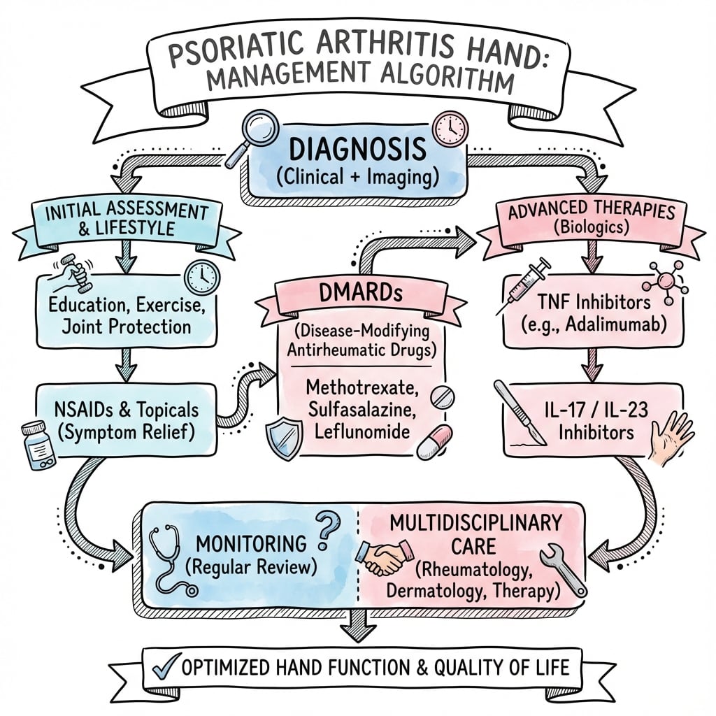
Management
Modern biologic therapy has revolutionized PsA treatment but creates perioperative management challenges. Understanding drug mechanisms and half-lives is essential for surgical planning.
- mechanism
- Folate antagonist DMARD
- halfLife
- 3-15 hours
- perioperativeGuidance
- Continue through surgery - reduces flares without increasing infection
- mechanism
- TNF-alpha inhibitor
- halfLife
- 10-20 days
- perioperativeGuidance
- Hold 2-4 weeks before surgery, resume when wound healed
- mechanism
- TNF-alpha receptor fusion
- halfLife
- 3-5 days
- perioperativeGuidance
- Hold 1 week before, shorter washout than adalimumab
- mechanism
- TNF-alpha antibody
- halfLife
- 8-10 days
- perioperativeGuidance
- Schedule surgery mid-cycle, hold until wound healed
- mechanism
- IL-12/23 inhibitor
- halfLife
- 15-45 days
- perioperativeGuidance
- Hold 4-6 weeks before major procedures
- mechanism
- IL-17A inhibitor
- halfLife
- 22-31 days
- perioperativeGuidance
- Hold 3-4 weeks before surgery, emerging agent
Perioperative Biologic Management: The ACR recommends holding biologics for one dosing interval before elective surgery and resuming when wound healing is adequate (typically 14 days). Methotrexate can continue through surgery. Document rheumatology clearance. Increased infection risk is modest (OR 1.3-1.5) but significant in immunocompromised patients.
Arthritis Mutilans Management
The most severe PsA pattern requires specialized reconstructive approach. Modern biologics have reduced prevalence but surgical salvage remains necessary.
Ray Resection
Removal of severely destroyed rays improves function by eliminating painful unstable digits and narrowing hand for better grip.
- Telescoping digit with bone loss exceeding 50 percent
- Loss of extensor function
- Painful unstable digit interfering with function
- Patient preference after understanding alternatives
- Ray resection at metacarpal base
- Preserve thumb, index, long when possible
- Address adjacent metacarpals to narrow hand
- Reconstruct first web space
- Intrinsic muscle balancing
- Pain relief: 85-90%
- Improved grip: 60-70%
- Patient satisfaction: 70-80%
- Return to ADLs: Variable
Arthrodesis with Bone Grafting
Attempted salvage of ray with structural bone graft and rigid fixation. Technically demanding with unpredictable outcomes.
- Key digit (thumb, index, long)
- Patient motivated for staged reconstruction
- Adequate soft tissue envelope
- Good medical control
- Structural iliac crest graft
- Rigid plate or external fixation
- Prolonged protected mobilization
- Consider BMP augmentation
- Union rate: 60-70% (lower than standard arthrodesis)
- Requires prolonged immobilization
- Staged procedures common
- Better for thumb than fingers
Tendon Problems in Psoriatic Arthritis
Enthesitis represents a hallmark PsA feature affecting tendon insertions. Flexor tenosynovitis contributes to dactylitis.
Flexor Tenosynovitis and Trigger Digits
Inflammatory tenosynovitis causes triggering and contributes to sausage digit appearance. Treatment parallels idiopathic trigger finger but with higher recurrence.
- Diffuse tendon sheath swelling
- Triggering at A1 pulley
- Dactylitis when combined with joint synovitis
- May affect multiple digits
- Optimize systemic therapy
- Corticosteroid injection (60-70% success)
- Splinting
- Activity modification
- Standard A1 pulley release
- Extended tenosynovectomy if needed
- Higher recurrence than idiopathic (20% vs 5%)
- Resume biologics when wound healed
Extensor Tendon Rupture
Less common than in rheumatoid arthritis but occurs from dorsal erosions and synovitis. EDC ruptures most frequent.
- Dorsal wrist synovitis
- Carpal bone erosions
- Lister tubercle destruction
- Failed medical therapy
- EIP to EPL transfer (EPL rupture)
- Side-to-side repair (single EDC)
- Tendon graft (chronic, retracted)
- Address underlying bony prominences
Management Algorithm
Medical therapy is rheumatology-led and disease-modifying; surgery treats the mechanical consequences of established structural damage. Key surgical-planning principles unique to PsA:
- Stage by symptom and biomechanics: address the most symptomatic joint first; DIP fusion often stabilises a secondary PIP swan-neck by restoring extensor balance, so re-assess the PIP before committing to a second fusion.
- Respect the skin: never incise through active plaques; the Koebner phenomenon can flare psoriasis along the wound. Optimise skin disease with dermatology first.
- Coordinate biologic timing with the prescribing rheumatologist - continue methotrexate, withhold biologics over the surgical window, and resume once the wound has healed.
Perioperative Management
Coordinated care with rheumatology optimizes outcomes and minimizes complications. Document comprehensive perioperative plan.
Preoperative Optimization
- Rheumatology clearance documented
- Biologic holding strategy confirmed
- Diabetes control (HbA1c less than 7.5%)
- Nutrition assessment (albumin greater than 3.5 g/dL)
- Smoking cessation 4 weeks minimum
- Active psoriasis over surgical field: Consider treatment delay
- Chronic plaque psoriasis: Acceptable if quiescent
- Pustular psoriasis: Defer surgery
- Document lesions with photographs
- Hold biologics per agent half-life
- Continue methotrexate
- Reduce corticosteroids to less than 10 mg prednisone equivalent
- Resume NSAIDs 24 hours post-op
- Plan biologic resumption (typically 14 days)
Intraoperative Considerations
- Regional anesthesia preferred (shorter PACU time)
- Avoid general anesthesia if C-spine involvement
- Document airway assessment
- Atraumatic tissue handling (friable skin)
- Adequate debridement of devitalized bone
- Rigid fixation for fusions
- Meticulous hemostasis
- Consider negative pressure wound therapy for high-risk wounds
- Standard cefazolin dosing
- Vancomycin if MRSA risk
- Extended prophylaxis not indicated
- Document allergy status
Postoperative Care
- Inspect at 5-7 days (earlier if concerns)
- Negative pressure therapy for high-risk wounds
- Extended antibiotics NOT routine
- Early detection of infection critical
- Protected mobilization per procedure
- Hand therapy when stable
- Gradual strengthening
- Monitor for flare
- Methotrexate: Continue throughout
- Biologics: Resume when wound sealed (typically 14 days)
- Corticosteroids: Taper to baseline
- NSAIDs: Resume 24 hours post-op
Complications
Functional Outcomes
Modern biologic therapy combined with appropriate surgical intervention improves long-term outcomes compared to historical cohorts.
- Ultrasound enthesopathy at one or more DIP joints in 32% of PsA and 17% of psoriasis patients
- Extensor tendon enthesopathy far more frequent in fingers with clinical nail involvement (60% vs 22% in PsA, p less than 0.0001)
- Provides imaging support for the nail-enthesis theory linking nail disease to DIP arthropathy
Surgical Outcome Factors:
- Arthrodesis: 85-95% union, 90% pain relief
- Synovectomy: 60-70% good results at 2 years
- Arthroplasty: 70-80% survival at 10 years (inferior to OA)
- Tendon surgery: Comparable to idiopathic if disease controlled
Complications
- Infection: 2-5% (higher with biologics)
- Wound healing problems: 5-10% (psoriatic skin)
- Disease flare: 10-15% perioperatively
- Stiffness: 10-20%
- Fusion nonunion: 5-15%
- Fusion malunion: 5-10%
- Arthroplasty loosening: 20-30% at 10 years
- Implant fracture: 5%
Long-term Disease Progression
Even with optimal medical and surgical therapy, PsA demonstrates progressive course in subset of patients.
- Polyarticular onset
- Elevated inflammatory markers
- Early radiographic erosions
- Arthritis mutilans pattern
- Inadequate response to biologics
- Clinical examination every 3-6 months
- Radiographs annually or with symptoms
- Functional assessment (DASH, MHQ)
- Rheumatology co-management
- Adjust therapy based on progression
The Spondyloarthropathy Patient on the Operating Table: C-Spine, Airway and Systemic Screening
The topic states that regional anaesthesia is preferred, that you should "avoid general anaesthesia if C-spine involvement" and "document airway assessment", and lists spondylitis (20%, HLA-B27-associated) as one of the five patterns - but never develops what this means for the hand surgeon. Because psoriatic arthritis is a spondyloarthropathy, the hand operation cannot be planned in isolation from the axial disease. (The axial disease itself is covered in the seronegative-spondyloarthropathy and ankylosing-spondylitis topics; this is the hand-surgery-perioperative angle.)
- Screen the neck before you anaesthetise the hand. A subset of PsA patients have cervical spine involvement - reduced neck extension, cervical fusion/ankylosis, and (less commonly than in rheumatoid arthritis) atlantoaxial or subaxial instability. Ask about neck pain, stiffness and neurological symptoms, assess cervical range of motion, and if there is any concern obtain cervical radiographs (including flexion-extension views) and involve anaesthesia early. A rigid, ankylosed cervical spine is brittle and difficult to intubate/position, and an unstable atlantoaxial joint risks cord injury on airway manipulation.
- This is a strong argument for regional anaesthesia. Most hand and wrist procedures for PsA can be done under a brachial-plexus block or wrist/digital block, which avoids airway manipulation and neck positioning altogether - the safest option in a patient with a stiff or unstable cervical spine, and the reason regional is preferred here beyond its usual advantages.
- Remember the rest of the spondyloarthropathy. PsA carries associated features and comorbidity that affect perioperative risk and consent: an increased cardiovascular and metabolic-syndrome burden (relevant to fitness for surgery), possible inflammatory bowel disease and acute anterior uveitis in the broader SpA spectrum, and the systemic effects of long-term immunomodulation. Optimise these with the physician/rheumatologist rather than treating the hand in isolation.
Q: What must the hand surgeon check before operating on a PsA patient, beyond the hand? A: The cervical spine and airway, because PsA is a spondyloarthropathy. Ask about neck pain/stiffness/neurology, assess cervical ROM, and where there is concern get flexion-extension cervical films and early anaesthetic input - the neck may be ankylosed/brittle or have atlantoaxial instability. This strongly favours regional anaesthesia (brachial-plexus/wrist/digital block) for the hand, avoiding airway manipulation and neck positioning. Also account for the cardiometabolic and other extra-articular SpA burden when assessing fitness and consenting.
Completing the Perioperative Drug Picture: JAK Inhibitors
The biologic perioperative table and management text cover the TNF, IL-12/23 and IL-17 agents and methotrexate, but omit the JAK-inhibitor class (tofacitinib, upadacitinib) - even though the topic's own cited perioperative guideline (the ACR/AAHKS 2017 recommendation, in the Evidence Base) explicitly addresses it. As oral JAK inhibitors are now an established PsA option, the hand surgeon must know that they follow a different perioperative rule from the injectable biologics.
- A different washout logic. For injectable biologic DMARDs the guidance is to operate at the end of the dosing cycle (the "hold one dosing interval" rule). For oral targeted small molecules the rule is time-based: withhold tofacitinib for about 7 days before surgery (per the ACR/AAHKS guideline), and apply the same "hold a short defined interval, then resume on wound healing" principle to newer JAK inhibitors such as upadacitinib. This contrasts with methotrexate, which is continued through surgery.
- Why it matters. JAK inhibitors broadly suppress cytokine signalling and are associated with infection risk (and class warnings around venous thromboembolism and cardiovascular events in at-risk groups), so a short preoperative hold balances flare risk against wound-healing and infection concerns. As with the biologics, resume once the wound has healed and coordinate the exact timing with the prescribing rheumatologist.
- The unifying perioperative framework. Continue methotrexate/csDMARDs; withhold biologic DMARDs and schedule surgery at the end of the dosing cycle; withhold JAK inhibitors for a short defined interval (tofacitinib ~7 days); minimise glucocorticoids to the lowest effective dose rather than stress-dosing; and restart all held agents when the wound shows healing (typically ~14 days) - always agreed jointly with rheumatology.
Q: How does perioperative management of a JAK inhibitor differ from an injectable biologic? A: Injectable biologics are managed by the dosing cycle (operate at the end of the interval); JAK inhibitors are held for a short fixed time - the ACR/AAHKS guideline says withhold tofacitinib ~7 days pre-operatively (same principle for upadacitinib), while methotrexate is continued. Minimise glucocorticoids, and resume held agents once the wound has healed (~14 days), coordinated with rheumatology.
Guidelines, Registries & Global Practice
Psoriatic arthritis affects roughly 0.1 to 0.25 percent of the general population worldwide, occurring in 6 to 25 percent of people with psoriasis depending on ascertainment. Prevalence is highest in populations of European descent and lower in East Asian and sub-Saharan African populations. Onset typically falls between 30 and 50 years with an approximately equal sex ratio. Hand and other peripheral joint involvement dominates the disease burden globally.
- focus
- Pharmacological management of PsA
- recommendation
- csDMARD (methotrexate) first for polyarthritis; escalate to bDMARD (TNF, IL-17, IL-23) on inadequate response; IL-17/IL-23 favoured with prominent skin disease
- focus
- Domain-based treatment across all PsA manifestations
- recommendation
- Treat-to-target across peripheral arthritis, axial disease, enthesitis, dactylitis, skin and nails; choose agent by dominant domain
- focus
- PsA treatment recommendations
- recommendation
- TNF inhibitor preferred as first biologic in treatment-naive active PsA over oral small molecules in most patients
- focus
- Perioperative antirheumatic drug management
- recommendation
- Continue methotrexate; withhold biologics, operate at end of dosing cycle, resume when wound healed; minimise glucocorticoids
- focus
- Biologic eligibility and monitoring
- recommendation
- Biologics after failure of at least two csDMARDs with active disease; structured response review and withdrawal criteria
Registry and Resource Considerations
- Registries: National biologics registries (BSRBR-RA/UK, DANBIO/Denmark, ATTRA/Czech, CORRONA/US) track drug survival, infection and malignancy signals for the bDMARDs used in PsA; dedicated hand-implant registry data in PsA are sparse, so survivorship is extrapolated from rheumatoid and osteoarthritis small-joint series.
- High-resource settings: Early biologic access has reduced the incidence of arthritis mutilans and severe erosive hand disease; small-joint arthrodesis with rigid internal fixation and hand therapy is standard.
- Limited-resource settings: Restricted biologic availability means more advanced erosive presentations; methotrexate remains the backbone, and K-wire arthrodesis or ray resection are pragmatic, low-cost reconstructive options where implants and theatre access are constrained.
Multidisciplinary care (universal principle): rheumatology leads disease-modifying therapy and monitoring; hand surgery addresses fixed deformity, instability and tendon failure; dermatology optimises skin and nail disease before incisions are made to reduce the Koebner phenomenon; and hand therapy drives postoperative rehabilitation. Perioperative biologic timing must be agreed jointly with the prescribing rheumatologist.
Controversies and Areas of Uncertainty
Surgical PsA evidence is dominated by small retrospective series; no randomised trials compare hand reconstruction techniques in this population. The main unresolved questions:
- Optimal biologic washout duration: society guidance (ACR/AAHKS) is based on low- to moderate-quality evidence and recommends operating at the end of the dosing cycle rather than a fixed number of weeks. The true infection trade-off for hand surgery specifically is unquantified, and flare risk from prolonged withdrawal must be weighed against a modest infection signal.
- Arthrodesis versus arthroplasty: fusion is favoured because of aggressive erosive bone loss and poor implant survival, but pyrocarbon and surface-replacement implants have not been studied with adequate follow-up in PsA, so the boundary for joint-preserving replacement remains opinion-based.
- Role of synovectomy: unlike rheumatoid arthritis, synovectomy is not disease-modifying in PsA because bone destruction, not synovial pannus, predominates; its value beyond temporary symptom relief is debated.
- Surgery in the biologic era: widespread early biologic therapy has reduced arthritis mutilans and severe erosive hand disease, shifting the surgical caseload toward lower-grade deformity - but long-term data on whether this durably prevents the need for hand surgery are still maturing.
- Timing of skin optimisation: how quiescent psoriasis must be over the surgical field to avoid the Koebner phenomenon is not defined by trial evidence.
Essential Mnemonics
SADDSFive Patterns of PsA - SADDS
Hook:Remember SADDS for the five clinical patterns - think 'SAD Disease States'
PENCILSRadiographic Features - PENCILS
Hook:PENCILS helps remember PsA radiographic findings - think of drawing the deformities
RAREMutilans Reconstruction - RARE
Hook:RARE procedures for rare but devastating arthritis mutilans
MCQ Practice Points
Q: What is the pathognomonic radiographic finding in psoriatic arthritis of the hand?
A: Pencil-in-cup deformity - caused by bone resorption creating a pointed proximal phalanx articulating with an expanded cup-shaped base. Results from aggressive osteolysis at articular margins. Most commonly seen in DIP joints. Associated with arthritis mutilans in severe cases.
Q: Which joint distribution pattern distinguishes psoriatic arthritis from rheumatoid arthritis in the hand?
A: Psoriatic arthritis preferentially affects DIP joints and shows a ray pattern (entire digit involved). Contrast with RA which affects MCP and PIP joints symmetrically and spares DIPs. Dactylitis (sausage digit) is characteristic of PsA due to flexor tenosynovitis combined with joint inflammation.
Q: What clinical feature is most specific for psoriatic arthritis versus other inflammatory arthropathies?
A: Nail changes occur in 80-90% of patients with hand PsA and include: pitting, onycholysis, oil drop discoloration, and subungual hyperkeratosis. Nail involvement correlates with DIP disease. Enthesitis (inflammation at tendon insertions) is another distinguishing feature.
Q: What are the key radiographic features that distinguish psoriatic arthritis from rheumatoid arthritis?
A: PsA features: proliferative new bone formation (periostitis), asymmetric distribution, DIP involvement, ankylosis, and pencil-in-cup deformity. RA features: periarticular osteopenia, symmetric erosions, MCP/PIP involvement, and ulnar deviation. Both show erosions but PsA has bone formation.
Q: What is the recommended first-line DMARD for psoriatic arthritis with hand involvement?
A: Methotrexate remains first-line DMARD for peripheral PsA, effective for both joint and skin disease. TNF inhibitors (adalimumab, etanercept) indicated for inadequate response or severe disease. IL-17 inhibitors (secukinumab) and IL-23 inhibitors increasingly used. Apremilast (PDE4 inhibitor) is an oral alternative.
Summary
Psoriatic arthritis of the hand presents unique diagnostic and therapeutic challenges. The disease exhibits five distinct clinical patterns with DIP predominance distinguishing it from rheumatoid arthritis. Diagnosis relies on CASPAR criteria with pathognomonic pencil-in-cup radiographic deformity.
Modern biologic therapy has revolutionized disease management, reducing arthritis mutilans prevalence and delaying surgical intervention. When surgery is required, arthrodesis provides more reliable outcomes than arthroplasty due to aggressive bone destruction. Coordination with rheumatology for perioperative biologic management is essential.
The hand surgeon must recognize the limited role of synovectomy compared to RA, understand appropriate fusion positioning, and master salvage techniques including ray resection for arthritis mutilans. Patient selection, realistic expectations, and optimization of medical therapy determine outcomes in this challenging condition.
Clinical Decision Scenarios
Practise clinical reasoning and management decisions out loud
“A 42-year-old female with known psoriatic arthritis presents with progressive deformity of her right middle finger. She has tried methotrexate and adalimumab with partial response. Examination shows a 'pencil-in-cup' deformity at the DIP joint with fixed flexion at the PIP creating a swan-neck posture. Radiographs demonstrate severe DIP erosions and early PIP changes. Her skin psoriasis is well-controlled. Discuss your management.”
“You are asked to see a 38-year-old man in rheumatology clinic with newly diagnosed psoriatic arthritis. He has painful swelling of his right index finger with involvement of the MCP, PIP, and DIP joints - a 'sausage digit'. Radiographs show early erosive changes at the DIP with soft tissue swelling. He is starting methotrexate. The rheumatologist asks your opinion on surgical management. What is your approach?”
“A 55-year-old man with long-standing psoriatic arthritis has arthritis mutilans of the right hand with telescoping of the ring and little fingers, fixed flexion and a painful unstable little ray. He is on secukinumab. He wants a stable, pain-free hand for manual work. How do you approach reconstruction and what do you tell him about his medication around surgery?”
CASPAR Criteria (Score ≥3)
- Current psoriasis (2 pts) or history/family (1 pt)
- Nail dystrophy - pitting, onycholysis (1 pt)
- Negative rheumatoid factor (1 pt)
- Current/past dactylitis (1 pt)
- Radiographic new bone formation (1 pt)
- Sensitivity 91.4%, Specificity 98.7%
Five Clinical Patterns - SADDS
- Symmetric polyarticular (30-40%) - RA-like
- Asymmetric oligoarticular (35%) - most common
- DIP predominant (5-10%) - classic pattern
- Destructive/mutilans (5%) - telescoping
- Spondylitis (20%) - axial involvement
Radiographic Features - PENCILS
- Pencil-in-cup (pathognomonic)
- Erosions with sclerosis
- New bone formation (periostitis)
- Calvarium sign (widened joint)
- Isolated DIP involvement
- Luxation/subluxation
- Soft tissue swelling
Surgical Principles
- Arthrodesis preferred over arthroplasty
- DIP fusion: 15-20° flexion (middle/ring)
- Synovectomy limited role vs RA
- Ray resection for mutilans most reliable
- Coordinate with rheumatology for biologics
- Hold TNF-inhibitors 2-4 weeks pre-op
Biologic Management
- Methotrexate: Continue through surgery
- Adalimumab: Hold 2-4 weeks (t½ 10-20d)
- Etanercept: Hold 1 week (t½ 3-5d)
- Infliximab: Hold 2-3 weeks (t½ 8-10d)
- Resume when wound healed (~14 days)
- Document rheumatology clearance
Distinguish from RA
- DIP involvement (RA spares)
- Asymmetric distribution
- Radiographic new bone (RA only erosions)
- Seronegative (RF negative)
- Nail dystrophy
- Dactylitis pathognomonic
Indications for Surgery
- Pain despite 6mo optimized medical Rx
- Progressive deformity affecting ADLs
- Joint instability
- Tendon rupture
- Arthritis mutilans salvage
- Contraindications: active psoriasis over field
Arthritis Mutilans - RARE
- Ray resection (most reliable)
- Arthrodesis with bone grafting
- Reconstruction extensor mechanism
- External fixation with distraction
- Prevalence reduced 16% to 5% with biologics
- Wrist fusion provides stable platform
Evidence Base
- ACR20 response in 58% with adalimumab vs 14% with placebo at week 12 (p less than 0.001)
- Mean change in modified total Sharp score -0.2 (adalimumab) vs +1.0 (placebo), inhibiting structural damage
- Landmark RCT establishing TNF-inhibitor efficacy in active PsA
- ACR20 at week 16: 38% (apremilast 30 mg) vs 19% placebo (p=0.0001)
- Sustained ACR20 response 63% at week 52 with continuous apremilast 20 mg
- Most common adverse events diarrhoea and nausea, generally self-limited; oral PDE4 inhibitor
- ACR20 at week 16: 62.6% (secukinumab 300 mg) vs 27.4% placebo (p less than 0.0001)
- Significant inhibition of radiographic progression (van der Heijde-modified total Sharp score) at week 24 across all secukinumab arms
- Adverse event rates comparable to placebo at week 24; no new safety signals
- Continue conventional synthetic DMARDs (including methotrexate) through elective arthroplasty
- Withhold biologic DMARDs and schedule surgery at the end of the dosing cycle; resume once the wound shows healing (typically about 14 days)
- Withhold tofacitinib for 7 days; continue glucocorticoids at the lowest current dose rather than giving supraphysiologic stress dosing
- Derived from 588 PsA cases and 536 controls across 30 international centres
- CASPAR criteria: established inflammatory articular disease plus 3 or more points from current psoriasis (2), history/family history of psoriasis, dactylitis, juxta-articular new bone, RF negativity, and nail dystrophy
- Sensitivity 91.4% and specificity 98.7% - simple and highly specific