Gout | Pseudogout (CPPD) | Tophi | Crystal Deposition
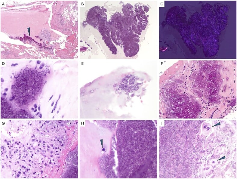
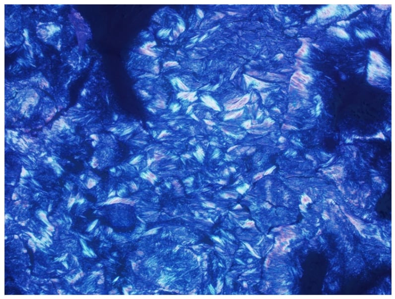
- Gout Crystals: Monosodium urate, negatively birefringent needles (yellow when parallel to polariser).
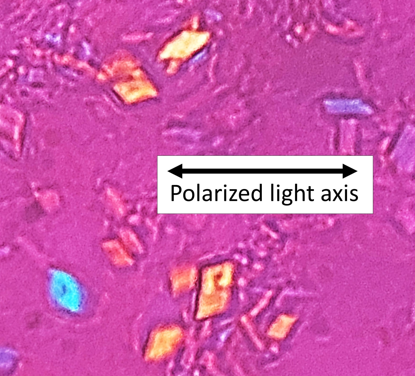
- CPPD Crystals: Calcium pyrophosphate, positively birefringent rhomboids (blue when parallel).
- Tophaceous Gout: Chronic deposits in tendons/joints → nerve compression, erosions, skin breakdown.
- CPPD Wrist: Triangular fibrocartilage (TFCC) calcification classic finding on x-ray.
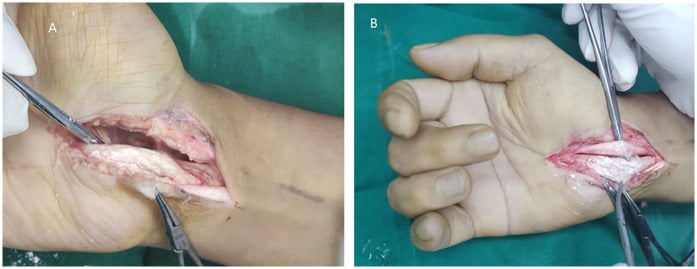
- Surgery Indications: Nerve compression (CTS), tendon rupture, skin ulceration, refractory pain.
- “Negative birefringence = gout (urate needles)
- “Positive birefringence = CPPD (rhomboids)
- “Tophaceous gout: carpal tunnel syndrome common
- “CPPD: wrist TFCC calcification on x-ray
Overview and Epidemiology
Crystalline arthropathy is joint inflammation caused by crystal deposition: monosodium urate (MSU) in gout, calcium pyrophosphate (CPP) in CPPD, or pseudogout.
Gout. It affects about 4% of the general population, a figure that is increasing, and men three times as often as women. Onset peaks at 40-60 years in men and comes after the menopause in women. The classic site is the first MTP joint (podagra), the presentation in 70% of first attacks, but the hand joints are also common: 10-15% of patients present with hand involvement of the wrist, MCP, PIP or DIP joints.
The risk factors for gout:
- Hyperuricaemia, a serum uric acid greater than 7 mg/dL
- Obesity and the metabolic syndrome
- Alcohol, particularly beer, and a purine-rich diet (red meat, seafood)
- Diuretics and chronic kidney disease
- A family history
CPPD. It is found in 5-10% of people over 65, equally in men and women, and its prevalence rises with age. The wrist is the commonest site in the hand.
The risk factors for CPPD:
- Advanced age, over 60 years
- Osteoarthritis
- Hyperparathyroidism and haemochromatosis
- Hypomagnesaemia and hypophosphatasia
- Prior joint trauma
Pathophysiology
Crystal-induced inflammation. Gout and CPPD both cause an acute inflammatory arthritis from crystal deposition. Neutrophils phagocytose the crystals and set off an inflammatory cascade, and the result is intense pain, swelling and erythema that mimic septic arthritis.
Gout. Hyperuricaemia, from overproduction or underexcretion of uric acid, comes first. Urate crystals then precipitate in synovial fluid and tissue (lower temperature in the extremities), and the acute attack follows when they trigger neutrophil activation and IL-1β release.
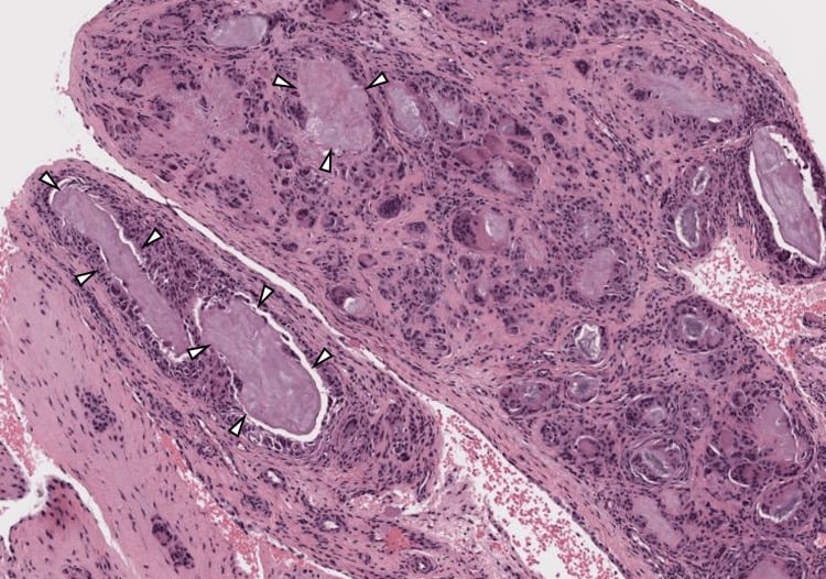
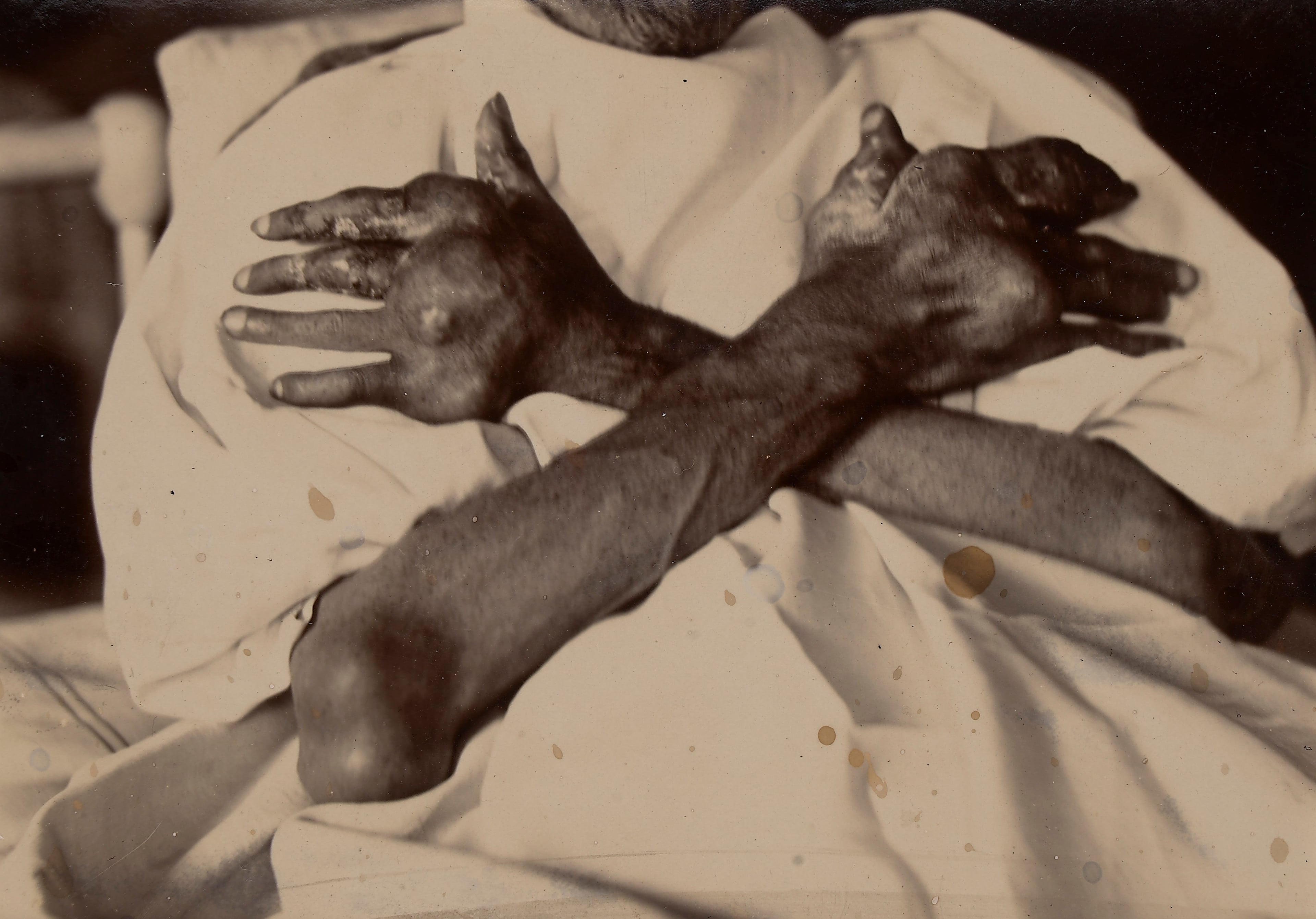
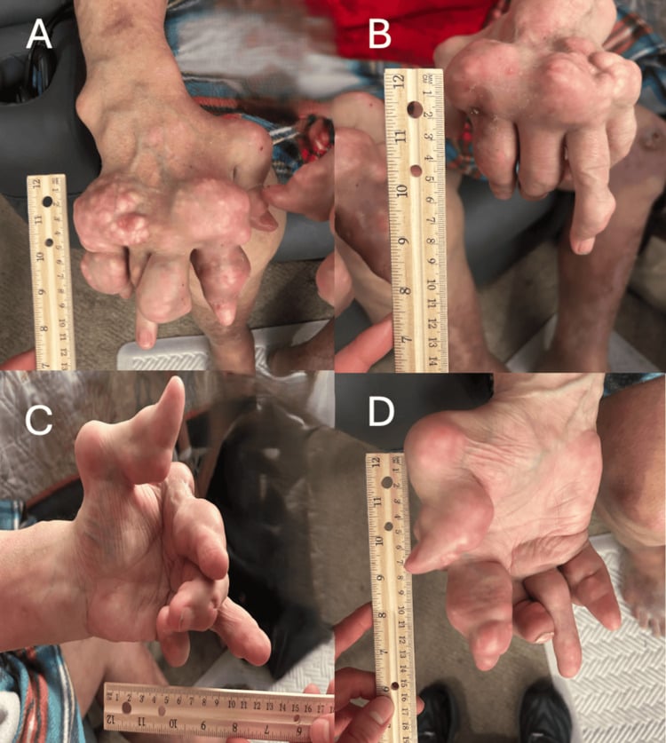
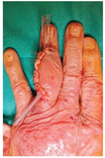
The tophaceous phase. Persistent hyperuricaemia builds tophi, aggregates of urate crystals with a granulomatous reaction: chalky white monosodium urate deposits surrounded by foreign-body giant cells. Tophi erode bone, weaken tendons, compress nerves and ulcerate skin.
CPPD. Abnormal cartilage metabolism, whether age-related, genetic or metabolic, allows pyrophosphate to accumulate, and calcium pyrophosphate crystals deposit in the cartilage (chondrocalcinosis). Crystals shed into the joint cause an acute attack of pseudogout. Over time, progressive cartilage and bone damage produces a chronic CPPD arthropathy in an OA-like pattern.
Hand sites. Each condition has its own distribution in the hand and wrist:
- Common hand and wrist sites
- Wrist, first MCP (thumb), finger MCPs, PIPs, DIPs
- Common hand and wrist sites
- Finger pulps, extensor tendons (dorsal hand and wrist), olecranon bursa, carpal tunnel
- Common hand and wrist sites
- Wrist (TFCC, radiocarpal), MCPs (less common)
Clinical Presentation
The attack. Severe joint pain begins suddenly, often overnight, and wakes the patient. It is monoarticular at first, although recurrent attacks can be polyarticular. The joint is red, hot and swollen, and so exquisitely tender that the patient cannot tolerate touch or even the bedsheets. Without treatment it is self-limited and resolves in 7-10 days.
In the hand. An affected wrist, MCP, PIP or DIP joint is swollen and erythematous, and pain reduces its range of movement.
Triggers. Ask about each in the history:
- An alcohol binge, particularly beer
- Dehydration
- Acute illness or surgery
- Starting allopurinol, which causes a paradoxical flare
Examination. The skin over the joint is red, oedematous, shiny and tense, and chronic cases may have visible tophi. Tenderness is marked enough that the patient withdraws; the joint is warm with an effusion, and pain severely limits movement. Septic arthritis is the differential that must be ruled out, by joint aspiration.
Investigations
Joint aspiration is diagnostic in both gout and CPPD. Aspirate the affected joint (wrist, MCP) under sterile conditions and send the fluid for a cell count and differential, a Gram stain and culture to rule out septic arthritis, and crystal analysis by polarised light microscopy.
- Shape
- Needle-shaped
- Birefringence
- Negatively birefringent
- Colour when parallel to polariser
- Yellow
- Shape
- Rhomboid or rectangular
- Birefringence
- Positively birefringent
- Colour when parallel to polariser
- Blue
Cell count. Gout and CPPD give an inflammatory fluid of 2,000-100,000 WBC/μL with a neutrophil predominance. Septic arthritis usually exceeds 50,000 WBC/μL, with a positive Gram stain or culture.
Crystal arthritis mimics septic arthritis, and crystals can coexist with infection. If clinical suspicion of septic arthritis remains, treat the joint as infected.
Blood tests. In gout the serum uric acid shows hyperuricaemia but can be normal during an acute attack. ESR and CRP are raised in acute gout and in acute pseudogout. In CPPD, serum calcium, phosphate and magnesium screen for the metabolic causes (hyperparathyroidism, haemochromatosis).
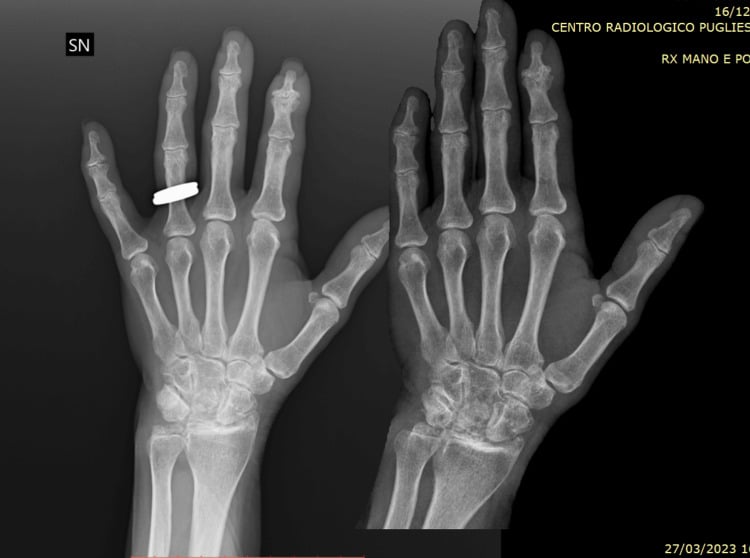
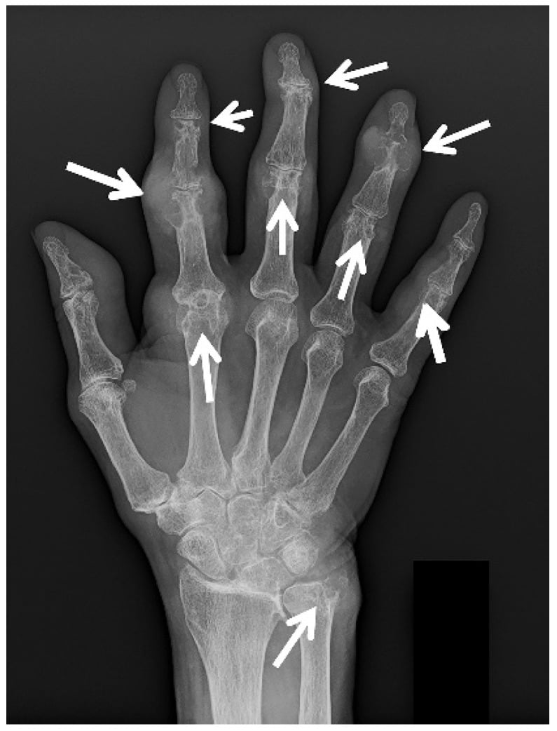
Radiographs in gout. Early films are normal apart from soft-tissue swelling. Chronic tophaceous gout produces punched-out "rat-bite" erosions, which are set against rheumatoid arthritis in the next section. Soft-tissue tophi are radiopaque if calcified, which is rare.
Radiographs in CPPD. The classic finding is chondrocalcinosis, linear or punctate calcification of cartilage, in the triangular fibrocartilage (TFCC) at the wrist. Degenerative change (joint-space narrowing, osteophytes, subchondral sclerosis) may mimic osteoarthritis or a SLAC wrist.
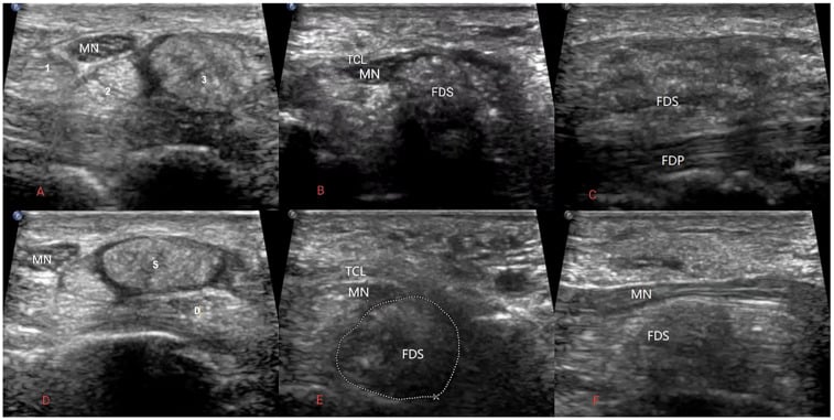
Ultrasound. In gout the double contour sign, a hyperechoic line on the cartilage surface, represents urate crystals. In CPPD the deposits are hyperechoic within the cartilage.
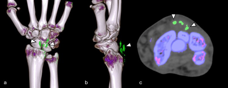
Dual-energy CT (DECT). DECT colour-codes monosodium urate, typically green, to show the urate burden, and it is useful for monitoring treatment response and the reduction in urate volume. In established gout its pooled sensitivity is about 81% and specificity about 91%; sensitivity is lower in disease of under 6 weeks.
MRI is rarely needed, and shows tophi as intermediate-signal masses.
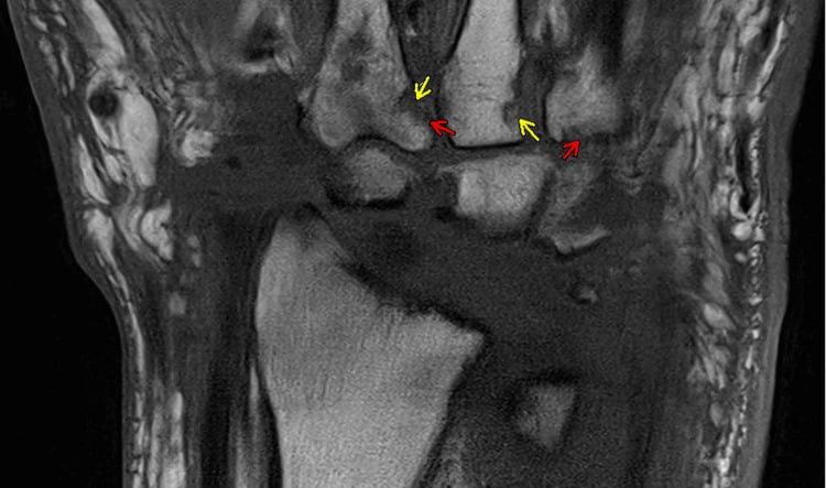
Imaging or aspiration. DECT and ultrasound are increasingly used, but sensitivity is low in early gout and a negative scan cannot exclude disease. Crystal identification on aspiration remains the reference standard, and over-reliance on imaging risks missing both early gout and coexistent septic arthritis.
Distinguishing Tophaceous Gout of the Hand from Rheumatoid Arthritis and Osteoarthritis
On the radiograph. Tophaceous gout and rheumatoid arthritis are told apart by the erosions, the joint space, the bone density, the distribution and the soft tissue:
- Tophaceous gout
- Punched-out, para- or juxta-articular, sclerotic margins with an overhanging edge (Martel's sign), set slightly away from the joint
- Rheumatoid arthritis
- Marginal erosions at the bare area, no sclerotic rim
- Tophaceous gout
- Preserved until late
- Rheumatoid arthritis
- Uniform early narrowing
- Tophaceous gout
- No periarticular osteopenia
- Rheumatoid arthritis
- Periarticular osteopenia early
- Tophaceous gout
- Asymmetric, can involve DIP joints
- Rheumatoid arthritis
- Symmetric, MCP/PIP/wrist, usually spares DIP
- Tophaceous gout
- Eccentric, lumpy tophi (MSU usually radiolucent, may calcify)
- Rheumatoid arthritis
- Symmetric fusiform swelling
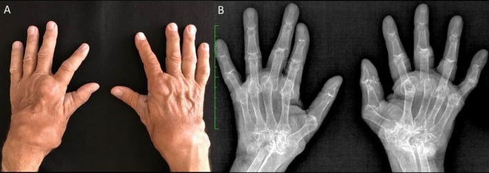
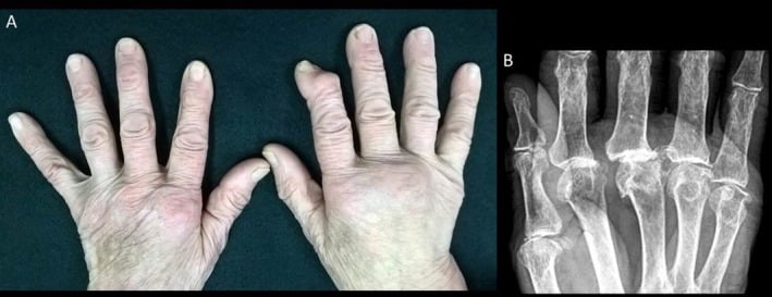
Tophus or rheumatoid nodule. Both are firm, but a tophus may ulcerate and discharge chalky white material, and aspiration or histology showing MSU crystals is definitive. Rheumatoid nodules favour extensor surfaces and are associated with seropositive erosive disease.
Serology. RA is associated with RF and anti-CCP. Gout is associated with hyperuricaemia and with negatively birefringent MSU crystals on aspiration.
Osteoarthritis. Nodal OA gives hard nodal enlargement, Heberden (DIP) and Bouchard (PIP) nodes, with osteophytes, subchondral sclerosis and non-uniform joint-space loss, and no overhanging erosions.
CPPD. Look for chondrocalcinosis (TFCC, cartilage), hook-like osteophytes at the second and third MCP joints, and a radiocarpal or SLAC-type pattern. The pseudogout/CPPD topic covers it in detail.
The trap. A deforming, asymmetric hand arthropathy with subcutaneous nodules is not automatically rheumatoid. "Pseudo-rheumatoid" tophaceous gout is a classic mimic, and joint aspiration for crystals settles it.
Q: A hand radiograph shows well-defined "punched-out" erosions with overhanging sclerotic edges and preserved joint space, involving DIP joints asymmetrically, with no periarticular osteopenia. Why is this gout rather than rheumatoid arthritis?
A: The combination of para-articular punched-out erosions with an overhanging edge (Martel's sign), preserved joint space until late, absent periarticular osteopenia, asymmetric distribution and DIP involvement is characteristic of tophaceous gout. Rheumatoid arthritis instead gives marginal erosions, early uniform joint-space loss, periarticular osteopenia, and a symmetric MCP/PIP/wrist distribution that spares the DIP joints. Confirm with joint aspiration showing negatively birefringent MSU crystals; RF/anti-CCP support RA. Remember tophaceous gout can cause ulnar drift and swan-neck deformity ("pseudo-rheumatoid" gout).
Medical Management
The aim is to reduce inflammation and pain rapidly. The first-line drugs:
- NSAIDs - indomethacin 50mg three times daily or naproxen 500mg twice daily, started immediately at diagnosis and continued until the attack resolves (7-10 days). Renal impairment, a history of GI bleeding and anticoagulation contraindicate them
- Colchicine - the low-dose regimen is 1.2mg as a loading dose, 0.6mg an hour later, then 0.6mg daily. It is effective if started early, within 24-48 hours. Diarrhoea and GI upset are dose-dependent, and severe renal or hepatic impairment contraindicates it
- Corticosteroids - used if NSAIDs or colchicine are contraindicated. Intra-articular triamcinolone 10-40mg if the attack is monoarticular and septic arthritis has been ruled out; oral prednisone 30-40mg daily for 5 days, then a taper; IM or IV methylprednisolone for hospitalised patients unable to take oral medication
Supportive care. Rest and elevate the joint, apply ice, and avoid weight-bearing if the attack is in the lower limb. Aspirating the joint relieves pain as well as confirming the diagnosis.
Do not start urate-lowering therapy during an attack. Starting allopurinol or febuxostat in the acute attack worsens the flare; wait until it has resolved, 2-4 weeks.
Surgical Management
Gout. The indications, each with its operation:
- Surgical procedure
- Carpal tunnel release + tophus excision
- Surgical procedure
- Tendon repair or reconstruction + tophus removal
- Surgical procedure
- Wound care, tophus excision, wound closure or skin graft
- Surgical procedure
- Nerve decompression + tophus excision
- Surgical procedure
- Arthroplasty or arthrodesis
- Surgical procedure
- Tophus excision (elective)
CPPD is usually managed without surgery. Severe wrist arthritis is the exception, covered under Arthritis Surgery.
Medical control first. Optimise medical management, ULT in gout, before elective surgery; perioperative uric acid control reduces wound complications. The conventional teaching is a serum urate under 6 mg/dL for 3-6 months before elective tophus excision. Severe carpal tunnel syndrome with motor signs, or an ulcerated or infected tophus, mandates earlier surgery.
The evidence behind the wait. No high-level evidence defines the exact urate threshold or duration that minimises wound complications; the practice is extrapolated from case series.
TENTSTophaceous Gout Surgical Indications
Hook:TENTS = indications for surgical tophus excision in chronic gout!
Complications and Outcomes
- Incidence
- 10-20%
- Prevention/Management
- Optimise uric acid control pre-op, meticulous closure, avoid tension
- Incidence
- 5-10%
- Prevention/Management
- Sterile technique, antibiotics if signs of infection, continue ULT
- Incidence
- 5%
- Prevention/Management
- Gentle dissection, preserve tendon, may need repair/reconstruction
- Incidence
- Less than 5%
- Prevention/Management
- Careful dissection if tophus adherent to nerve
- Incidence
- 5-10%
- Prevention/Management
- Adequate ULT post-op (uric acid less than 6 mg/dL)
Wound dehiscence is the most common complication. Gout impairs wound healing, and the skin is often friable or atrophic from the chronic tophus. Prevention is uric acid control before surgery, no tension on the skin, and delayed closure if needed; management is local wound care and healing by secondary intention, with delayed closure or a skin graft if the defect is large.
Tendon rupture occurs where the tophus has weakened the tendon, and nerve injury where it is densely adherent to the nerve.
Infection is a higher risk than in standard hand surgery, and draining tophi may be colonised. Treat it with antibiotics covering Staphylococcus aureus, debridement if there is an abscess, and continued ULT.
Outcomes. After tophus excision for skin breakdown the wound heals in 70-80%, and delayed healing is common. Recurrence follows inadequate uric acid control after surgery, so success depends on perioperative and long-term urate control with allopurinol or febuxostat.
Guidelines, Registries & Global Practice
Global Epidemiology:
- Gout is the most common inflammatory arthritis worldwide; prevalence is rising with ageing populations and metabolic syndrome
- Age-standardised prevalence in adults 65+ was ~3,100 per 100,000 (GBD 2021), with burden in older men nearly double that of women and projected to keep rising to 2050
- Marked ethnic variation: highest prevalence in Māori and Pacific Island populations of Oceania (genetic urate transporter variants); lower rates historically reported in parts of Africa and East Asia
- Hand/wrist involvement complicates roughly 10-15% of established gout; CPPD prevalence rises steeply with age
Side-by-Side Guidelines:
- Diagnosis
- MSU crystals; supports DECT/US when aspiration unavailable
- First-line ULT
- Allopurinol (incl. CKD), low-dose start
- Serum urate target
- under 6 mg/dL, treat-to-target
- Notable position
- Strong: ULT for tophi/erosions/frequent flares; prophylaxis 3-6 months
- Diagnosis
- Crystal confirmation preferred; imaging adjuncts
- First-line ULT
- Allopurinol, titrate to target
- Serum urate target
- under 6 mg/dL (under 5 for severe/tophaceous)
- Notable position
- Endorses treat-to-target and patient education
- Diagnosis
- Clinical plus crystal/imaging
- First-line ULT
- Allopurinol first-line
- Serum urate target
- under 6 mg/dL (under 5 for tophi)
- Notable position
- Emphasises ULT offered to all after a diagnosis is confirmed
- Diagnosis
- Classification: clinical + lab + imaging
- First-line ULT
- n/a (classification)
- Serum urate target
- n/a
- Notable position
- Sensitivity 92%, specificity 89%
- No dedicated arthroplasty registry stratum exists for crystalline hand arthropathy; surgical evidence is limited to case series, so consent must reflect this
- Pharmacovigilance data (FAST trial, EU regulator-mandated) reshaped febuxostat cardiovascular guidance globally
- High-resource: ready access to polarised microscopy, DECT, biologic uricase (pegloticase) for refractory tophaceous disease, and elective hand surgery
- Limited-resource: diagnosis often clinical; allopurinol remains the affordable backbone; DECT and pegloticase frequently unavailable; patients may present later with advanced tophaceous deformity requiring surgery
- Universal priorities: confirm crystals where possible, rule out septic arthritis, and achieve sustained serum urate under 6 mg/dL before and after any surgery
- Document joint aspiration and crystal analysis before committing to a diagnosis
- Always exclude septic arthritis (Gram stain, culture) - a missed joint sepsis is a key litigation risk
- Consent for tophus excision must cover wound dehiscence (10-20%), infection (5-10%), possible skin graft, and recurrence if urate control fails
Exam Viva Scenarios
Practise clinical reasoning and management decisions out loud
“A 55-year-old man presents with sudden onset severe pain and swelling in his right wrist. The wrist is red, hot, and exquisitely tender. He cannot move it. He has a history of gout affecting his great toe. What is your diagnosis and initial management?”
“A 60-year-old man with long-standing gout presents with progressive numbness in his thumb, index, and middle fingers. He has visible chalk-like deposits on his fingers and wrist. EMG shows severe carpal tunnel syndrome. His uric acid is 9 mg/dL despite allopurinol. What is your management?”
“A 70-year-old woman presents with acute wrist pain and swelling. X-ray shows calcification in the triangular fibrocartilage. You aspirate the joint and see rhomboid-shaped crystals under polarized microscopy. They appear blue when parallel to the polarizer. What is your diagnosis and management?”
Crystal Types
- Gout: Monosodium urate (MSU)
- CPPD: Calcium pyrophosphate (CPP)
- Birefringence distinguishes them
- Joint aspiration is diagnostic
Crystal Identification
- Gout: Negatively birefringent needles (yellow parallel)
- CPPD: Positively birefringent rhomboids (blue parallel)
- Polarized light microscopy essential
Acute Gout Attack
- Sudden severe pain, red/hot/swollen joint
- Wrist, MCP, PIP involvement possible
- Mimics septic arthritis (must rule out)
- Self-limited 7-10 days
Acute Management
- NSAIDs: Indomethacin, naproxen (first-line)
- Colchicine: 1.2mg load, 0.6mg 1h later, 0.6mg daily
- Steroids: Intra-articular or oral if NSAID contraindicated
- Do NOT start allopurinol during attack
Chronic Gout (ULT)
- Allopurinol 100-800mg daily (first-line)
- Febuxostat 40-80mg (alternative)
- Target: Uric acid less than 6 mg/dL (less than 5 for tophi)
- Colchicine prophylaxis 3-6 months during ULT start
Tophaceous Gout
- Chalky white deposits in joints/tendons
- Complications: CTS, tendon rupture, skin ulceration
- Punched-out erosions on x-ray (rat-bite)
- Surgery for nerve compression, skin breakdown
Surgical Indications
- Carpal tunnel syndrome (most common)
- Tendon rupture (extensor/flexor)
- Skin ulceration/draining tophus
- Nerve compression (median, ulnar)
- Severe erosive arthropathy
CTR in Tophaceous Gout
- Standard CTR + tophus excision
- Dissect tophi off median nerve carefully
- Debulk extensively, irrigate
- Wound dehiscence risk 10-20%
- Continue ULT perioperatively
Tophus Excision Complications
- Wound dehiscence: 10-20%
- Infection: 5-10%
- Tendon rupture: 5%
- Nerve injury: Less than 5%
- Recurrence: 5-10% if poor uric acid control
CPPD / Pseudogout
- Wrist most common hand site
- TFCC calcification (chondrocalcinosis) on x-ray
- Acute: NSAIDs, colchicine, steroids
- No disease-modifying therapy (no ULT equivalent)
Imaging
- Gout: Punched-out erosions, overhanging edges
- CPPD: Chondrocalcinosis (TFCC calcification)
- Ultrasound: Double contour sign (gout)
- X-ray normal in early gout
Exam Pearls
- Rule out septic arthritis (joint aspiration mandatory)
- Negative birefringence = gout (yellow parallel)
- Positive birefringence = CPPD (blue parallel)
- CTS in tophaceous gout: CTR + tophus excision
- Target uric acid less than 6 mg/dL with ULT
Evidence Base
Neogi et al (2015 ACR/EULAR Gout Classification Criteria)
- Data-driven classification criteria; MSU crystals in symptomatic joint/bursa or tophus is a sufficient criterion (no further scoring needed)
- Clinical, laboratory and imaging (ultrasound double-contour sign, urate on dual-energy CT, radiographic erosion) domains
- Overall sensitivity 92% and specificity 89%
- Developed when MSU crystal identification was the diagnostic gold standard
FitzGerald et al (2020 ACR Guideline for the Management of Gout)
- Strong recommendation to start urate-lowering therapy for tophaceous gout, radiographic damage, or frequent flares
- Allopurinol preferred first-line ULT, including in moderate-to-severe CKD (stage 3 or higher)
- Treat-to-target strategy with serum urate target under 6 mg/dL (under 0.36 mmol/L)
- Anti-inflammatory flare prophylaxis for at least 3 to 6 months when starting ULT
Mackenzie et al (FAST trial)
- Multicentre randomised non-inferiority trial; 6128 gout patients aged 60+ with a CV risk factor
- Febuxostat non-inferior to allopurinol for the composite CV endpoint (adjusted HR 0.85, 95% CI 0.70 to 1.03) against a PRE-SPECIFIED NON-INFERIORITY MARGIN OF HR 1.3 - the upper confidence bound of 1.03 sits well inside it, and the point estimate favours febuxostat
- No increase in all-cause death or serious adverse events with febuxostat
- Tempered the cardiovascular signal previously raised by the CARES trial
Gamala et al (DECT diagnostic performance, systematic review/meta-analysis)
- Ten studies pooled; person-based sensitivity 0.81 and specificity 0.91 for diagnosing gout
- Joint-based sensitivity 0.83 and specificity 0.88
- Sensitivity falls to ~0.55 in recent-onset gout (6 weeks or less)
- DECT most useful in established disease, not early presentation
Doscher et al (en masse excision and curettage of hand tophi)
- 12 patients, 24 periarticular tophi excised from MCP/IP joints (some wrist and elbow), all on urate-lowering therapy
- Restoration of tendon excursion and joint mobility in all patients
- No wound complications and no recurrence over follow-up up to 15 years
- Highlights that meticulous technique plus medical urate control optimises results
Tang et al (Global Burden of Disease, gout in older adults)
- Age-standardised prevalence in adults 65+ was ~3111 per 100,000 in 2021
- Disease burden in older men nearly twice that of women
- Burden projected to rise further by 2050, driven by ageing and metabolic risk
- Greatest increases in high-SDI/high-income regions