Immune-Mediated Cartilage Inflammation
- Relapsing polychondritis is a rare, IMMUNE-MEDIATED disease characterised by recurrent episodes of INFLAMMATION and progressive DESTRUCTION of CARTILAGE and other proteoglycan-rich connective tissue, affecting the EARS, NOSE, LARYNGOTRACHEAL airway, JOINTS, eyes, audiovestibular system and cardiovascular system.
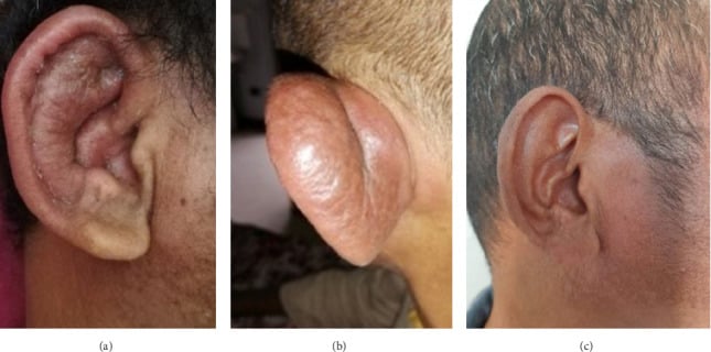
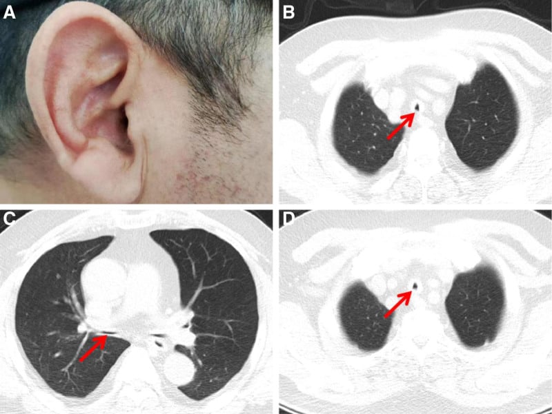
- The clinical HALLMARK is BILATERAL AURICULAR CHONDRITIS - a painful, red, swollen pinna - that characteristically AFFECTS the cartilaginous ear but SPARES the non-cartilaginous EARLOBE; recurrent attacks produce a floppy, deformed ('cauliflower') ear, and this lobe-sparing pattern is a key examination clue.
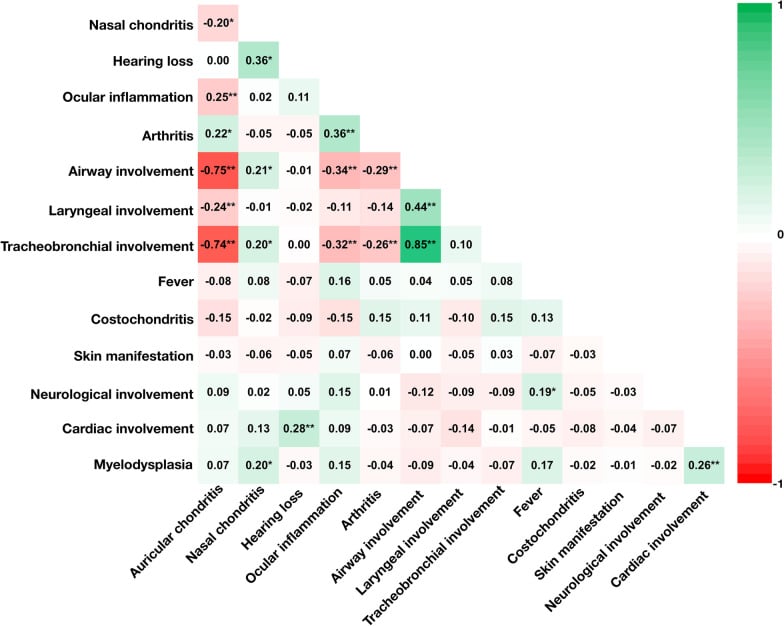
- BUT THE HALLMARK IS ABSENT IN THE PATIENTS WHO MATTER MOST. In a 126-patient cohort, airway involvement and auricular chondritis were strongly NEGATIVELY correlated (r = -0.75, P less than 0.001): an Ear pattern in 50.8%, an AIRWAY pattern in 38.9%, an Overlap pattern in only 4.8%. Patients with the Ear pattern had the HIGHEST survival. So a normal pinna does not exclude the disease - it is characteristic of the airway-dominant group with the worse prognosis, and patterns can evolve over time.
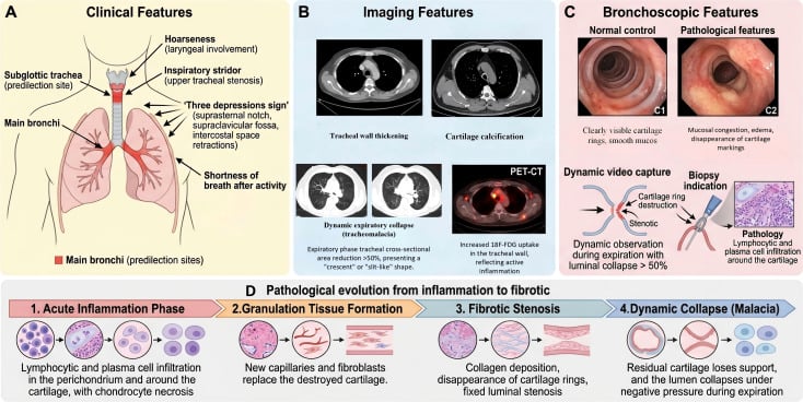
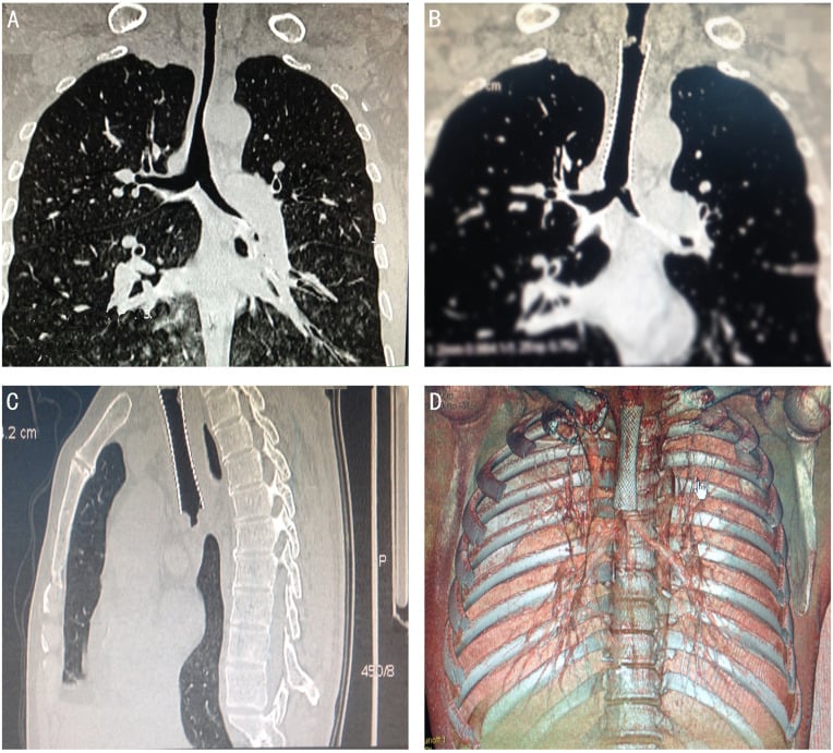
- NASAL chondritis can cause a SADDLE-NOSE deformity, and LARYNGOTRACHEAL chondritis is the most DANGEROUS manifestation - inflammation and softening/collapse or stenosis of the airway causing hoarseness, cough, stridor and potentially LIFE-THREATENING airway obstruction; airway involvement is a leading cause of morbidity and mortality. In a 232-patient referral cohort, 146 (63%) had laryngeal involvement and 21 needed TRACHEOSTOMY; the independent risk factors were age 25 or under or 65 and over, laryngotracheal OEDEMA, and PULMONARY INFECTION - the last being modifiable and directly relevant to any immunosuppressed patient facing an anaesthetic.
- TRACHEOSTOMY MARKS SEVERITY, IT DOES NOT CAUSE THE DEATHS. In that cohort tracheostomy was associated with significantly higher IN-HOSPITAL mortality but made NO difference to long-term survival (P = 0.706) - so the association must not be used as an argument for withholding it.
- AORTIC DISEASE IS UNCOMMON BUT LETHAL. With systematic CT screening of 172 patients, aortic involvement was found in 6.4% (11 patients) at a median of 2 years after diagnosis; 5 relapsed and 1 died of a ruptured abdominal aortic aneurysm. Aortitis on CT and a higher ESR predicted aortic death; aortitis and abdominal involvement predicted relapse - though with only 11 events these are signals, not validated risk factors.
- The ARTHRITIS is typically a SERONEGATIVE, NON-EROSIVE, episodic and often migratory poly/oligoarthritis affecting peripheral and parasternal (costochondral/sternoclavicular) joints - it does not usually cause the erosive joint destruction of rheumatoid arthritis - so the orthopaedic relevance is mainly recognising the systemic disease behind a seronegative arthritis rather than reconstructing destroyed joints.
- DIAGNOSIS is clinical, using the McADAM (and modified MICHET) criteria (e.g. bilateral auricular chondritis, non-erosive seronegative inflammatory arthritis, nasal chondritis, ocular inflammation, respiratory-tract chondritis, audiovestibular damage), supported by inflammatory markers and imaging of the airway; classical features such as auricular chondritis can appear LATE, so it must be considered in unexplained cartilage-related or ocular inflammation, and biopsy is occasionally needed.
- There are important ASSOCIATIONS - other autoimmune disease, ANCA-ASSOCIATED VASCULITIS, and haematological disease including MYELODYSPLASTIC syndrome and the recently described VEXAS syndrome (in older men) - and MANAGEMENT is medical and multidisciplinary: CORTICOSTEROIDS for flares with steroid-sparing IMMUNOSUPPRESSION (methotrexate, azathioprine; biologics/avacopan in selected/overlap cases), plus urgent airway management for laryngotracheal disease.
- “Relapsing polychondritis = recurrent inflammation/destruction of CARTILAGE (ear, nose, airway, joints). HALLMARK: bilateral auricular chondritis SPARING the (non-cartilaginous) earlobe; saddle-nose deformity.
- “THE TRAP: the ear sign and airway disease are strongly NEGATIVELY correlated (r = -0.75). Ear pattern 50.8%, AIRWAY pattern 38.9%, overlap only 4.8% - and the Ear pattern has the BEST survival. A normal pinna points towards the dangerous group, not away from it.
- “Laryngotracheal involvement = life-threatening (airway softening/collapse/stenosis) - a leading cause of death. 63% had laryngeal involvement in a 232-patient cohort; tracheostomy risk factors were age 25 or under / 65 or over, laryngotracheal oedema and pulmonary infection. Arthritis is SERONEGATIVE and NON-EROSIVE.
- “Diagnosis = McAdam/Michet criteria (clinical). Associations: ANCA-vasculitis, myelodysplasia/VEXAS. Treat with corticosteroids + immunosuppression; urgent airway care.
Bilateral auricular chondritis - red, painful, swollen pinna that spares the non-cartilaginous earlobe; recurrent attacks → floppy/'cauliflower' ear. Nasal chondritis → saddle nose.
Laryngotracheal chondritis → airway softening/collapse/stenosis, stridor and potentially fatal obstruction - a leading cause of death. 63% had laryngeal involvement in a 232-patient cohort. But the ear sign and airway disease are negatively correlated (r = -0.75) - normal ears do not reassure.
The Diagnostic Criteria and the Immunopathology
- The McAdam criteria (need three of six). Diagnosis by ≥3 of 6 clinical features: (1) bilateral auricular chondritis, (2) non-erosive seronegative inflammatory arthritis, (3) nasal chondritis, (4) ocular inflammation (scleritis/episcleritis/uveitis), (5) respiratory-tract chondritis (laryngeal/tracheal), (6) audiovestibular damage (sensorineural hearing loss/vertigo/tinnitus).
- The modifications. Damiani & Levine allow the diagnosis with fewer criteria if there is histological confirmation or chondritis at ≥2 sites responding to steroids/dapsone. Michet requires proven chondritis in ≥2 of the 3 (auricular/nasal/laryngotracheal) cartilages, or 1 of those plus ≥2 other features (ocular, audiovestibular, arthritis).
- The immunopathology. An autoimmune response to type II collagen and other cartilage matrix proteins (matrilin-1); anti-type-II-collagen antibodies appear in acute flares (correlating with activity but not sensitive/specific enough to diagnose), there is a T-cell-mediated attack, and an association with HLA-DR4. Biopsy shows perichondrial inflammation with loss of the basophilic cartilage matrix (proteoglycan depletion) and chondrocyte destruction.
Q: What are the McAdam criteria and the immunopathology of relapsing polychondritis?
A: McAdam = ≥3 of 6: bilateral auricular chondritis, non-erosive seronegative arthritis, nasal chondritis, ocular inflammation, respiratory-tract chondritis, audiovestibular damage. Damiani & Levine allow fewer criteria + biopsy/steroid-response; Michet needs ≥2 of 3 cartilages (or 1 + ≥2 other features). Immunopathology = autoimmune attack on type II collagen (± matrilin-1); anti-type-II-collagen antibodies in flares (activity marker, not diagnostic); T-cell-mediated; HLA-DR4-associated; biopsy shows loss of the basophilic cartilage matrix.
VEXAS and MAGIC: The Key Overlap Syndromes
- VEXAS syndrome. VEXAS (Vacuoles, E1 enzyme, X-linked, Autoinflammatory, Somatic) is caused by a somatic mutation in UBA1 (the X-linked E1 ubiquitin-activating enzyme) in myeloid precursors, giving an adult-onset (older men) severe autoinflammatory disease with fever, cytopenias, cutaneous and chondritic features, and a strong link to myelodysplastic syndrome; the marrow shows characteristic cytoplasmic vacuoles. Some older men labelled with relapsing polychondritis - particularly those with cytopenias, fever and a raised MCV - in fact have VEXAS, so UBA1 testing is now recommended in that phenotype; it is often treatment-refractory with a poor prognosis. The proportion varies widely between reported series and no agreed figure exists, so quote the phenotype that should trigger the test rather than a percentage.
- MAGIC syndrome. The overlap of relapsing polychondritis with Behçet disease is MAGIC syndrome - Mouth And Genital ulcers with Inflamed Cartilage - combining recurrent oral/genital aphthous ulcers with cartilage inflammation.
Q: What is VEXAS syndrome, and what is MAGIC syndrome, in relation to relapsing polychondritis?
A: VEXAS (Vacuoles, E1 enzyme, X-linked, Autoinflammatory, Somatic) = a somatic UBA1 mutation (X-linked E1 ubiquitin-activating enzyme) in myeloid precursors → adult-onset autoinflammation in older men with fever/cytopenias/chondritis + myelodysplastic syndrome; marrow shows cytoplasmic vacuoles. A significant minority of older men labelled with RP (esp. with cytopenias) actually have VEXAS → test UBA1; refractory/poor prognosis. MAGIC syndrome = RP overlapping with Behçet (Mouth And Genital ulcers with Inflamed Cartilage).
Presentation & Diagnosis
Relapsing polychondritis is an immune-mediated disease of recurrent cartilage inflammation and destruction. The hallmark is bilateral auricular chondritis that spares the earlobe (which lacks cartilage); nasal chondritis causes a saddle-nose deformity; and laryngotracheal chondritis causes hoarseness, stridor and potentially life-threatening airway collapse/stenosis. The arthritis is seronegative, non-erosive, episodic and often migratory (peripheral and parasternal joints). Ocular inflammation, audiovestibular damage and cardiovascular disease (aortitis, valve disease) also occur. Diagnosis is clinical (McAdam/Michet criteria); classical features can appear late, so it must be considered in unexplained cartilage-related or ocular inflammation.
The Trap: The Ear Sign Is Missing in the Patients Most at Risk
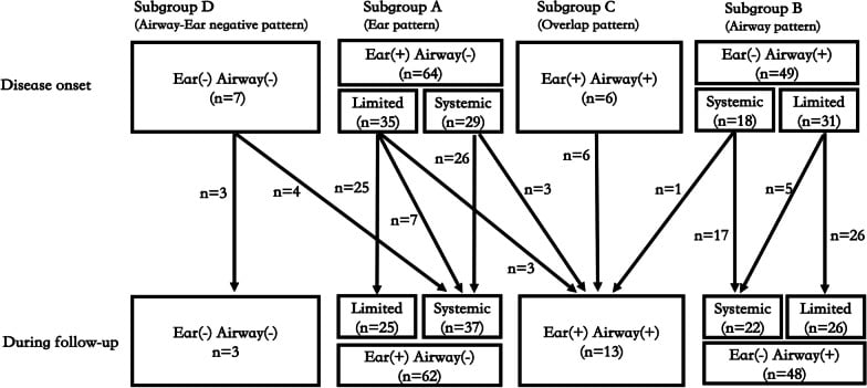
The lobe-sparing ear is the sign every candidate is taught to look for, and relying on it is exactly how the dangerous cases are missed. In a retrospective cohort of 126 patients, airway involvement and auricular chondritis were strongly NEGATIVELY correlated (r = -0.75, P less than 0.001) - the two rarely appear in the same patient.
- Four clinical patterns emerged. An Ear pattern in 50.8%, an Airway pattern in 38.9%, an Overlap pattern in only 4.8%, and an Airway-Ear negative pattern in 5.6%. So close to two in five patients declare themselves through the airway, and in those the pinna is typically normal.
- The pattern predicts survival. Patients with the Ear pattern had the highest survival rate and a relatively lower inflammatory burden. The reassuring sign and the good prognosis travel together, which is precisely why its absence must not be reassuring.
- Patterns evolve. A minority of Ear-pattern and Airway-pattern patients progressed to Overlap during follow-up, and some Airway-Ear negative patients later developed the ear sign - so a normal pinna today does not settle the question, and the disease should be re-evaluated over time.
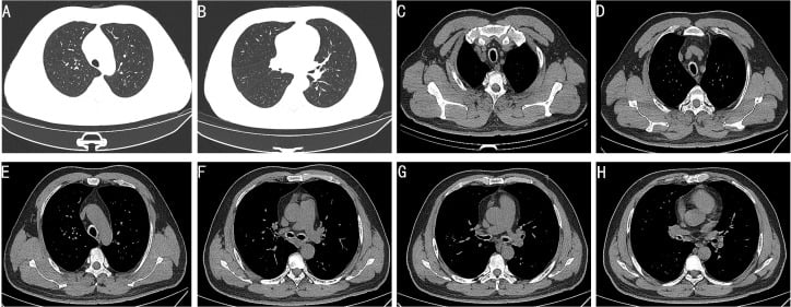
- The practical rule. Do not require auricular chondritis to consider the diagnosis. Unexplained hoarseness, cough, stridor or tracheal wall thickening in an adult with systemic inflammation deserves the thought even with entirely normal ears.
In 232 patients with relapsing polychondritis, 146 (63%) had laryngeal involvement and 21 underwent tracheostomy. Three independent risk factors emerged on multivariate analysis: age 25 or under, or 65 and over (OR 24.6, 95% CI 5.31-113.8), laryngotracheal oedema (OR 26.7, 95% CI 4.21-169.2) and pulmonary infection (OR 18.8, 95% CI 3.17-111.9). Note how wide those confidence intervals are - the direction is clear, the magnitude is not. A prediction nomogram achieved a C-index of 0.936, internally validated by bootstrapping at 0.926, but it has not been externally validated, so it is a promising tool rather than one to act on outside its source population. Crucially, tracheostomy was associated with a significant rise in in-hospital mortality yet made no difference to long-term survival (P = 0.706) - it marks a sick patient rather than causing the harm, and should not be withheld on the basis of that mortality association.
Associations & Management
- Associations: other autoimmune disease, ANCA-associated vasculitis, and haematological disease - myelodysplastic syndrome and VEXAS syndrome (older men) - screen as appropriate.
- Corticosteroids for flares, with steroid-sparing immunosuppression (methotrexate, azathioprine), and biologics/avacopan in refractory or overlap (ANCA-vasculitis) cases.
- Airway disease (the priority): urgent assessment and management of laryngotracheal involvement - this is the leading cause of morbidity/mortality (may need stenting/tracheostomy and intensive immunosuppression).
- Orthopaedic role: recognise the systemic disease behind a seronegative, non-erosive arthritis; coordinate any care with rheumatology.
Two features make relapsing polychondritis recognisable and dangerous. The diagnostic clue is the pattern of ear involvement: bilateral, painful, red auricular chondritis that affects the cartilaginous pinna but spares the non-cartilaginous earlobe - a pattern that, with a saddle-nose deformity and a seronegative non-erosive arthritis, points strongly to the disease (formalised in the McAdam and Michet criteria). The danger is the airway: laryngotracheal chondritis softens and can collapse or stenose the airway, producing stridor and potentially fatal obstruction, and it is a leading cause of death - so respiratory symptoms in a patient with chondritis must be assessed urgently. There is a trap in that sequence. The ear sign and airway involvement are strongly negatively correlated, and close to two in five patients present with an airway-dominant pattern in which the pinna looks entirely normal; the ear pattern carries the best survival, so the absence of the hallmark should raise concern rather than settle it. Because classical features can appear late and because the disease associates with ANCA-vasculitis and with myelodysplasia/VEXAS, it should be considered in unexplained cartilage-related or ocular inflammation and managed with rheumatology, with corticosteroids and immunosuppression and prompt airway care.
Mnemonics & Memory Aids
CHONDRO
Hook:CHONDRO: Cartilage destruction, Hearing/eyes, Otic (lobe-sparing), Nose (saddle), Danger (airway), Rheumatic (seronegative), Old men/VEXAS + steroids.
Clinical Decision Scenarios
Practise clinical reasoning and management decisions out loud
“A patient has recurrent painful, red, swollen ears that spare the earlobes, a saddle-nose deformity and a seronegative arthritis. What is the diagnosis and what is the main danger?”
Recognise it
- Bilateral auricular chondritis SPARING the non-cartilaginous earlobe
- Saddle-nose deformity (nasal chondritis)
- Seronegative, non-erosive, episodic arthritis
The danger
- Laryngotracheal chondritis → airway softening/collapse/stenosis
- Hoarseness, stridor, potentially fatal obstruction (leading cause of death)
- 63% laryngeal involvement, 21 tracheostomies in a 232-patient cohort
- Tracheostomy risk: age 25 or under / 65 or over, laryngotracheal oedema, pulmonary infection
- Tracheostomy raises in-hospital mortality but NOT long-term survival - a severity marker
- Aortic involvement 6.4% on systematic CT (11/172), median 2 yrs after diagnosis; 1 died of AAA rupture
- Also ocular inflammation, audiovestibular damage, valve disease
The pattern trap
- Ear sign and airway involvement strongly NEGATIVELY correlated (r = -0.75, n = 126)
- Ear pattern 50.8%, Airway pattern 38.9%, Overlap only 4.8%, Airway-Ear negative 5.6%
- Ear-pattern patients had the HIGHEST survival - normal ears mark the worse-prognosis group
- Patterns evolve on follow-up; a normal pinna today does not settle the diagnosis
Diagnosis & associations
- Clinical: McAdam / modified Michet criteria (features can appear late)
- Associations: other autoimmune disease, ANCA-vasculitis
- Haematological: myelodysplastic syndrome, VEXAS (older men)
Management
- Corticosteroids for flares + steroid-sparing immunosuppression (methotrexate/azathioprine)
- Biologics / avacopan for refractory or overlap cases
- Urgent airway management; multidisciplinary with rheumatology
Evidence & Key Studies
Clinical patterns and the evolution of relapsing polychondritis based on organ involvement: a Chinese retrospective cohort study
- 126 patients (66 male, 60 female), mean age at onset 47.1 (SD 13.8) years, median follow-up 18 months at a single Chinese centre.
- THE CENTRAL FINDING: airway involvement and auricular chondritis were strongly NEGATIVELY correlated (r = -0.75, P less than 0.001) - the hallmark ear sign is largely absent in the patients with airway disease.
- Four patterns: Ear 50.8%, Airway 38.9%, Overlap only 4.8%, Airway-Ear negative 5.6%. Each of the Ear and Airway patterns further divided into limited and systemic forms (27.8% limited Ear, 24.6% limited Airway at presentation).
- Patients with the Ear pattern had the HIGHEST survival rate and relatively lower inflammatory status. Patterns evolved on follow-up: a minority progressed to Overlap, some Airway-Ear negative patients later developed the ear sign, and a minority of limited disease became systemic.
- Limitations: single centre, retrospective, and a median follow-up of only 18 months - far too short to characterise a relapsing lifelong disease, so the rates of pattern evolution and the survival difference are almost certainly incomplete. Survival is reported comparatively, not as an absolute rate.
Predictors and prognosis of tracheostomy in relapsing polychondritis
- 232 patients reviewed at Peking Union Medical College Hospital; 146 (63%) had laryngeal involvement, of whom 21 underwent tracheostomy.
- Independent risk factors for tracheostomy on multivariate logistic regression: age 25 or under or 65 and over (OR 24.584, 95% CI 5.310-113.815), laryngotracheal oedema (OR 26.685, 95% CI 4.208-169.228) and pulmonary infection (OR 18.834, 95% CI 3.172-111.936).
- A predictive nomogram achieved a C-index of 0.936 (95% CI 0.894-0.977), with internal bootstrap resampling giving 0.926 - but there is NO external validation, so it should not yet be applied outside this population.
- Tracheostomy was associated with a significant increase in IN-HOSPITAL mortality (P = 0.021) but did NOT affect long-term survival (P = 0.706) - an association marking disease severity, not evidence that the procedure causes harm.
- Limitations: single tertiary referral centre (selecting for severe disease), retrospective, and only 21 tracheostomy events supporting a three-variable model - which is why the confidence intervals span more than an order of magnitude.
Aortic involvement in relapsing polychondritis
- 172 patients across three French centres, all imaged with aortic CT - so this is a prevalence figure from systematic imaging rather than from symptomatic presentation.
- Aortic involvement in 11 patients (6.4%), appearing a median of 2 years after the diagnosis of relapsing polychondritis: isolated aortitis in 2, and in the remaining 9 combinations of aortitis with aneurysm or ectasia, or isolated aneurysm or ectasia. Thoracic in 6, abdominal in 2, both in 4.
- Outcome varied: resolution in 3, improvement in 3, stabilisation in 4, deterioration in 1. FIVE patients had a recurrence of the aortic lesion and ONE died of ruptured abdominal aortic aneurysm.
- Predictors of aortic-related death were aortitis on CT and a higher median ESR; predictors of aortic relapse were aortitis on CT and abdominal aortic involvement - so the aortitis phenotype, rather than aneurysm alone, marks the dangerous group.
- Limitations: 11 events in total, so every predictor rests on single-figure numbers and none should be treated as a validated risk factor.
Relapsing polychondritis with late auricular chondritis - a diagnostic challenge
- A SINGLE CASE REPORT - it illustrates a diagnostic sequence and can carry no frequency.
- Relapsing polychondritis is a rare autoimmune disease; classical features such as auricular chondritis (perichondritis) can manifest late, creating diagnostic challenges, and other (e.g. ocular) signs may precede them.
- The diagnosis was confirmed using the Modified Michet's criteria once auricular chondritis developed, after systemic features (fever, sensorineural hearing loss, nasal tenderness, hoarseness) and ocular inflammation.
- Management with corticosteroids and azathioprine, in a multidisciplinary setting, was required for the relapsing systemic disease.
Relapsing polychondritis overlapping with ANCA-associated vasculitis
- A SINGLE CASE REPORT - it establishes that the overlap occurs, not how often.
- Relapsing polychondritis (presenting with auricular cartilage inflammation) can overlap with ANCA-associated vasculitis, which can cause severe organ involvement including necrotizing/crescentic glomerulonephritis and renal failure.
- Treatment in the overlap case required high-dose corticosteroids with cyclophosphamide induction, and avacopan was added for the ANCA-associated vasculitis component.
- The case highlights relapsing polychondritis as part of a systemic autoimmune spectrum that can require intensive immunosuppression.
The ear-airway negative correlation (r = -0.75), the four clinical patterns with their percentages, and the higher survival of the Ear pattern come from Zhang (DOI), 126 patients with a median follow-up of only 18 months. The tracheostomy figures, the three risk factors with their odds ratios, the nomogram and the in-hospital versus long-term mortality contrast come from Yin (DOI), 232 patients at a single referral centre with 21 tracheostomy events. The 6.4% aortic prevalence on systematic CT, the median 2-year latency, the 5 relapses and the single aneurysm-rupture death come from Le Besnerais (DOI). The late appearance of auricular chondritis comes from Rana (DOI) and the ANCA-vasculitis overlap from Nguyen (DOI) - both single case reports. The lobe-sparing pattern, the saddle-nose deformity, the seronegative non-erosive arthritis, the McAdam and Michet criteria, the type-II-collagen immunopathology, and the VEXAS and MAGIC overlaps are standard, well-established teaching.
What is not established: there is no randomised trial of any treatment in relapsing polychondritis, no validated disease-activity index in routine use, no agreed airway-surveillance interval, and no externally validated risk score - the tracheostomy nomogram has only internal bootstrap validation. All three cohorts above are retrospective single-country series from tertiary centres, which select for severe disease and may overstate the frequency of airway and aortic involvement relative to the whole population of patients.