Orthopaedic Emergency | Urgent Washout | Staph aureus
- Orthopaedic emergency - joint destruction within 24-48 hours
- Aspirate joint before antibiotics if possible
- Staph aureus is most common organism
- WCC greater than 50,000 highly suggestive (greater than 90% sensitivity)
- Surgical washout + IV antibiotics is standard treatment
- “Aspirate: WCC, Gram stain, crystals, culture
- “Kocher criteria for paediatric hip septic arthritis
- “Risk factors: RA, DM, immunosuppression, recent joint procedure
- “Gonococcal: Young, migratory arthralgia, skin lesions
Joint aspiration is essential. Send for: WCC and differential, Gram stain, Culture, Crystals (rule out gout). WCC greater than 50,000 with greater than 90% PMNs highly suggestive.
Most common organism in adults. Consider MRSA in at-risk patients. Gonococcus in young sexually active. Strep, GNBs less common. Consider IV drug users (unusual organisms).
Joint destruction occurs rapidly (24-48 hours). Cartilage damage from enzymes and inflammatory response. Do not delay washout. Repeated washouts may be needed.
Surgical washout (arthroscopic or open) + IV antibiotics (4-6 weeks). Choice until cultures: Flucloxacillin (or vancomycin if MRSA risk). Consult microbiology.
WGCCAspirate Analysis
Hook:WGCC = Aspirate essentials (WCC, Gram, Culture, Crystals)!
Overview
Septic arthritis is a bacterial infection of a joint. It is an orthopaedic emergency because cartilage destruction occurs rapidly (within 24-48 hours).
Pathophysiology
Bacteria enter the joint via haematogenous spread (most common), direct inoculation (injection, surgery), or spread from adjacent osteomyelitis.
Inflammatory response and bacterial enzymes cause rapid cartilage destruction.
Risk Factors
- Rheumatoid arthritis
- Diabetes mellitus
- Immunosuppression
- Recent joint injection or surgery
- IV drug use
- Prosthetic joint
Pathophysiology
Routes of Infection
Bacteria reach the synovial space through three main routes:
-
Haematogenous spread (most common, 70-80%)
- Bacteraemia from distant focus (skin, UTI, pneumonia)
- Synovium is highly vascular with no basement membrane
- Bacteria lodge and proliferate rapidly
-
Direct inoculation (15-20%)
- Joint injection or aspiration
- Arthroscopy or open surgery
- Penetrating trauma
-
Contiguous spread (5-10%)
- Adjacent osteomyelitis
- Soft tissue infection spreading to joint
Pathological Cascade
- Bacterial proliferation triggers inflammatory response
- Neutrophil infiltration releases proteolytic enzymes (collagenase, elastase)
- Cytokines (IL-1, TNF-alpha) amplify destruction
- Cartilage matrix degradation begins
- Proteoglycan loss impairs load-bearing capacity
- Chondrocyte death from hypoxia and enzyme damage
- Irreversible cartilage damage
- Pannus formation
- Bone erosion at joint margins
Microbiology
Staphylococcus aureus - Most common overall (60-70%)
- Both MSSA and MRSA
- Produces adhesins, toxins, biofilm
Streptococci - Second most common (15-20%)
- Group A, B, and viridans streptococci
- Group B common in diabetics, elderly
Gram-negative bacilli (10-15%)
- E. coli, Pseudomonas, Klebsiella
- More common in elderly, immunocompromised, IVDU
Consider organism based on patient risk factors and presentation.
HIDRoutes of Joint Infection
Hook:Bacteria HID in joints via blood, injection, or direct spread!
PRISMSeptic Arthritis Risk Factors
Hook:Think of PRISM to identify high-risk patients for septic arthritis!
Clinical Features and Diagnosis
Clinical Features
- Painful, swollen joint - acute onset over hours to days
- Unable to bear weight (if lower limb)
- Limited ROM (held in position of comfort - flexion for knee, abduction/ER for hip)
- Warmth and erythema over joint
- Fever (may be absent in elderly, immunocompromised)
- Usually monoarticular (knee most common, then hip, shoulder)
Joint Distribution
- Frequency
- 40-50%
- Key Points
- Most common, easily aspirated
- Frequency
- 15-20%
- Key Points
- Children especially, can be missed
- Frequency
- 10-15%
- Key Points
- May present as pseudoparalysis
- Frequency
- 5-10%
- Key Points
- Must exclude osteomyelitis
- Frequency
- 5%
- Key Points
- Consider gonococcal, IVDU
Differential Diagnosis
The acute hot, swollen joint has a wide differential. Crystals and infection can coexist, so identifying crystals NEVER excludes sepsis if the clinical picture fits.
- Key distinguishing features
- Acute monoarticular, systemically unwell, rapid progression
- Synovial fluid
- Turbid; WCC often over 50,000, PMN over 90%
- Discriminating test
- Gram stain and culture; synovial WCC/PMN
- Key distinguishing features
- Recurrent, podagra, tophi, hyperuricaemia
- Synovial fluid
- Negatively birefringent needle-shaped urate crystals
- Discriminating test
- Polarised microscopy (crystals can coexist with sepsis)
- Key distinguishing features
- Older patients, knee/wrist, chondrocalcinosis on X-ray
- Synovial fluid
- Positively birefringent rhomboid CPPD crystals
- Discriminating test
- Polarised microscopy; X-ray chondrocalcinosis
- Key distinguishing features
- Recent GI/GU infection, HLA-B27, oligoarticular, enthesitis
- Synovial fluid
- Inflammatory, sterile culture
- Discriminating test
- History; sterile cultures; serology
- Key distinguishing features
- Known RA, symmetrical polyarthritis
- Synovial fluid
- Inflammatory, sterile
- Discriminating test
- History; but RA patients are high-risk for true sepsis
- Key distinguishing features
- Trauma or coagulopathy/anticoagulation, very rapid swelling
- Synovial fluid
- Frank blood, possible fat globules (fracture)
- Discriminating test
- History; coagulation screen; imaging
- Key distinguishing features
- Afebrile or low-grade, weight-bears, recent viral illness
- Synovial fluid
- Mild effusion, low WCC
- Discriminating test
- Kocher criteria; serial review; aspiration if uncertain
Polyarticular Septic Arthritis: The Dangerous Exception
The clinical section notes septic arthritis is "usually monoarticular", and the prognosis list flags "polyarticular involvement (often haematogenous)" as a poor sign — but the polyarticular form is never developed, and it is the presentation most likely to kill the patient.
- It is the minority but the more lethal form. Most septic arthritis is monoarticular; roughly 10-20% is polyarticular (two or more joints). Polyarticular disease reflects overwhelming bacteraemia, so its mortality is much higher — quoted around 30-50% in some series versus roughly 10% for monoarticular disease.
- The host and the organism are characteristic. It is almost always haematogenous, usually Staphylococcus aureus (sometimes streptococci, pneumococcus or gonococcus), and is strongly associated with pre-existing joint disease — rheumatoid arthritis is the classic host — and immunosuppression (diabetes, steroids, malignancy).
- It mandates a hunt for the source. Two or more septic joints means blood-borne seeding, so you must find the focus of bacteraemia — above all infective endocarditis (echocardiography, repeated blood cultures, examine for a new murmur and peripheral stigmata), plus skin, intravascular lines, the urinary tract and the spine. Treat each affected joint (aspiration or washout) and the systemic source.
A polyarticular flare in a known rheumatoid patient is easily mislabelled an "RA flare" — yet RA patients are the highest-risk group for true (and polyarticular) sepsis. A flaring, systemically unwell RA patient must be aspirated, not just given more steroid.
Q: A patient has septic arthritis in three joints. What is the priority beyond washing them out? A: Recognise polyarticular septic arthritis — a marker of haematogenous S. aureus bacteraemia with much higher mortality. Hunt the source: echocardiography for infective endocarditis, repeated blood cultures, and a search for skin/line/urinary/spinal foci. Treat every joint and the systemic source, and never dismiss it as an RA flare in a rheumatoid patient.
Septic Arthritis of the Unusual Joints: Sternoclavicular and Sacroiliac
The microbiology section flags that IV drug users present with sepsis in "unusual joints — sternoclavicular, SI joint", but these are never developed — and they are exactly the joints that get missed, because they are deep, hard to examine and present insidiously.
- Who and what. Septic arthritis of the sternoclavicular (SC) joint, sacroiliac (SI) joint, symphysis pubis and spine is characteristic of IV drug users (and the immunosuppressed). The organism is still most often S. aureus, but with a notably higher rate of Pseudomonas and other Gram-negatives in IVDU.
- Sternoclavicular joint
- Anterior chest / medial-clavicle pain, swelling and tenderness; insidious
- Sacroiliac joint (pyogenic sacroiliitis)
- Buttock, low-back or hip pain, often unilateral, with fever; mimics mechanical back pain
- Sternoclavicular joint
- Localised SC swelling and tenderness
- Sacroiliac joint (pyogenic sacroiliitis)
- FABER/Patrick test, direct SI tenderness, pelvic compression
- Sternoclavicular joint
- CT or MRI to define a retrosternal/mediastinal abscess and osteomyelitis
- Sacroiliac joint (pyogenic sacroiliitis)
- MRI (bone-marrow oedema, fluid, abscess)
- Sternoclavicular joint
- Abscess can track into the mediastinum (mediastinitis)
- Sacroiliac joint (pyogenic sacroiliitis)
- Easily missed/mistaken for mechanical pain or hip pathology
- Sternoclavicular joint
- Aspiration/culture; often en-bloc joint resection (± soft-tissue coverage) for abscess/osteomyelitis
- Sacroiliac joint (pyogenic sacroiliitis)
- CT-guided aspiration; antibiotics, drain an abscess; surgery less often needed
- The take-home. These axial joints are deep, hard to examine and aspirate and present insidiously, so they are under-recognised. Maintain a high index of suspicion in IVDU and the immunosuppressed, use cross-sectional imaging (CT/MRI) early, and remember the SC joint's capacity for retrosternal/mediastinal spread, which raises the threshold from a simple washout toward joint resection.
Q: An IV drug user has fever and anterior chest-wall pain over the medial clavicle. What do you suspect and how do you proceed? A: Sternoclavicular joint septic arthritis — image with CT/MRI to look for a retrosternal/mediastinal abscess, aspirate for culture (expect S. aureus or Pseudomonas), and be prepared for en-bloc joint resection rather than simple washout if there is abscess or osteomyelitis. The sacroiliac joint is the other classic IVDU "unusual joint", diagnosed on MRI.
Investigations
Joint Aspiration (Gold Standard)
Aspiration technique:
- Aseptic technique essential
- Mark anatomical landmarks
- Aspirate BEFORE antibiotics if possible (but do not delay treatment)
Synovial fluid analysis:
- Septic Arthritis
- greater than 50,000
- Normal
- less than 200
- Inflammatory
- 2,000-50,000
- Septic Arthritis
- greater than 90%
- Normal
- less than 25%
- Inflammatory
- 50-75%
- Septic Arthritis
- Positive 50-75%
- Normal
- Negative
- Inflammatory
- Negative
- Septic Arthritis
- Positive 80-90%
- Normal
- Negative
- Inflammatory
- Negative
Important: WCC greater than 50,000 has 90% sensitivity but crystals do NOT exclude infection - can coexist.
Blood Tests
- WCC - elevated in 50-60%
- CRP - elevated in greater than 90% (most sensitive)
- ESR - elevated but slow to change
- Blood cultures - positive in 40-50%
- Procalcitonin - may help differentiate from crystal arthropathy
Imaging
- Often normal early
- Soft tissue swelling, joint effusion
- Late: joint space narrowing, erosions, destruction
- Detects effusion (especially hip, shoulder)
- Guides aspiration
- Cannot differentiate septic from sterile effusion
- Most sensitive for early changes
- Shows bone marrow oedema, soft tissue involvement
- Useful for deep joints (hip, SI joint)
Management
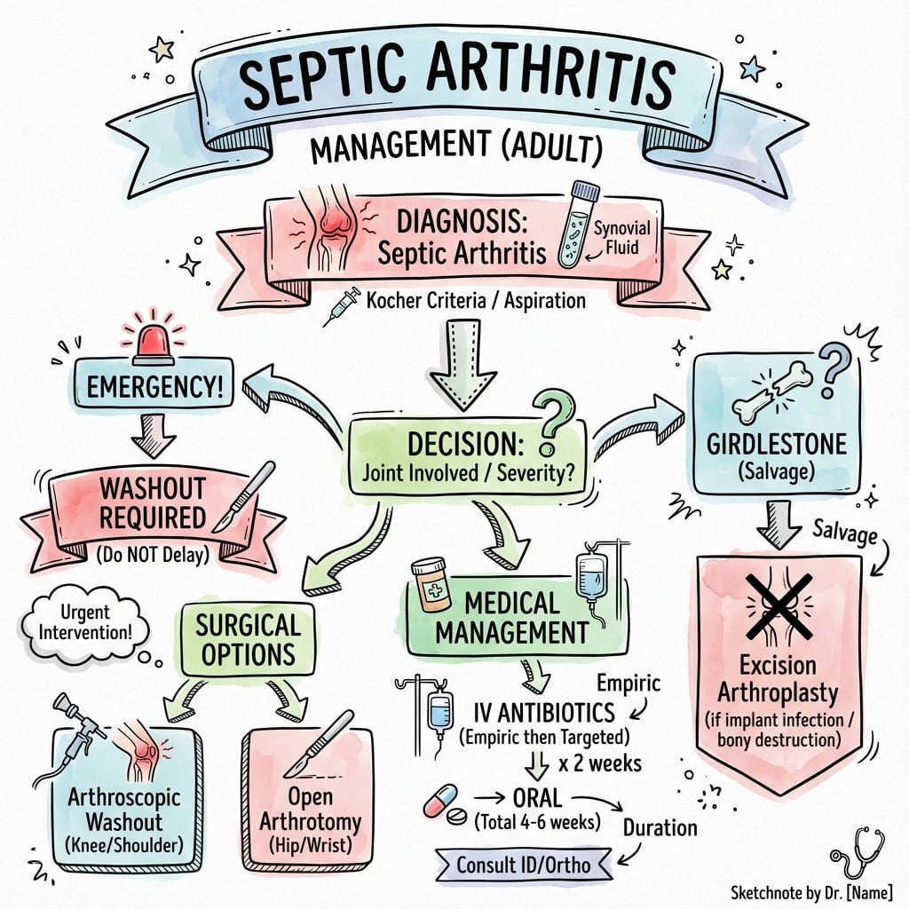
Principles: Urgent washout + IV antibiotics.
Surgical Washout
- Knee, shoulder, ankle, wrist
- Advantages: Less soft tissue trauma, better visualisation, shorter recovery
- Technique: Thorough lavage with 9+ litres saline, debridement of infected tissue
- Remove fibrin clots and debris
- Hip (difficult arthroscopic access)
- Failed arthroscopic washout
- Complex cases with extensive infection
- Tissue samples for culture (at least 3-5 samples)
- Copious lavage (minimum 9 litres)
- May need repeated washouts every 48-72 hours if ongoing sepsis
- Consider leaving drain in situ
Antibiotic Therapy
- Start after aspiration (do not delay for culture results)
- Flucloxacillin 2g IV QID - first-line for most cases
- Vancomycin if MRSA risk (recent hospitalisation, IVDU, diabetes)
- Add gentamicin or ceftriaxone if Gram-negative suspected
- Adjust based on cultures and sensitivities
- Involve infectious diseases/microbiology team
- Duration: IV 2-4 weeks then oral step-down to complete 4-6 weeks total
- Flucloxacillin 1g QID
- Cephalexin 1g TDS
- Clindamycin (if penicillin allergy)
Post-operative Management
- Early active and passive ROM exercises
- Weight bearing as tolerated
- Physical therapy involvement
- Splinting in position of function if needed
- Repeat inflammatory markers (CRP, WCC) every 2-3 days
- Clinical assessment for resolution
- Repeat aspiration if persistent effusion
Post-operative care crucial for functional outcome.
Guidelines, Registries & Global Practice
Global epidemiology:
- Annual incidence of native-joint septic arthritis is roughly 4-10 per 100,000 in the general population, rising to 30-70 per 100,000 in patients with rheumatoid arthritis or prosthetic joints.
- S. aureus dominates worldwide; the proportion that is MRSA varies widely by region (low in much of Scandinavia and the Netherlands, considerably higher in parts of the USA, South Asia and among community strains in remote and Indigenous populations).
- Gonococcal arthritis remains an important cause in young sexually active adults globally; tuberculous and fungal joint sepsis are disproportionately common in high-TB-burden and resource-limited settings and in the immunocompromised.
Side-by-side guidance:
- Diagnostic emphasis
- Aspirate before antibiotics; synovial WCC, Gram, crystals, culture
- Empirical antibiotics
- Flucloxacillin IV; vancomycin/teicoplanin if MRSA risk or penicillin allergy
- Drainage
- Repeated needle aspiration or surgical washout; surgical if large/loculated
- Diagnostic emphasis
- Multiple deep tissue/synovial cultures; hold antibiotics if stable
- Empirical antibiotics
- Tailored once cultured; broad cover (anti-staph + Gram-negative) if septic
- Drainage
- DAIR for acute PJI with stable implant; staged revision for chronic
- Diagnostic emphasis
- MSIS/ICM criteria, synovial alpha-defensin, leucocyte esterase
- Empirical antibiotics
- ID-directed
- Drainage
- Stage selection by chronicity and host factors
- Diagnostic emphasis
- Standardised PJI definition; synovial WCC and PMN thresholds
- Empirical antibiotics
- Pathogen-directed, biofilm-active agents (rifampicin combinations for staphylococci)
- Drainage
- DAIR within ~3 weeks; one- or two-stage revision
Regional empirical regimens differ chiefly in how aggressively they cover MRSA — driven by local resistance data rather than true clinical disagreement. The universal principles (aspirate first, urgent drainage, source control, ID/microbiology involvement) are consistent across all bodies.
Registry and resource notes:
- Arthroplasty registries (NJR, AJRR, AOANJRR, the Swedish and Norwegian registers) track PJI as a leading cause of revision and inform implant- and fixation-specific infection risk; deep infection is among the top three reasons for revision knee arthroplasty in most registries.
- High-resource settings favour arthroscopic washout, antibiotic-loaded cement spacers and multidisciplinary bone-infection units. In limited-resource settings, open arthrotomy with serial saline lavage, longer empirical courses, and a lower threshold for considering tuberculous or fungal aetiology are appropriate adaptations.
Septic arthritis is an orthopaedic emergency. Joint cartilage is destroyed within 24-48 hours. Aspirate the joint, start antibiotics, and proceed to surgical washout urgently.
Complications

Early Complications
- Inadequate debridement - failure to remove all infected tissue
- Resistant organism - MRSA, multi-drug resistant GNB
- Biofilm formation (especially PJI) - bacteria protected from antibiotics
- May require repeated washouts (every 48-72 hours until resolved)
- Consider changing antibiotic regimen based on sensitivities
- Can progress to septicaemia with bacteraemia
- Multi-organ failure in severe cases (ARDS, AKI, DIC)
- Mortality 10-15% overall
- Higher mortality in elderly (up to 30%) and immunocompromised patients
- Requires ICU admission and aggressive resuscitation
- Wound dehiscence after open washout
- Sinus tract formation with chronic drainage
- Skin necrosis requiring plastic surgery input
Late Complications
- Cartilage loss is irreversible once proteoglycans depleted
- Occurs within 24-48 hours without treatment
- Results in secondary osteoarthritis requiring arthroplasty
- Worse outcomes in weight-bearing joints (hip, knee)
- Particularly hip joint in children
- Septic arthritis can damage blood supply to femoral head
- May develop Perthes-like changes
- Long-term surveillance required
- Fibrous or bony fusion of joint surfaces
- More common with delayed treatment beyond 7 days
- May require arthrodesis for pain relief
- Consider arthroplasty if bone stock adequate
- Physeal damage if infection crosses growth plate
- Limb length discrepancy (can be several centimetres)
- Angular deformity requiring corrective osteotomy
- Growth arrest lines visible on X-ray
- Post-infectious arthritis even after eradication
- Reduced range of motion from fibrosis
- May require prolonged rehabilitation
Outcomes and Prognosis
Functional outcomes:
- Good to excellent outcome in 70-80% if treated promptly
- Poor outcome associated with delayed diagnosis
- Hip and shoulder have worse functional prognosis
Prognostic Factors
- Delay in treatment more than 7 days (single most important factor)
- Age more than 65 years
- Pre-existing joint disease (RA, OA)
- Polyarticular involvement (often haematogenous)
- Virulent organisms (S. aureus worse than streptococci)
- Prosthetic joint involvement
- Immunocompromised state (diabetes, HIV, malignancy, steroids)
- Axial joint involvement (hip, shoulder)
- Treatment within 24-48 hours
- Single joint involvement
- Streptococcal or gonococcal infection
- Young healthy patient
- Peripheral joint (knee, ankle)
Controversies & Areas of Uncertainty
- Antibiotic duration. The Gjika RCT (PMID 30992295) showed 2 weeks non-inferior to 4 weeks after surgical drainage, but the cohort was dominated by hand/wrist joints. How far this extends to large weight-bearing joints, S. aureus bacteraemia, or immunocompromised hosts remains unsettled, and most units still use 3-6 weeks for large-joint or staphylococcal disease.
- Needle aspiration vs surgical washout for native joints. Serial closed aspiration achieves source control in selected accessible joints (notably the knee) in some series, while many surgeons regard urgent arthroscopic or open washout as standard. No RCT settles this; decision rests on joint, organism, loculation and response.
- Arthroscopic vs open drainage. Meta-analytic data (PMID 33939020) favour arthroscopy for knee and shoulder, but the evidence is low-quality and retrospective; the hip is still generally opened.
- DAIR vs early revision in acute PJI. Success of DAIR depends heavily on a short symptom duration, well-fixed implant, susceptible organism and exchange of modular parts. The exact time threshold (often quoted as ~3 weeks) and the role of rifampicin combinations for staphylococci are debated.
- Diagnostic biomarkers. Synovial alpha-defensin, leucocyte esterase and serum/synovial procalcitonin add specificity but are imperfect; none replaces aspiration, culture and clinical judgement. The "WCC over 50,000" threshold is a guide, not an absolute cut-off (sensitivity is well under 100%).
- Crystals plus infection. Demonstrating gout or CPPD does not exclude concurrent sepsis; missed co-existent infection is a recognised pitfall.
Exam Viva Scenarios
Practise clinical reasoning and management decisions out loud
“A 60-year-old diabetic presents with a hot, swollen, painful knee. He cannot bear weight. Temperature is 38.5°C. How do you manage?”
“A 72-year-old woman presents 3 weeks after total knee replacement with increasing pain, wound drainage, and low-grade fever. Her wound looks erythematous with some purulent discharge. How would you assess and manage this?”
“A 55-year-old man with known gout presents with a hot, swollen, exquisitely tender first metatarsophalangeal joint and a temperature of 38.2°C. Aspiration shows negatively birefringent crystals. The on-call doctor wants to discharge him on colchicine. What is your approach?”
Key Points
- Orthopaedic emergency
- Staph aureus most common
- Aspirate: WCC, Gram, culture, crystals
Aspirate
- WCC greater than 50,000 (greater than 90% PMN) suggestive
- Gram stain for quick ID
- Crystals to rule out gout
Treatment
- Surgical washout (arthroscopic/open)
- IV antibiotics 2-4 weeks
- Total 4-6 weeks antibiotics
Special
- PJI: DAIR vs 2-stage revision
- Gonococcal: Young, may respond to antibiotics alone
Evidence Base
Does This Adult Patient Have Septic Arthritis? (Rational Clinical Examination)
- Systematic review of 14 studies, 6242 patients (653 with septic arthritis)
- Synovial WCC drives diagnosis: LR 7.7 if over 50,000/mcL, LR 28 if over 100,000/mcL
- WCC under 25,000/mcL lowers probability (LR 0.32) but does NOT exclude sepsis
- PMN of at least 90% gives LR 3.4; clinical signs and serum tests are weak alone
Kocher Criteria for Paediatric Hip
- 4 independent predictors: history of fever, non-weight-bearing, ESR of at least 40 mm/hr, serum WCC over 12,000/mcL
- 0 predictors gives under 0.2% probability of septic arthritis
- 3 predictors 93.1%, 4 predictors 99.6% probability
- Retrospective derivation; later external validation showed lower probabilities, so it supplements rather than replaces aspiration
Arthroscopy vs Open Arthrotomy - Systematic Review and Meta-analysis
- 20 studies, 10,249 patients with septic arthritis of any joint
- Arthroscopy: lower re-infection (OR 1.35, 95% CI 1.16-1.58) and complications (OR 1.32)
- Benefit strongest for knee and shoulder; shorter hospital stay with arthroscopy
- Authors caution overall evidence quality is low (no RCTs)
Two vs Four Weeks of Antibiotics After Surgical Drainage (RCT)
- Randomised non-inferiority trial, 154 adults (77 per arm) after surgical lavage
- Cure: 99% (2-week arm) vs 97% (4-week arm); only 3 recurrences overall
- No difference in adverse events or sequelae; shorter hospital stay with 2 weeks
- Majority were hand/wrist joints; median IV duration only 1-2 days
Serum Procalcitonin as a Diagnostic Marker (Meta-analysis)
- 10 studies, 838 patients
- Pooled sensitivity 0.54, specificity 0.95 for septic arthritis
- Positive LR 10.97 (good rule-in), negative LR 0.49 (poor rule-out)
- More specific than CRP but cannot exclude infection when negative