Myelomeningocele and Orthopaedic Care
- Neurological Level: Determines ambulatory potential.
- Hip Dysplasia: Common but treatment controversial.
- Clubfoot: Often rigid; Ponseti first-line, but relapse is far more frequent than in idiopathic feet.
- Scoliosis: Congenital and neuromuscular types.
- Fractures: Insensate limbs prone to pathological fractures.
- “Level determines function
- “Hip surgery controversial
- “Clubfoot: Ponseti first, expect relapse
- “Insensate = fracture risk
Overview and Epidemiology
Spina bifida is a neural tube defect: the caudal neural tube fails to close, which it should do by about day 28 of embryonic development. It is diagnosed prenatally or at birth. Orthopaedic problems are universal, in the hips, spine, feet and knees and as fractures, and the neurological level determines ambulatory potential.
The spectrum. The forms differ in what herniates through the posterior arch defect:
- Myelomeningocele (95% of cases) - the most severe, open form. Spinal cord and meninges herniate through the defect, the neural placode is exposed and there is significant neurological deficit. It requires immediate surgical closure after birth and is nearly always associated with a Chiari II malformation.
- Meningocele (4%) - the meninges herniate but the spinal cord is intact, and neurological function is usually better. It may present as a lumbosacral mass at birth, and surgical closure is still required.
- Spina bifida occulta (1%) - a minor posterior arch defect without herniation, often asymptomatic and found incidentally. The skin markers are a hair tuft, dimple, lipoma or haemangioma, and it may be associated with a tethered cord.
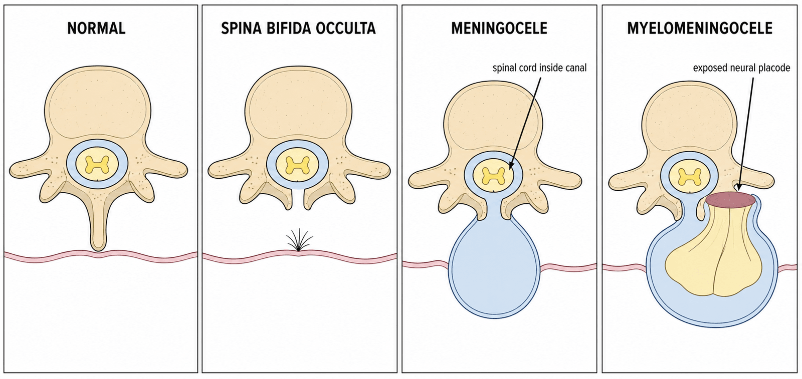
Epidemiology. The incidence is 0.5-1 per 1000 live births in folate-fortified populations, against 2-4 per 1000 historically. Rates are highest in Celtic/British Isles populations and parts of China and Latin America and lowest where flour is mandatorily fortified, and there is a slight female predominance.
Risk factors. Maternal folate deficiency, anticonvulsants (valproate, carbamazepine), maternal diabetes, obesity, hyperthermia, a prior affected pregnancy and MTHFR polymorphisms.
Prevention. Periconceptional folic acid, 400-800 mcg daily, is advised for all women planning pregnancy, starting at least a month before conception and continuing through the first trimester; high-risk women (previous NTD pregnancy, antiepileptic drugs, diabetes, obesity) take 5 mg daily. At 400-800 mcg it prevents most NTDs: the MRC Vitamin Study RCT showed a 72% reduction in NTD recurrence, and fortification programmes reduce population NTD prevalence by roughly 30-50%.
Fortification. Mandatory flour fortification was adopted in the USA (1998), Canada, Australia/New Zealand (2009) and over 80 countries, but is not yet mandatory across most of Europe. There it is debated over the theoretical masking of B12 deficiency and cancer concerns, though large analyses have not confirmed harm.
Associated conditions. The neurosurgical, urological and cognitive problems that accompany the defect:
- Chiari II malformation - nearly 100% with myelomeningocele
- Hydrocephalus - 80-90% require a VP shunt
- Tethered cord - may develop progressively
- Syringomyelia - a common finding on MRI
- Neurogenic bladder - universal below the lesion level
- Neurogenic bowel - requires a bowel management programme
- Cognitive impairment - variable, with intelligence often preserved
Pathophysiology, Anatomy & Pathomechanics
Level predicts function. The level of the lesion determines muscle function below it, and motor function is predictable from the spinal level. Sensation is typically absent below the lesion, and the motor level often differs from the sensory level.
- Key Muscle
- Iliopsoas
- Function
- Hip flexion
- Clinical Test
- Observe hip flexor power
- Key Muscle
- Iliopsoas, Adductors
- Function
- Hip flexion/adduction
- Clinical Test
- Hip adduction against resistance
- Key Muscle
- Quadriceps
- Function
- Knee extension
- Clinical Test
- Knee extension against gravity
- Key Muscle
- Tibialis Anterior
- Function
- Ankle dorsiflexion
- Clinical Test
- Walk on heels
- Key Muscle
- EHL, Peroneals
- Function
- Great toe extension, eversion
- Clinical Test
- Great toe dorsiflexion
- Key Muscle
- Gastrocnemius
- Function
- Ankle plantarflexion
- Clinical Test
- Single leg heel raise
- Key Muscle
- Bladder/Bowel
- Function
- Continence
- Clinical Test
- Urodynamics
Muscle imbalance. Active muscles without functioning antagonists cause progressive deformity, and this drives most of the deformities in spina bifida. Active hip flexors (L1-L2) without hip extensors (L5-S1) produce a hip flexion contracture; active quadriceps (L3) without hamstrings produce knee hyperextension. Gravity and positioning add unopposed forces of their own, so prolonged sitting also creates a hip flexion contracture.
The insensate limb. The lower limbs are insensate below the lesion level. Without protective sensation, trauma goes unrecognised, pressure sores form under braces and casts, and a pathological fracture presents with swelling rather than pain. Insensate, non-weight-bearing limbs also become osteoporotic, so fractures occur with minimal trauma.
Deformity by level.
- Thoracic - no lower-limb motor function. Paralytic dislocation of both hips, scoliosis in 100%, and a severe kyphosis that may interfere with sitting.
- High lumbar (L1-L2) - hip flexors act without extensors (flexion contracture) and adductors without abductors (adduction contracture), and the imbalance produces progressive hip subluxation or dislocation. Knee flexion contractures are common.
- Mid lumbar (L3-L4) - active quadriceps make these children community ambulators. Hip instability is less severe; the knee can extend but may hyperextend, and foot deformities (clubfoot, calcaneus) are common.
- Low lumbar (L5-S1) - near-normal motor function and good ambulatory potential, with mild foot deformities (pes cavus, clawing) and less severe spinal problems.
Classification Systems
Classification by neurological level ties the working muscles to what the child can expect to do and what orthosis it will take.
- Motor Function
- No lower-limb motor function
- Ambulatory Potential
- Wheelchair only
- Orthotic Needs
- Standing frame (therapeutic)
- Motor Function
- Hip flexion only
- Ambulatory Potential
- Standing frames; wheelchair primary
- Orthotic Needs
- HKAFO
- Motor Function
- Quadriceps active
- Ambulatory Potential
- Community ambulation with AFOs
- Orthotic Needs
- KAFO or AFO
- Motor Function
- Ankle dorsiflexion
- Ambulatory Potential
- Community, minimal aids; good ambulators
- Orthotic Needs
- AFO, often supramalleolar
- Motor Function
- Ankle plantarflexion
- Ambulatory Potential
- Near-normal walking
- Orthotic Needs
- Shoe inserts only
Clinical Assessment
History. Start with the documented motor and sensory level and the ambulatory status, both current and the best function achieved. Then bladder and bowel management (clean intermittent catheterisation, bowel programme), prior surgery (closure, shunt, orthopaedic procedures) and the shunt itself: its type, the last revision and any symptoms of malfunction.
Red flags. Four things to identify:
- Shunt malfunction - headache, vomiting, irritability, decreased consciousness
- Tethered cord - deteriorating gait, new weakness, increasing scoliosis, change in bladder function
- Skin breakdown - location, duration, prior wounds
- Recent fractures - often missed because of the lack of pain
Neurological examination. Test each myotome systematically for the motor level, and light touch and pinprick for the sensory level; reflexes may be variable depending on the level. Document it as the baseline for later comparison.
Spine. Assess scoliosis with Adam's forward bend test and trunk shift. Lumbar kyphosis is common and is judged rigid or flexible; look at the skin over the spine for scars and sinus tracts, and assess sitting balance, which is essential in wheelchair users.
Hip. Document any flexion contracture with the Thomas test, and test stability with Barlow and Ortolani in infants and by assessing it with motion. Observe the gait pattern if the child walks, and the sitting posture for pelvic obliquity from hip problems.
Knee. Flexion contractures are common and limit the ability to use orthoses. Hyperextension may occur where the quadriceps function, and an extension lag assesses quadriceps strength.
Foot. Name the deformity (clubfoot, vertical talus, calcaneus, cavus), assess its rigidity by whether it corrects, and look for pressure points, calluses and ulcers. Then ask whether it is braceable: can the foot fit in an AFO without pressure problems?
Skin. Insensate skin is very vulnerable, so this is a critical part of the examination. Look under braces and orthoses, over bony prominences and prior wound sites, and for signs of infection.
Investigations
Radiographs.
- Spine - scoliosis, kyphosis, vertebral anomalies
- Hip - DDH, subluxation
- Foot - clubfoot, talus position
MRI of the spine looks for a tethered cord, and is obtained before scoliosis surgery.
Other tests. EMG if the level is unclear, and urodynamics for bladder function.
Differential Diagnosis
The differential is for the flaccid lower-limb paralysis, foot deformities and neurogenic bladder seen in a child, plus the imaging differential of a lumbosacral mass or midline skin lesion.
- Distinguishing Features
- Open defect, neural placode exposed, Chiari II, hydrocephalus
- Key Discriminator
- Visible open lesion at birth, flaccid paralysis below level
- Distinguishing Features
- Skin-covered; cutaneous markers (lipoma, hair tuft, dimple, sinus)
- Key Discriminator
- Progressive (not static) deficit; MRI shows low conus
- Distinguishing Features
- Absent sacral segments, popliteal webbing, maternal diabetes
- Key Discriminator
- Lateral radiograph shows absent sacrum/lumbar vertebrae
- Distinguishing Features
- Multiple rigid joint contractures, normal sensation, intact bladder
- Key Discriminator
- Sensation PRESERVED; symmetric rigid contractures
- Distinguishing Features
- Symmetric flaccid weakness, fasciculations, normal sensation/sphincters
- Key Discriminator
- Genetic (SMN1); sensation and continence preserved
- Distinguishing Features
- Acquired progressive deficit, back pain, level on MRI
- Key Discriminator
- History of trauma or enhancing lesion on MRI
The cardinal feature separating spina bifida (and other dysraphisms) from arthrogryposis and SMA is absent sensation below the level.
Management Algorithm
Prenatal repair. The MOMS RCT showed that in-utero repair of the myelomeningocele halves shunt rates and improves motor and ambulation outcomes, but at the cost of preterm birth and uterine dehiscence. Not all fetuses are candidates, and counselling must balance the maternal risk.
The controversy. Hip dysplasia is common at high lumbar levels, where the hip flexors work without extensors or abductors, and its treatment is controversial. The traditional view is that reduction does not improve walking, and Wright's evidence-based review (Clin Orthop Relat Res 2011) supports it at Grade B; some still advocate reduction for sitting balance. Current thinking is more individualised, operating for specific indications rather than routinely, and surgery is often avoided unless the hip is painful.
By level. At the thoracic level surgery brings no benefit and is avoided. At high lumbar levels (L1-L2) treatment is controversial and many do not operate. At mid and low lumbar levels a hip may benefit from reduction if there is ambulatory potential.
Indications for surgery. The specific indications are:
- A painful hip (rare, because of insensitivity)
- Sitting imbalance or pelvic obliquity from a unilateral dislocation
- L3-L4 level with ambulatory potential
- Skin breakdown over the prominence
When to leave the hip alone. Avoid surgery in bilateral painless dislocations and where function is good despite the imaging.
Surgical Techniques
Indications. Beyond a progressive curve past the 40-50 degree threshold, fusion is indicated for:
- Declining sitting balance affecting function
- Trunk imbalance causing pain or skin breakdown
- Pulmonary compromise (rare)
Before the operation.
- MRI spine to exclude a tethered cord, releasing a symptomatic tethered cord first
- Neurosurgical consultation to assess the shunt
- Latex-free theatre, with all equipment pre-ordered
- Blood typing, because blood loss is high
- Nutritional optimisation, since many are malnourished
The fusion. Posterior spinal fusion is the standard approach, from T2 to the pelvis in wheelchair users, with pelvic fixation by iliac screws or the Galveston technique. Short fusions have a high failure rate and are avoided, and allograft supplementation of the bone graft is worth considering.
Pitfalls. Poor bone quality needs larger, longer screws, and skin closure may be difficult over a kyphosis. Position the shunt carefully to avoid kinking, and expect that custom seating may be needed after surgery.
Complications
Pressure sores are very common: skin breaks down over bony prominences that the child cannot feel. The high-risk sites:
- Under braces and orthoses
- Ischial tuberosities in wheelchair users
- Sacrum and coccyx
- Heels and malleoli
- Over prominent hardware
Preventing them. Check the skin regularly, daily for at-risk areas, and teach patients and families to do it. Use pressure-relieving wheelchair cushions and regular weight shifts and position changes.
When a sore is present. Remove all pressure from the area. Wound care may need plastic surgery, underlying osteomyelitis may require debridement, and healing can take months.
Tethered cord. Radiological tethering is near-universal, but the clinical picture is progressive: new weakness, scoliosis progression, pain or urological change. It is investigated with MRI and treated by neurosurgical release, which is indicated only for symptomatic progression, not for imaging findings alone.
Shunt malfunction. Its frequency is variable, and it needs urgent neurosurgery. For any operation, inform neurosurgery of the plan, position the child to avoid pressure on the shunt, monitor for signs of malfunction afterwards, and be aware the shunt may need adjustment for positioning.
Wound healing. Poor skin quality is common and the risk of infection is increased. Consider plastic surgery involvement for complex closures; extended antibiotics are often required.
Latex Allergy in Spina Bifida — Why and What To Do
Mechanism. Latex allergy is a Type I, IgE-mediated immediate hypersensitivity to soluble proteins in natural rubber latex (from Hevea brasiliensis), not to the finished rubber itself.
Why spina bifida is high-risk. Spina bifida is the archetypal cause of clinically significant natural-rubber-latex allergy. These patients are sensitised by early, repeated and mucosal/parenteral exposure: multiple operations from birth (closure, shunt, orthopaedic) and lifelong clean intermittent catheterisation with latex catheters. The cumulative number of operations is the strongest risk factor; myelomeningocele (versus lower dysraphism) and atopy add further risk.
Sensitisation is not anaphylaxis. Latex sensitisation (specific IgE) is quoted at 30-70%, and was 47% in Pittman's series; clinical anaphylaxis is far rarer, one event across 646 operations in that same cohort. Sensitisation can be silent, and a positive specific IgE without prior clinical reaction still mandates avoidance, because the first clinical event may be intra-operative anaphylaxis.
Latex-fruit syndrome. Latex proteins (notably hevein/prohevein and class-I chitinases) cross-react with plant foods. Ask specifically about reactions to banana, avocado, kiwi and chestnut (the classic quartet), and also passionfruit, fig and papaya; a history of oral itch or swelling with these foods is a clinical clue to occult sensitisation.
Perioperative protocol.
- Latex-free from birth - primary prevention is the highest-yield step. Raising every child with spina bifida in a latex-free environment from birth measurably lowers sensitisation rates, so avoidance is not only a theatre issue.
- Latex-free (latex-safe) theatre for all procedures - synthetic (nitrile/neoprene) gloves, and latex-free catheters, tourniquets, drains, syringe plungers, medication vial bungs and anaesthetic circuit.
- First on the list - to minimise airborne latex particulate from prior cases.
- Premedication - pre-operative antihistamines may be given in known cases, but routine antihistamine/steroid premedication does not reliably prevent latex anaphylaxis and does not replace strict avoidance.
- Recognise anaphylaxis - unexplained intra-operative cardiovascular collapse, bronchospasm or urticaria, often appearing minutes after induction or skin or mucosal contact, is latex anaphylaxis until proven otherwise. Management is standard anaphylaxis care: remove the trigger, adrenaline, fluids, airway support.
Postoperative Care
Skin and casts. Skin monitoring is critical in the first 48-72 hours, because insensate limbs cannot report problems. Casts are windowed for skin inspection, bivalved when possible, well padded over bony prominences and not too tight. Nursing staff check the skin daily, parents check the cast edges daily and are taught to inspect before discharge, and the cast is kept on only briefly for skin tolerance before a transition to a carefully moulded orthosis when safe.
Positioning. Avoid pressure on the shunt, use a pressure-relieving mattress and turn the child regularly, at least every 2 hours. Heel protection is mandatory.
Pain. Pain perception may be diminished, but central processing is intact and these children still need adequate analgesia. Watch for signs of discomfort such as irritability and changes in vital signs; regional blocks can be effective.
Rehabilitation goals by level.
- Thoracic - maximise upper-body strength and wheelchair skills
- L1-L2 - standing programme if appropriate; transfers
- L3-L4 - gait training with appropriate orthoses
- L5-sacral - optimise gait efficiency and minimise energy expenditure
Orthoses. Pressure sores are common, so careful orthotic fitting is essential. An experienced orthotist fits them, the fit is checked at every clinic visit, regular adjustments anticipate growth, and they are replaced when worn or outgrown.
Outcomes/Prognosis
- Community Ambulation Rate
- 0%
- Community Ambulation Rate
- 0-10%
- Community Ambulation Rate
- 50-80%
- Community Ambulation Rate
- 80-100%
Prognostic factors. Outcome is better with a lower neurological level (L4-sacral), preserved cognition, strong family support, access to multidisciplinary care and early intervention for deformities. It is worse with a higher lesion level, significant cognitive impairment, multiple shunt revisions, severe scoliosis and recurrent pressure sores.
Life expectancy has improved significantly with modern care, and most patients with myelomeningocele reach adulthood. The main causes of death are shunt-related, renal failure and respiratory, and quality of life can be excellent with appropriate support.
Transition to adult care. Plan the transition from age 14-16 years and identify an adult orthopaedic surgeon experienced with spina bifida. Surveillance continues for skin breakdown, progressive deformity and new weakness (tethered cord), and the Spina Bifida Foundation offers support services.
Guidelines, Registries & Global Practice
Global Epidemiology
- Prevalence: 0.5-1 per 1000 live births in folate-fortified countries; 2-4 per 1000 historically and in unfortified regions
- Highest rates: British Isles/Celtic populations, northern China, parts of Latin America
- Lowest rates: USA, Canada, Australia/New Zealand and other countries with mandatory flour fortification
- Mandatory fortification (now in over 80 countries) reduces population NTD prevalence by roughly 30-50%
Folic Acid Guidelines - Side by Side
- Recommendation
- 400 mcg/day periconceptionally; advocates mandatory staple-food fortification
- Recommendation
- 400 mcg/day for all; 5 mg/day if previous NTD, diabetes, BMI of 30 or more, or on antiepileptics
- Recommendation
- 400-800 mcg/day for all women capable of pregnancy (Grade A); mandatory grain fortification since 1998
- Recommendation
- 400-500 mcg/day; 5 mg/day high-risk; mandatory bread-making flour fortification since 2009
Orthopaedic & Surgical Practice Consensus
- AAOS / POSNA & EPOS (European Paediatric Orthopaedic Society): deformity management individualised to functional level; preserve a plantigrade, braceable foot; avoid routine hip reduction in high-level lesions
- AO/Scoliosis Research Society: pre-operative spinal MRI to exclude symptomatic tethered cord; long posterior fusion to the pelvis for neuromuscular curves affecting sitting balance
- Fetal therapy consensus (post-MOMS): in-utero repair offered in selected centres meeting MOMS eligibility criteria, balanced against preterm birth and uterine risk
Registries & Surveillance
- EUROCAT (European congenital anomaly surveillance) and the ICBDSR track NTD prevalence and the impact of fortification policy internationally
- National Spina Bifida Patient Registry (USA) captures longitudinal multidisciplinary outcomes
- No dedicated implant registry exists for spina bifida; arthroplasty/instrumentation outcomes are drawn from neuromuscular subgroups within general registries
High- vs Limited-Resource Practice Variation
- High-resource: multidisciplinary clinics (paediatric orthopaedics, neurosurgery, urology, rehabilitation, orthotics), urodynamics-guided bladder care, fetal surgery programmes, custom orthoses and seating
- Limited-resource: late presentation of unrepaired/neglected defects, higher infection and pressure-sore burden, reliance on serial casting and simpler bracing; fortification programmes are the highest-yield public-health intervention
MCQ Practice Points
Q: A patient with L3 spina bifida has which muscle function? A: Quadriceps (knee extension). Can be a community ambulator.
Q: Does hip reduction improve walking in spina bifida? A: Controversial - traditional view is no improvement.
Q: What is the goal of foot surgery in spina bifida? A: Plantigrade foot that can be braced.
Q: What allergy is common in spina bifida? A: Latex allergy.
Q: What must be excluded before scoliosis surgery in spina bifida? A: Tethered cord - requires pre-operative MRI and neurosurgical release if present.
Q: How does a pathological fracture present in spina bifida? A: Swelling and warmth without pain - mimics infection but occurs due to insensate osteoporotic bone.
Self-Assessment Quiz
Viva Scenarios
Practise clinical reasoning and management decisions out loud
“5-year-old with L3 level spina bifida. Ambulant with AFOs. Presents with bilateral hip subluxation.”
“Same child also has rigid bilateral clubfoot. How would you manage?”
“10-year-old with thoracic level spina bifida. Wheelchair-bound. Thoracolumbar scoliosis 60 degrees with pelvic obliquity.”
“8-year-old with L2 level spina bifida presents with swelling and warmth of the left thigh. No history of trauma. Parents worried about infection.”
LEVELS
- L1-L2: Hip flexors
- L3-L4: Quads (ambulator)
- L5: Ankle dorsiflexion
- Thoracic: Wheelchair
ORTHOPAEDIC
- Hips: Controversial
- Spine: Scoliosis/Kyphosis
- Feet: Clubfoot
- Fractures: Insensate
SURGERY ISSUES
- Latex allergy
- Tethered cord
- Pressure sores
- Shunt malfunction
GOALS
- Maximize function
- Plantigrade feet
- Sitting balance
- Prevent sores
Evidence Base
MOMS Trial (Adzick et al)
- RCT of prenatal vs postnatal MMC repair, stopped early for efficacy (183 patients)
- Shunt placement by 12 months: 40% prenatal vs 82% postnatal (RR 0.48)
- Improved motor function and ambulation at 30 months; reduced hindbrain herniation
- Increased preterm delivery and uterine dehiscence
MOMS Full Cohort (Farmer et al)
- Full 183-patient cohort, 30-month outcomes
- Independent ambulation 44.8% prenatal vs 23.9% postnatal (p=0.004)
- Functional level at least 2 better than anatomic level in 26.4% vs 11.4%
- Lesion at L3 or below and in-utero leg movement predicted independent walking