Anterior Rotator Cuff | Internal Rotation | Lesser Tuberosity
- Subscapularis = internal rotator, anterior cuff, inserts on lesser tuberosity
- Upper fibres tear most commonly (within biceps sheath)
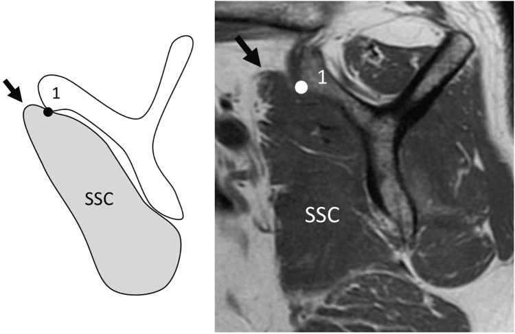
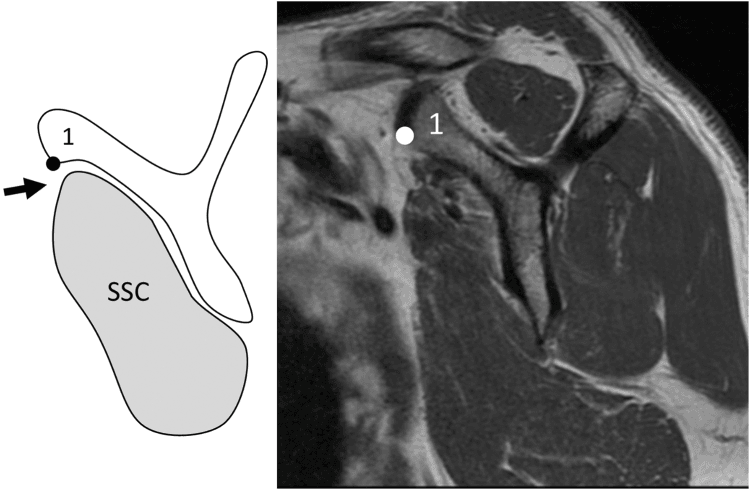
- Comma sign = SGHL/coracohumeral ligament complex indicates superior edge
- Lift-off and bear-hug tests for clinical diagnosis
- Biceps pathology frequently associated
- “Napoleon and lift-off tests assess subscapularis function
- “Fox & Romeo classification based on tear extent
- “No single percentage threshold mandates repair; correlate symptoms, weakness, footprint, mobility, fatty infiltration and associated cuff disease
- “Biceps subluxation/dislocation is a strong clue to pulley/subscapularis injury and requires its own treatment decision
Overview and Epidemiology
The subscapularis accounts for 10-25% of rotator cuff tears and was historically underdiagnosed. A tear costs internal-rotation strength and anterior shoulder function, and associated biceps problems are common. Recognising the tear and treating it appropriately improves outcomes.
Who. Males predominate, which is attributed to occupational factors, and heavy labourers are at higher risk. The fifth and sixth decades are the common ages. The tear often comes with other cuff tears, as part of an anterosuperior pattern.
Causes. The aetiology is traumatic or degenerative. The tendon also fails after surgery and in massive cuff tears, and subcoracoid stenosis contributes:
- Traumatic - hyperextension or forced external rotation
- Degenerative - anterior impingement
- Iatrogenic - after shoulder surgery, arthroplasty in particular
- Massive cuff tears - the subscapularis tear is associated with them
- Subcoracoid stenosis - a contributing factor
Anatomy and Biomechanics
The muscle. The subscapularis is the only internal rotator of the rotator cuff, and inserts on the lesser tuberosity through a broad tendon. The long head of biceps runs in the groove between it and the supraspinatus.
- Subscapularis
- Lesser tuberosity
- Supraspinatus
- Greater tuberosity
- Infraspinatus
- Greater tuberosity
- Subscapularis
- Internal rotation
- Supraspinatus
- Abduction
- Infraspinatus
- External rotation
- Subscapularis
- Anterior
- Supraspinatus
- Superior
- Infraspinatus
- Posterior
- Subscapularis
- 10-25%
- Supraspinatus
- Most common
- Infraspinatus
- Common with SSP
Tendon above, muscle below. The upper 60% of the insertion is tendinous and holds sutures, so it can be repaired to anchors. The lower 40% is muscular, holds sutures poorly, and may need margin convergence.
Why the upper fibres fail first. The upper fibres are intra-articular, within the biceps sheath, and they tear first; full-thickness tears then extend from superior to inferior. The lower fibres are extra-articular and better protected.
What the muscle does. Internal rotation is its primary function. It is also an anterior stabiliser of the humeral head, a humeral head depressor with the rest of the cuff, and it forms a force couple with the posterior cuff, infraspinatus and teres minor, the external rotators. Losing it disrupts that force-couple balance, and repair restores the anterior restraint and the balance with it.
Landmarks. Five structures frame the subscapularis:
- Lesser tuberosity - the insertion
- Biceps groove - the lateral border
- Comma sign - the SGHL complex at the superior edge
- Coracoid process - anterior
- Biceps pulley - at the junction with supraspinatus
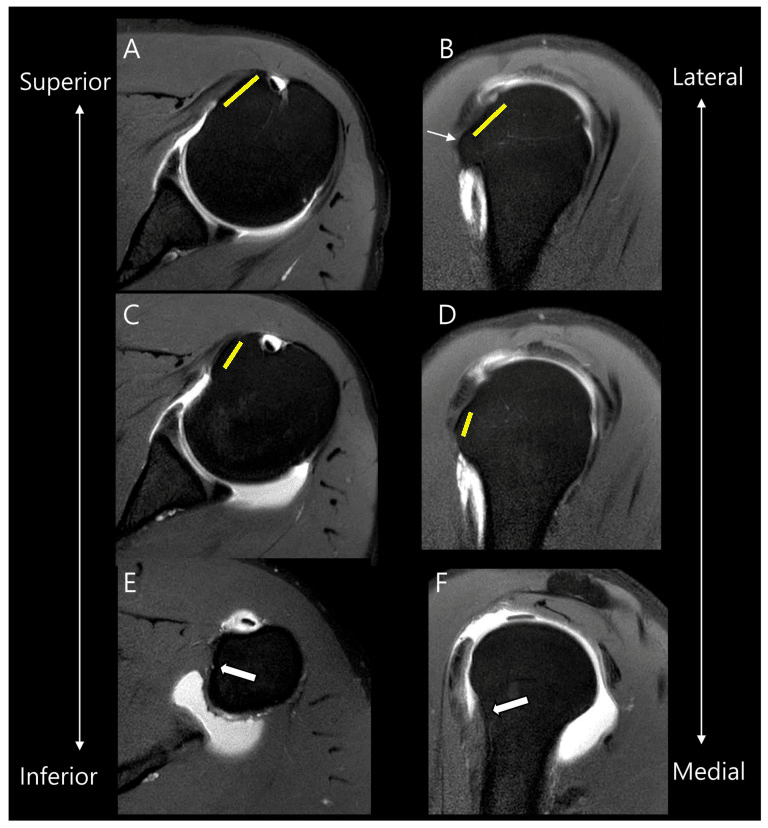
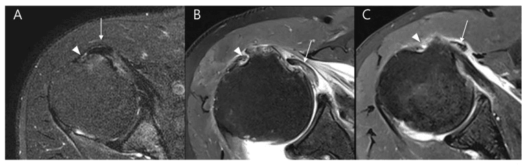
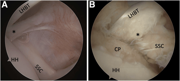
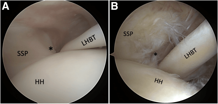
Classification Systems
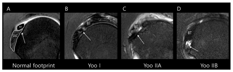
Fox and Romeo grade the tear by how much of the tendon has failed, from a partial articular-surface lesion to complete detachment. Lafosse grades extent as well and adds fatty infiltration as a prognostic factor, with a global anterosuperior tear at the top of the scale.
- Tear Extent
- Partial articular-surface tear
- Planning Use
- Describe depth, facet involvement and pulley/biceps pathology
- Tear Extent
- Complete tear of superior 25%
- Planning Use
- Assess mobility and repairable footprint
- Tear Extent
- Complete tear of superior 50%
- Planning Use
- Assess retraction, atrophy and associated cuff tear
- Tear Extent
- Complete tear of entire tendon
- Planning Use
- Determine primary reparability versus reconstruction/transfer
What the grade does not decide. The Fox and Romeo grade has been linked to treatment: debridement with or without repair for Type 1, arthroscopic repair for Types 2 and 3, and repair with or without tendon transfer for Type 4. Classification communicates morphology, though; it does not replace the clinical indication or the assessment of reparability.
Clinical Assessment
History. Anterior shoulder pain and internal-rotation weakness, noticed in tasks such as tucking in a shirt. Ask about:
- Trauma - hyperextension or forced external rotation
- Prior shoulder surgery - arthroplasty carries a particular risk
- Symptoms of associated biceps pathology
Examination. Tests that target the subscapularis:
- Lift-off (Gerber) - the hand behind the back is lifted off it against resistance; positive if the patient cannot
- Bear-hug - the hand on the opposite shoulder resists the examiner pushing the arm into external rotation; positive if weak
- Napoleon (belly-press) - the hand presses into the abdomen; the wrist flexing rather than staying straight is positive, the patient compensating for a weak subscapularis
- Internal-rotation lag sign
Assess the biceps as well, with Speed's and Yergason's tests.
Barth's prospective series against arthroscopy is the reference for these tests, and its headline result is a failure rate: 40% of the subscapularis tears found at arthroscopy were not predicted by preoperative examination using all four tests together. Sensitivities are low across the board - bear-hug 60%, belly-press 40%, Napoleon 25%, lift-off 17.6% - while specificities are high (91.7-100%).
That asymmetry sets how the battery should be used. A positive test is close to diagnostic. A negative examination is nearly uninformative and never rules out a tear; if the history and the imaging suggest subscapularis pathology, a normal examination is not the answer.
The tests are more useful for sizing a tear than for finding one, because each becomes positive only past a threshold of tendon involvement: roughly 30% for bear-hug and belly-press, 50% for Napoleon, and 75% for lift-off. That last figure resolves what looks like a contradiction in the evidence below - Gerber's original series found the lift-off test reliable because every tear in it was a complete traumatic rupture, whereas most subscapularis tears in ordinary practice are partial upper-third tears the test cannot detect.
Investigations
Radiographs come first: AP, axillary and outlet views. They are of limited value for soft tissue, but may show lesser tuberosity changes or abnormalities of the biceps groove, and they rule out arthritis or fracture.
Ultrasound is optional. It assesses the subscapularis dynamically and can visualise tears, but it is operator-dependent and less reliable than MRI for the subscapularis specifically.
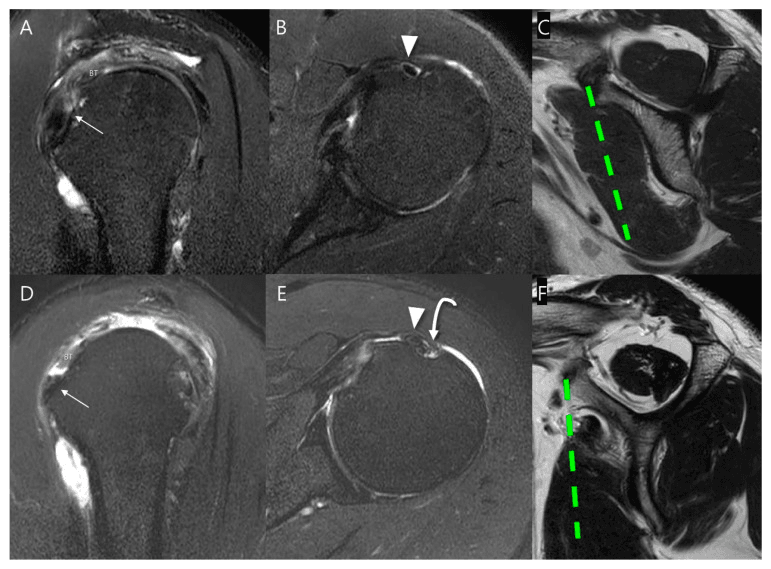
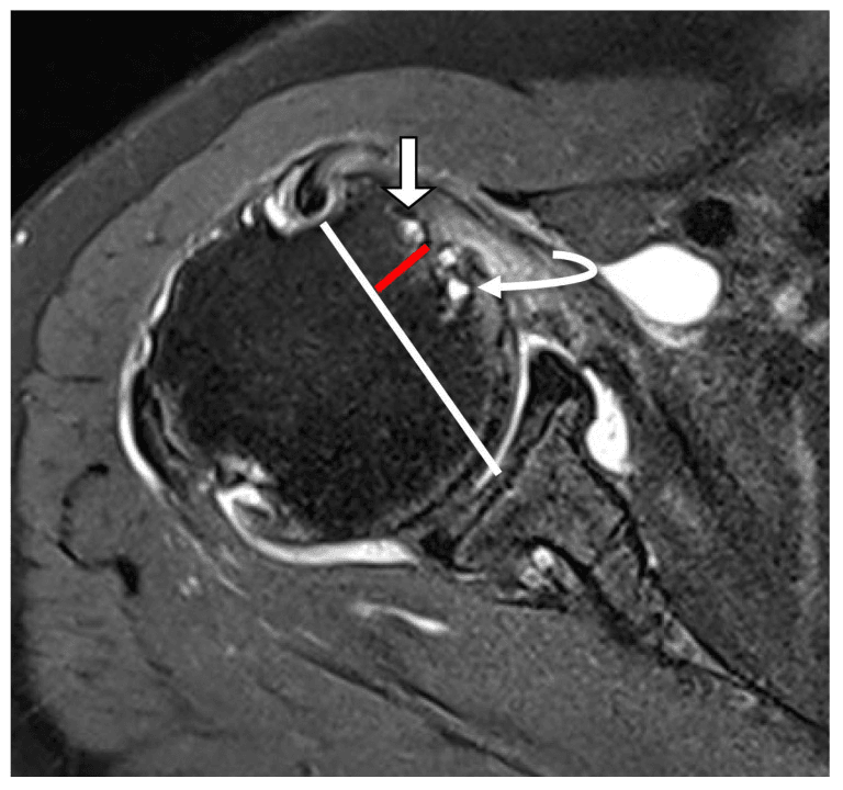
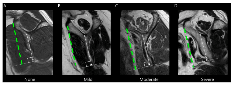
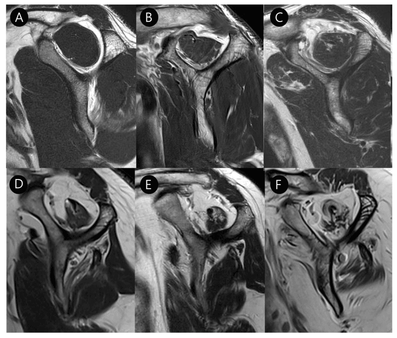
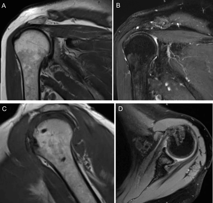
MRI is the best imaging modality and the essential one. Axial views show the subscapularis and its lesser tuberosity insertion: look for tendon discontinuity at the footprint and biceps subluxation medial to the groove. Sagittal views assess fatty infiltration, graded by Goutallier, and the scan should also be read for associated supraspinatus pathology, the anterosuperior cuff tear.
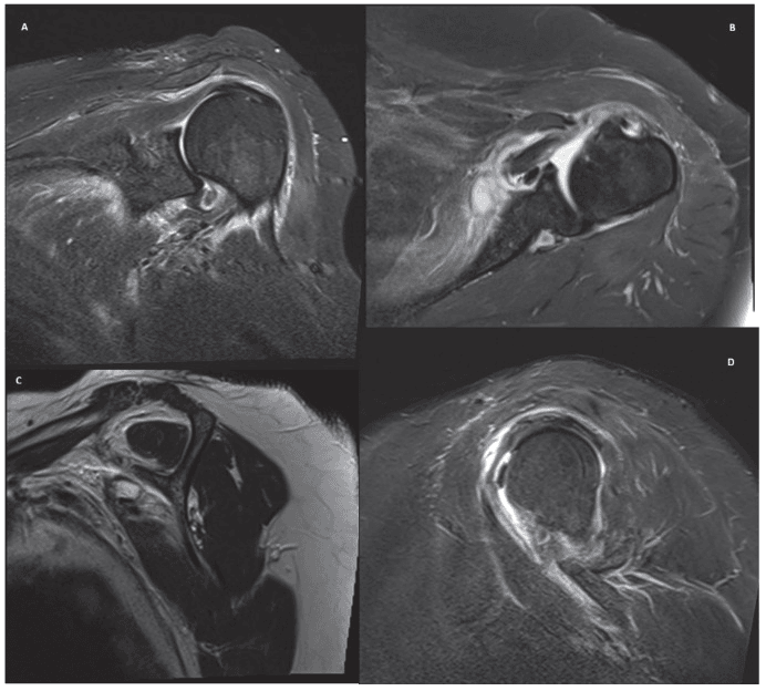
Muscle quality. The sagittal series below runs from a normal muscle through the grades of atrophy and fatty infiltration.
Differential Diagnosis
- Distinguishing Feature
- IR weakness, increased passive ER, anterior pain
- Key Test / Imaging
- Positive bear-hug/lift-off; axial MRI tendon gap
- Distinguishing Feature
- Bicipital groove pain, painful arc, often normal IR strength
- Key Test / Imaging
- Speed/Yergason positive; MRI medial biceps subluxation, intact subscap
- Distinguishing Feature
- Combined IR and abduction weakness, pseudoparalysis if massive
- Key Test / Imaging
- MRI both tendons; sagittal fatty grading
- Distinguishing Feature
- Pain on flexion/IR/adduction, no true weakness
- Key Test / Imaging
- Coracohumeral interval under 6 mm on axial MRI
- Distinguishing Feature
- Apprehension, history of dislocation, age under 30
- Key Test / Imaging
- Apprehension/relocation; MR-arthrogram labral lesion
- Distinguishing Feature
- Global loss of passive ROM (esp. ER), stiffness over weakness
- Key Test / Imaging
- Restricted passive ER; MRI capsular/rotator-interval thickening
Management Algorithm
Non-operative care is worth considering when:
- Symptoms and the functional deficit are acceptable
- A low-grade lesion is not clearly the pain generator
- Host risk or chronic irreparability outweighs the expected benefit of surgery
Repair is worth considering when:
- A traumatic or progressive tear causes meaningful internal-rotation weakness, pain or anterior force-couple failure
- The tendon can reach the lesser tuberosity with acceptable tension and tissue quality
- Associated cuff or biceps-pulley pathology is being addressed and repair improves the global construct
The irreparable complete tear. The choice lies among compensation, arthroplasty and tendon transfer, according to glenohumeral arthritis, humeral-head centring, deltoid and cuff function, age, goals and the available donor.
Debridement-versus-repair cannot be reduced to “under or over 50%”. Facet involvement, footprint detachment, biceps instability, symptoms, tissue quality and associated cuff disease all modify the decision.
The non-operative programme. Activity modification, analgesia, restoration of range and progressive cuff and scapular strengthening. Painful resisted internal rotation is modified early, then function is rebuilt. An injection may control symptoms but does not heal the tendon.
When it is not working. Reassess persistent weakness, instability or progression, rather than declaring non-operative treatment a failure after a fixed duration.
Pre-operative Planning
The imaging review confirms the tear extent on MRI and records the Goutallier grade, the position of the biceps, any associated supraspinatus tear and the coracoid morphology.
The surgical plan covers:
- Beach chair or lateral position
- Be prepared for a biceps procedure
- Plan the number and placement of anchors
- Consider subcoracoid decompression if there is stenosis
- Have a pectoralis major transfer as the backup if the tear proves irreparable
Surgical Technique
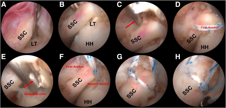
Visualisation. From the standard posterior viewing portal, rotate the arm into external rotation to see the subscapularis insertion; complete visualisation of the subscapularis requires it. Identify the lesser tuberosity, assess the biceps tendon and pulley, and in a complete tear look for the comma sign at the superior edge.
Repair Steps
Release adhesions. Mobilise tendon from coracoid base if needed. Ensure tendon reaches lesser tuberosity without tension.
Prepare lesser tuberosity with shaver/burr. Create bleeding bone surface for tendon healing.
Place suture anchors on lesser tuberosity. Typically 1-3 anchors depending on tear size. Position at medial edge of footprint.
Pass sutures through tendon using penetrating grasper or suture passer. Ensure adequate tissue bite.
Tie sutures to reduce tendon to footprint. Confirm secure repair with probe testing.
Pitfalls. Do not repair under tension, and do not place the anchors too laterally. Use the comma sign as the guide, consider subcoracoid decompression, and do not forget to assess the posterior cuff or to evaluate fatty infiltration. The repair is then protected after surgery.
Inspect the LHB and pulley in every subscapularis repair, and do not ignore biceps subluxation. A frankly unstable or structurally diseased biceps usually requires tenotomy or tenodesis, selected by tendon quality, desired cosmesis, rehabilitation, fixation risk and patient preference—not age alone. Preserve a stable healthy tendon when appropriate.
Pectoralis Major Transfer for the Irreparable Subscapularis
Indication. A symptomatic irreparable anterior cuff, with advanced fatty infiltration, retraction or attritional tissue, in a patient with functional demand, preserved passive motion and no significant glenohumeral arthritis. Arthritis or a massive combined tear points instead to reverse arthroplasty.
The subcoracoid (Resch) technique. The superior half-to-two-thirds of the pectoralis major is detached and ideally routed deep to (behind) the conjoint tendon to the lesser tuberosity. Passing behind the conjoint tendon, which then acts as a pulley, redirects the muscle's line of pull to better approximate the subscapularis vector.
The supracoracoid alternative. Transfer superficial to (in front of) the conjoint tendon is technically simpler but more non-anatomic, a straighter transverse pull, and it risks tethering the musculocutaneous nerve. The trade is ease against a less physiological vector.
Its ceiling, and the alternatives. Pectoralis major pulls in a different plane from the subscapularis, so the transfer reliably relieves pain and restores anterior stability but only partially restores internal-rotation strength. A lower-trapezius or latissimus dorsi transfer matches the line of pull more closely and some now prefer it, though there are no head-to-head trials to separate them.
Complications
- Incidence
- 10-30%
- Risk Factors
- Large tears, fatty infiltration
- Prevention/Management
- Careful patient selection, good technique
- Incidence
- 5-15%
- Risk Factors
- Prolonged immobilisation
- Prevention/Management
- Appropriate rehab protocol
- Incidence
- Rare
- Risk Factors
- Anterior instrumentation
- Prevention/Management
- Anatomic awareness
- Incidence
- Variable
- Risk Factors
- Associated pathology, CRPS
- Prevention/Management
- Address all pathology
Nerves at risk. The axillary nerve runs anteroinferiorly and the musculocutaneous nerve enters coracobrachialis near the coracoid, so anterior instrumentation should be cautious. Subcoracoid decompression must avoid overly aggressive bone removal near the coracoid tip.
Postoperative Care and Rehabilitation
The protocol exists to keep internal-rotation load off the repair, because internal rotation loads it directly.
Recovery Timeline
Sling immobilisation in neutral rotation. Avoid internal rotation. Passive external rotation only. Elbow and hand exercises.
Active-assisted ROM. Begin internal rotation. Avoid resisted IR until 10-12 weeks.
Progress active ROM. Begin isotonic strengthening. Internal rotation strengthening begins.
Progressive strengthening. Sport-specific activities. Return to full activity 4-6 months.
The restrictions that sit alongside it:
- No active internal rotation for 6-8 weeks
- Avoid behind-the-back movements initially
- Sleep in the sling for 4-6 weeks
- Progress gradually to full activity
Outcomes and Prognosis
- Repair Success
- Excellent
- Prognosis
- Best outcomes
- Repair Success
- Good
- Prognosis
- Favourable with repair
- Repair Success
- Variable
- Prognosis
- Depends on tissue quality
- Repair Success
- Reduced
- Prognosis
- Consider transfer
Who does well. Smaller tears (Types 1-2), acute tears, minimal fatty infiltration, younger patients and an isolated subscapularis tear predict a good result. Larger tears (Type 4), chronic tears, significant fatty infiltration (Goutallier 3-4), older patients and a combined anterosuperior tear predict a worse one.
A caution on the Goutallier grade. The grading was derived on the posterosuperior cuff, as the evidence card below sets out, so applying its thresholds to the subscapularis is an extrapolation, and the widely taught rule that grade 3 to 4 means irreparable, transfer instead, has never been validated against subscapularis outcomes in the Goutallier paper or by it.
Guidelines, Registries & Global Practice
Global epidemiology. Subscapularis involvement is found in roughly 10-25% of surgically treated rotator cuff tears, but arthroscopic series report a far higher rate of upper-fibre lesions (up to 27-30% of all shoulder arthroscopies in some cohorts) once surgeons actively look for them. The classic isolated traumatic tear (forced hyperextension/external rotation) affects predominantly middle-aged men; degenerative anterosuperior tears affect both sexes in the 6th-7th decades.
- Imaging / Diagnosis
- MRI/MR-arthrogram for cuff integrity; clinical tests emphasised
- Surgical Stance
- Repair symptomatic full-thickness tears; address biceps pulley
- Imaging / Diagnosis
- Ultrasound or MRI per local access; structured shoulder pathway
- Surgical Stance
- Repair in active patients; conservative trial for low-demand/degenerate
- Imaging / Diagnosis
- Define tear extent and reparability pre-op
- Surgical Stance
- Anchor repair of upper fibres; transfer if irreparable
- Imaging / Diagnosis
- Goutallier (CT) or Fuchs (MRI) fatty grading routine
- Surgical Stance
- Grade 3 to 4 infiltration shifts decision toward transfer/reverse
- No dedicated cuff registry; evidence is from cohort series and reverse-arthroplasty registries (NJR, AOANJRR, AJRR)
- Reverse total shoulder registries show subscapularis status influences stability and internal-rotation outcome
- Re-tear rates after subscapularis repair reported 10-30%, higher with fatty infiltration
- High-resource: MRI/MR-arthrogram, arthroscopic anchor repair, intra-op biceps tenodesis
- Limited-resource: ultrasound-led diagnosis, open repair, biceps tenotomy preferred (cheaper, no implant)
- Tendon transfer and reverse arthroplasty availability drives salvage choice globally
Related pages: Rotator Cuff Tears is the parent topic and carries the posterosuperior cuff on which the Goutallier grading carded here was actually derived; Massive Rotator Cuff Tears for the anterosuperior escape pattern that a subscapularis tear produces when the supraspinatus has also failed, which is the situation none of the transfer series above studied; Biceps Tendon Pathology and Proximal Biceps Ruptures for the long head, whose medial subluxation out of the groove is the single most reliable imaging clue to a subscapularis tear and whose pulley is the 'hidden lesion' this page describes; Subcoracoid Impingement for the narrowed coracohumeral interval that both causes and accompanies upper-border tears, and for the coracoplasty decision; Lesser Tuberosity Fractures for the bony equivalent of the same avulsion, which is repaired as a fracture rather than as a tendon and is the diagnosis to exclude in the adolescent; Pectoralis Major Anatomy and Pectoralis Major Rupture for the donor in the Resch transfer, including the sternal-versus-clavicular head distinction that determines the vector; Musculocutaneous Nerve Anatomy for the structure at risk when the transfer is routed behind the conjoint tendon, and Axillary Nerve Anatomy for the nerve Gerber warns must be protected during open repair; Anterior Shoulder Instability for the differential in a patient whose dominant complaint is giving way rather than weakness; Reverse Total Shoulder Arthroplasty for the endpoint when the anterosuperior cuff is unreconstructable; and Shoulder Arthroplasty Complications for subscapularis failure after anatomic replacement, which is now the commonest setting in which this tendon is found deficient.
Controversies & Areas of Uncertainty
No clear superiority for function or pain; tenotomy is faster and cheaper but risks Popeye deformity and cramping in younger/active patients. Choice remains age- and demand-driven, not evidence-mandated.
Threshold for repairing partial upper-fibre tears (debride vs anchor) is unsettled; many Fox-Romeo Type 1 lesions are treated by addressing the biceps pulley alone with good results.
Lesser-tuberosity footprint is small; whether double-row/anchor density improves subscapularis healing over single-row is not established and extrapolated from posterosuperior cuff data.
Pectoralis major (subcoracoid vs supracoracoid) vs latissimus/lower-trapezius transfer vs reverse arthroplasty: no head-to-head trials; selection is guided by age, arthritis and remaining cuff.
MCQ Practice Points
Q: Where does the subscapularis insert? A: Lesser tuberosity - The subscapularis is the only rotator cuff muscle to insert on the lesser tuberosity. The other three cuff muscles (supraspinatus, infraspinatus, teres minor) insert on the greater tuberosity.
Q: What is the primary function of the subscapularis? A: Internal rotation - The subscapularis is the ONLY internal rotator of the rotator cuff. It also provides anterior stability and contributes to the humeral head depressor function as part of the force couple.
Q: What is the comma sign? A: The Superior Glenohumeral Ligament (SGHL) and coracohumeral ligament complex - This tissue runs adjacent to the superior edge of the subscapularis and becomes visible as a "comma" in complete tears. It marks where repair should begin.
Q: In Fox & Romeo classification, what is a Type 3 subscapularis tear? A: Complete tear of the upper 50% of the tendon - Type 1 = partial articular, Type 2 = upper 25%, Type 3 = upper 50%, Type 4 = entire tendon.
Q: What clinical tests assess subscapularis function? A: Lift-off test (Gerber), Bear-hug test, Napoleon (belly-press) test - Lift-off tests ability to internally rotate against resistance with hand behind back. Bear-hug resists ER with hand on opposite shoulder. Napoleon assesses wrist position during belly-press.
Q: What pathology is commonly associated with subscapularis tears? A: Biceps pathology (subluxation, dislocation, tears) - The biceps pulley is at the junction of subscapularis and supraspinatus. Subscapularis tears often disrupt the pulley, causing biceps instability. Always address biceps at surgery.
Exam Viva Scenarios
Practise clinical reasoning and management decisions out loud
“A 55-year-old man has anterior shoulder pain and weakness with internal rotation after a fall. How do you assess for subscapularis tear?”
“During shoulder arthroscopy, you identify a complete tear of the upper 50% of subscapularis with biceps subluxation. Describe your management.”
“A 60-year-old man has complete subscapularis tear with Goutallier grade 4 fatty infiltration. MRI shows significant muscle atrophy. What are your options?”
Definition
- Tear of subscapularis tendon
- Inserts on lesser tuberosity
- Only internal rotator of rotator cuff
- Upper fibers (intra-articular) tear first
Fox & Romeo Classification
- Type 1: Partial articular surface
- Type 2: Upper 25% complete
- Type 3: Upper 50% complete
- Type 4: Entire tendon complete
Clinical Tests
- Lift-off test (Gerber) - hand behind back
- Bear-hug - hand on opposite shoulder
- Napoleon (belly-press) - hand on abdomen
- Positive = weakness/compensation
Key Anatomy
- Lesser tuberosity insertion
- Comma sign = SGHL complex (superior edge)
- Upper 60% tendinous (repairable)
- Lower 40% muscular (poor suture holding)
Associated Pathology
- Biceps subluxation/dislocation common
- Anterosuperior cuff tears
- Biceps pulley disruption
- Always address biceps at surgery
Outcomes
- 70-90% good/excellent if repairable
- Fatty infiltration reduces success
- Pec major transfer if irreparable
- Return to activity 4-6 months
Evidence Base and Key Trials
Arthroscopic Repair of Isolated Subscapularis Tears (Lafosse)
- Prospective series of 17 all-arthroscopic isolated subscapularis repairs
- Relative Constant score improved 58% to 96% (p under 0.05)
- UCLA score improved 16 to 32 points; repair intact on CT arthrography in 15 of 17
- No progression of fatty infiltration after durable repair
Fox and Romeo Classification of Subscapularis Tears
- Four-grade scheme: partial articular, upper 25%, upper 50%, complete
- Tear extent guides debridement vs anchor repair vs transfer
- Most widely cited subscapularis classification in exams