OCD | Juvenile vs Adult | Stable vs Unstable | MFC
- Lateral aspect of medial femoral condyle is the most common location (75%) - classic site
- Juvenile (open physis) has significantly better prognosis than adult form
- Stable lesions in juveniles can heal conservatively with activity modification (50-70%)
- Unstable or adult lesions usually require surgery (drilling, fixation, or excision)
- MRI is gold standard to assess stability (fluid behind fragment = unstable), but the classic criteria are only 11% specific in juveniles
- “Wilson sign: knee flexed 90° and tibia internally rotated, pain at ~30° on extension, relieved with ER (tibial spine impinges on MFC lesion)
- “Classic location: lateral aspect of MFC
- “T2 MRI shows fluid under fragment ('rim sign') = unstable
- “Juvenile stable: rest 3-6 months first line
Overview and Epidemiology
Osteochondritis dissecans (OCD) is an acquired, idiopathic lesion of subchondral bone that can affect the overlying articular cartilage. It ranges from softening of the cartilage to complete detachment of a bony fragment as a loose body. The knee is the most common location, accounting for 75% of all OCD.
Who. The classic patient is an active adolescent male athlete aged 10-20 with vague knee pain. Males outnumber females 2:1, and the incidence is 15-30 per 100,000. The lesion is bilateral in 20-30%, so always X-ray the other knee.
Juvenile or adult. OCD is divided by the state of the physis. Juvenile OCD occurs with the physis open and adult OCD with it closed, and the distinction decides both prognosis and treatment.
Physis status is the most important prognostic factor. An open physis (juvenile OCD) has much better healing potential due to vascularity; a closed physis (adult OCD) rarely heals without intervention.
Cause. The cause is uncertain and multifactorial. Repetitive microtrauma in active adolescents is the most accepted theory, with overuse in sport. The other proposed contributors:
- Vascular insufficiency: ischaemia of the subchondral bone in a watershed area
- Genetic factors: familial clustering is reported, as are epiphyseal anomalies
- Aberrant ossification: an abnormal ossification centre
- Discoid meniscus, associated with lateral lesions
Anatomy and Pathophysiology
Where. The medial femoral condyle carries 75% of lesions, classically on its lateral aspect: the lateral wall of the intercondylar notch, at the femoral origin of the PCL. The other sites:
- Lateral femoral condyle: 20%
- Patella: 5%
- Trochlea: less than 1%
- Tibial plateau: rare
Why there. During knee motion the tibial spine or the PCL impinges against the lateral aspect of the medial femoral condyle, which is the site of the classic lesion.
The sequence. The disease starts in bone and reaches the joint last, and stability is lost at the step where the cartilage breaks:
- A subchondral bone event: ischaemia or stress fracture of the subchondral bone
- The overlying cartilage is initially intact, and the lesion is stable
- The bone fragment separates from the parent bone
- A cartilage breach allows fluid in, and the lesion is unstable
- The fragment detaches completely as a loose body
Classification Systems
Lesions are classified by their location on the condyle, by their arthroscopic appearance and by MRI. The arthroscopic and MRI systems both sort lesions into stable and unstable, which is the distinction that drives treatment.
Anatomic location on the condyle. Location helps predict prognosis.
- Location
- Central
- Location
- Lateral (classic, aligned with the tibial spine)
- Location
- Inferocentral
- Location
- Central anterior
- Location
- Anterior lateral
Type 2 corresponds to the classic lateral aspect of the MFC.
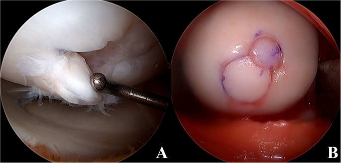
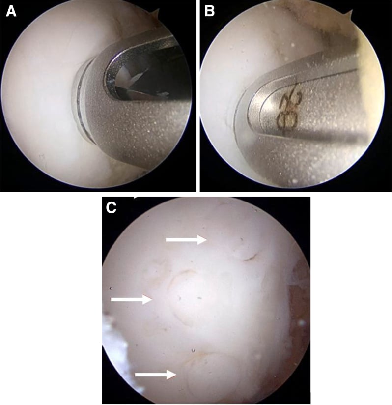
The ROCK classification. The Research in OsteoChondritis of the Knee (ROCK) group's arthroscopic classification is a modern, reproducible system worth knowing alongside the older Guhl/ICRS and Cahill schemes. It divides lesions by probing into immobile (stable) and mobile (unstable) families, because mobility on probing, not appearance alone, drives the choice between drilling and fixation.
- Pattern
- Cue ball
- Finding at arthroscopy
- Intact, normal-looking cartilage, no demarcation
- Pattern
- Shadow
- Finding at arthroscopy
- A subtle demarcation visible through intact cartilage
- Pattern
- Wrinkle in the rug
- Finding at arthroscopy
- A raised fissure or buckle of the cartilage surface, still stable
- Pattern
- Locked door
- Finding at arthroscopy
- A demarcated lesion that cannot be hinged open on probing
- Pattern
- Trapdoor
- Finding at arthroscopy
- A fragment that hinges open on probing
- Pattern
- Crater
- Finding at arthroscopy
- A detached fragment or loose body leaving an empty crater
Why ROCK matters. It gives a shared vocabulary that correlates with intra-operative stability better than MRI alone, and probing is more reliable than a rim on a juvenile MRI. It also maps directly to treatment: immobile lesions that fail non-operative care are drilled, and mobile lesions need fixation of a salvageable fragment or restoration of an unsalvageable crater.
Clinical Assessment
History. The pain is vague, poorly localised and activity-related, worse with impact and sport, and its onset is insidious and lasting. An intermittent effusion follows activity. Catching or locking suggests an unstable flap or a loose body.
Examination. Look for quadriceps atrophy if the problem is chronic and a mild to moderate effusion. The gait is antalgic, with the tibia externally rotated to avoid impingement. Tenderness lies over the classic site on the medial femoral condyle with the knee flexed 90°.
Wilson's sign is specific but has low sensitivity.
Flex the knee to 90°, internally rotate the tibia and extend. Pain at about 30° of flexion, as the tibial spine impinges on the MFC lesion, is positive, and external rotation relieves it by moving the spine away from the lesion. A classic sign, but notoriously unreliable.
Investigations
Radiographs are the initial screening tool. The views:
- AP
- Lateral
- Tunnel (notch): the most sensitive for classic MFC lesions, and the view to obtain when standard AP films are equivocal
- Skyline
What to look for. A crescent-shaped radiolucency, with a rim of sclerosis that suggests chronicity. Look for a calcified loose body in the joint, and record whether the physis is open or closed.
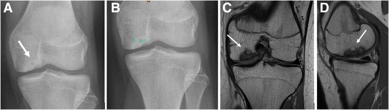
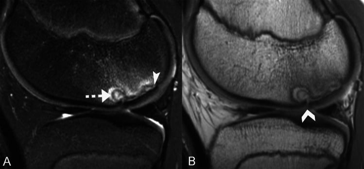
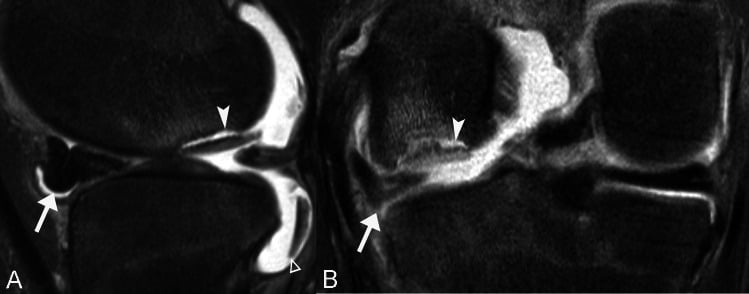
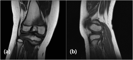
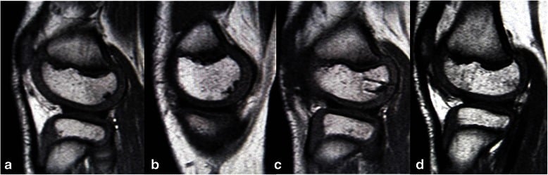
MRI confirms the diagnosis, assesses stability and plans surgery. The report should give size, location, physeal status, interface fluid or cysts, cartilage breach and loose bodies. Gadolinium can assess blood flow to the fragment but is rarely needed.
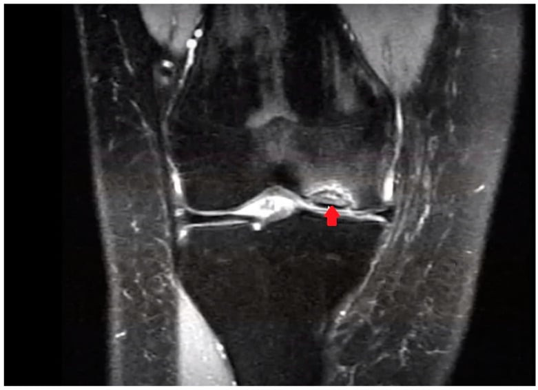
Signs of instability (Hefti criteria).
- Rim sign: high T2 fluid signal behind the fragment, greater than 1mm
- Multiple cysts underlying the bed
- A breach of the cartilage surface allowing fluid contrast
- The fragment elevated or displaced
Adult or child. The rim sign is the most specific criterion in adults, where the classic criteria together are 100% specific. In juveniles it is unreliable: the same criteria are only 11% specific (Kijowski), because a bright T2 rim in a child commonly reflects vascular granulation at a healing interface rather than fluid behind a mobile fragment. In a skeletally immature knee a single high-signal line is not enough; correlate interface fluid, cysts, cartilage breach and displacement.
Differential Diagnosis
The trap is the distal femoral ossification variant in a child under 10. It resolves with growth, so do not over-treat it.
- Key Distinguishing Features
- Adolescent athlete, vague pain, classic lateral MFC site
- Imaging Clue
- Crescent lucency + sclerotic rim; T2 rim if unstable
- Key Distinguishing Features
- Asymptomatic, posterior LFC/MFC, child under 10
- Imaging Clue
- Irregular ossification, normal cartilage, resolves with growth
- Key Distinguishing Features
- Acute trauma/patellar dislocation, haemarthrosis
- Imaging Clue
- Acute defect, no sclerotic rim, fat-fluid level
- Key Distinguishing Features
- Older adult, sudden medial pain, weight-bearing MFC
- Imaging Clue
- Subchondral oedema/collapse, no fragment rim
- Key Distinguishing Features
- Risk factors, often bilateral/multifocal
- Imaging Clue
- Serpiginous double-line sign on MRI
- Key Distinguishing Features
- Lateral pain/clunk, may coexist with lateral OCD
- Imaging Clue
- Bow-tie sign on multiple sagittal cuts
Management
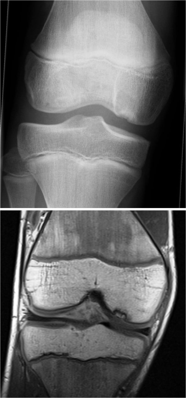
The decision. Management turns on the physis and on stability. Diagnose on radiographs and MRI, decide from the physis whether the lesion is juvenile or adult, and assess stability on MRI fluid signal, remembering that the MRI criteria overcall instability in juveniles. The stable juvenile lesion is treated conservatively first. Surgery is indicated for unstable lesions, adult lesions and failed conservative treatment.
Conservative treatment. For the stable juvenile lesion: activity modification, with no impact, for 3-6 months, and serial radiographs or MRI. Success is 50-70%.
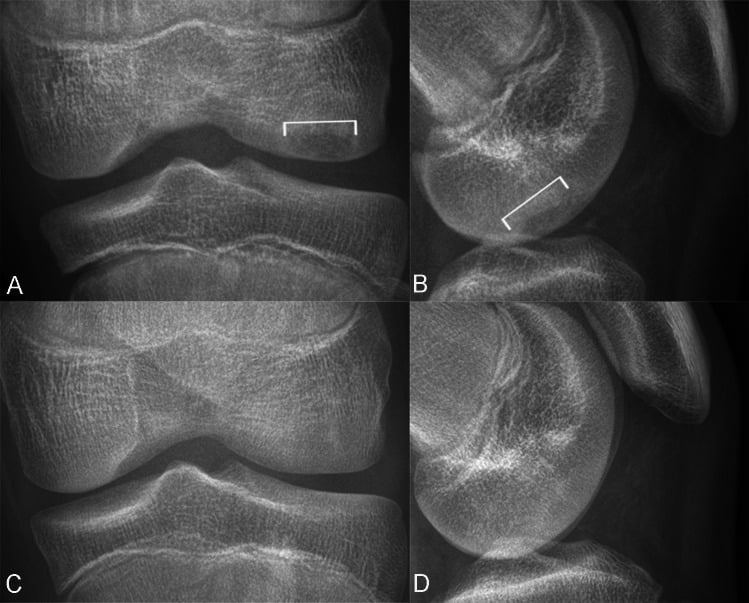
Which stable lesions fail. With a standardised non-operative protocol, roughly two-thirds of stable juvenile lesions heal by six months and about one-third fail, so a stable lesion justifies a non-operative trial but does not guarantee healing. Smaller lesions, less than 2cm, heal better. Size is best expressed relative to the femoral-condyle width, as a normalised ratio, rather than as an absolute millimetre value, because a given lesion is more significant in a smaller condyle; larger normalised lesions are less likely to heal.
Predictors of failure. Beyond a larger normalised size, an effusion and mechanical symptoms (giving-way, catching, locking) at presentation predict failure to heal. Chronological age alone is a weaker predictor than physeal status and these lesion features.
In practice. A small, quiet, stable lesion in an open-physis knee can be watched with serial imaging and activity restriction. A large lesion with an effusion or mechanical symptoms should be counselled as high-risk for failing conservative care and moved earlier toward drilling.
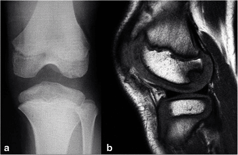
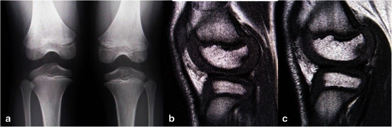
Surgery: can the fragment be saved? Salvageability is assessed arthroscopically. With good bone stock and cartilage the aim is preservation: retrograde drilling for a stable lesion, screw or pin fixation for an unstable one. A necrotic or comminuted fragment is removed as a loose body and the defect treated by marrow stimulation (microfracture), or by OATS or ACI for large defects.
DFSSurgical Indications
Hook:Operate for DFS - Detached, Failed conservative, Skeletal maturity.
- Physis
- Open
- Stability
- Stable
- Treatment
- Conservative (3-6mo)
- Prognosis
- Good (50-70% heal)
- Physis
- Open
- Stability
- Unstable
- Treatment
- Surgery (Fixation)
- Prognosis
- Good if fixed
- Physis
- Closed
- Stability
- Stable
- Treatment
- Surgery (Drilling/Fix)
- Prognosis
- Fair
- Physis
- Closed
- Stability
- Unstable
- Treatment
- Surgery (Fix/Excision)
- Prognosis
- Poor (OA risk)
Surgical Technique
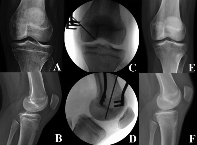
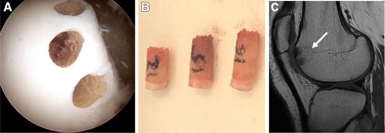
Drilling. Indicated for a stable lesion that has failed conservative treatment, usually in a juvenile. Drilling may be retrograde or transarticular.
Retrograde drilling. The goal is to create vascular channels for healing without breaching the cartilage. Under fluoroscopy, a K-wire is drilled from the extra-articular femur into the lesion, avoiding penetration of the articular surface. The channels bring vascularity to the lesion and stimulate a healing response.
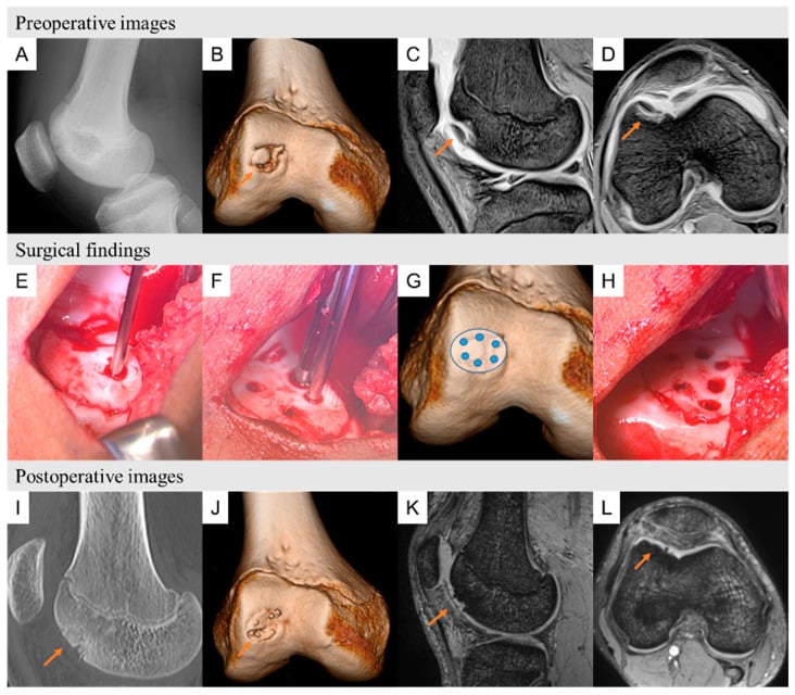
Internal fixation. Indicated for an unstable but salvageable fragment, a flap. The goal is to compress the fragment onto its bed to allow osseous union. At arthroscopy the fibrous tissue is debrided from the base with a curette and the fragment reduced and fixed, with the heads countersunk below the cartilage surface. The implant is a trade between compression and removal:
- Bioabsorbable nails or pins: no removal needed, lower compression
- Compression screws (Herbert): high compression, may need removal
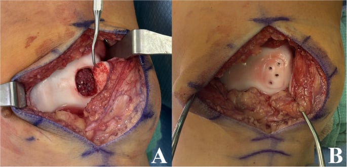
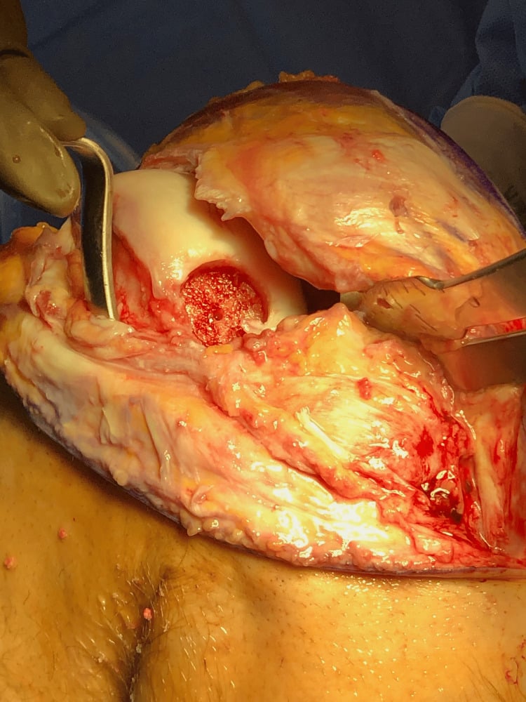
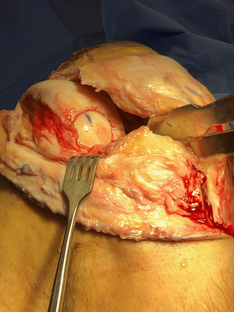
Excision and restoration. Indicated for a loose body or a fragmented, necrotic fragment that cannot be saved. The loose body is removed and the defect restored according to its size:
- Microfracture: small defects, less than 2cm²
- OATS (mosaicplasty): medium defects, with cylindrical plugs
- ACI/MACI: large defects, greater than 2-4cm²
Excision creates fibrocartilage, which is less durable.
Complications
- Risk
- 30-50% in conservative
- Prevention/Management
- Select patients carefully, ensure compliance
- Risk
- If fragment detaches
- Prevention/Management
- Surgical removal
- Risk
- Long-term risk
- Prevention/Management
- Preserve meniscus, restore articular surface
- Risk
- Prominent screws
- Prevention/Management
- Countersink well, use bioabsorbable
Postoperative Care
Weight bearing depends on what was done:
- Drilling: immediate weight bearing as tolerated, as the lesion is stable
- Fixation: touch-down for 6 weeks to protect the repair
- Grafting: non-weight-bearing for 6 weeks
Motion. Early passive range of motion prevents stiffness, and CPM is often used after cartilage repair.
Return to sport. After conservative treatment, at 3-6 months once MRI shows healing; after surgery, at 6-9 months once CT or MRI shows bone union. The criteria are no pain, full range of motion and radiographic healing.
Outcomes and Prognosis
Prognostic factors. Physeal status matters most, and lesion size is discussed under Management. Classic MFC lesions heal better than patellar or trochlear lesions, and stable lesions heal better than unstable ones.
Success rates after surgery.
- Drilling: 80-90% success in the failed conservative juvenile lesion
- Fixation: 80-90% success if the bone quality is good
- Excision: good short-term pain relief but poor long-term results, with a risk of OA
Guidelines, Registries & Global Practice
Global Epidemiology
- Incidence roughly 15-30 per 100,000 children and adolescents; rising with year-round single-sport participation worldwide.
- Peak age 10-15 years (juvenile, open physis); a smaller adult cohort presents with closed physes.
- Male predominance approximately 2:1; bilateral in 20-30% (image the contralateral knee).
- Classic site is the lateral aspect of the medial femoral condyle; lateral femoral condyle lesions associate with discoid meniscus.
Side-by-Side Guidance
- Stance
- Mostly inconclusive/consensus
- Emphasis
- MRI to characterise; nonoperative trial for stable immature lesions; surgery if unstable/failed
- Stance
- Arthroscopic classification + treatment algorithm
- Emphasis
- Probe-based stability assessment guides drilling vs fixation
- Stance
- Lesion- and skeletal-maturity-based
- Emphasis
- Preserve fragment where possible; restoration for unsalvageable defects
- Stance
- Skeletal maturity + stability driven
- Emphasis
- Open physis and stable = conservative first; closed/unstable = surgery
Registry and Restoration Data
- OCD is a leading indication for cartilage restoration registries (e.g. German KnorpelRegister DGOU, and ICRS-aligned datasets) tracking microfracture, OAT/mosaicplasty, ACI/MACI and osteochondral allograft outcomes.
- Registry signals mirror the literature: fragment preservation and osteochondral restoration outperform excision for long-term joint survival.
High- vs Limited-Resource Practice
MRI staging, arthroscopic probing, headless/bioabsorbable fixation and advanced restoration (OAT, ACI/MACI, allograft) are routinely available, enabling fragment-preserving and biologic strategies.
Where MRI and arthroscopy are scarce, diagnosis leans on tunnel-view radiographs and clinical judgement; treatment favours activity modification, open fixation, or excision when restoration is unavailable. Late presentation increases osteoarthritis risk.
Related pages: Osteochondral Defects of the Knee for the cartilage restoration techniques used when the fragment cannot be preserved, and Articular Cartilage Injuries for the underlying repair biology; Osteochondritis Dissecans of the Capitellum and Osteochondral Lesion of the Talus for the same disease at the two other classic sites — note that the talus uses entirely separate size thresholds and that neither site's evidence transfers to the knee; Physeal Injuries: Salter-Harris and Distal Femoral Physeal Injuries for the skeletal maturity that is the single strongest prognostic factor here; Discoid Meniscus and Patellofemoral Instability for the paediatric knee differentials; and Knee Osteoarthritis for the endpoint that fragment excision accelerates.
Controversies and Areas of Uncertainty
Retrograde (extra-articular) drilling spares cartilage but is technically harder and needs fluoroscopy; transarticular drilling is simpler but breaches the surface. Studies show similar healing, so the choice is largely surgeon preference.
A bright T2 rim is non-specific in juveniles (only ~11% specific per Kijowski). Many "rim-positive" paediatric lesions are stable on arthroscopy, so imaging alone should not mandate surgery.
Bioabsorbable implants avoid removal and MRI artefact but give less compression and can cause synovitis/sterile effusion. Headless metal screws give strong compression but may need removal. No clear winner.
For unsalvageable lesions, the best restoration (microfracture vs OAT/mosaicplasty vs ACI/MACI vs osteochondral allograft) is debated and size-dependent. Microfracture produces less durable fibrocartilage and is falling out of favour for larger defects.
MCQ Practice Points
Q: What is the classic location of OCD knee? A: Lateral aspect of the Medial Femoral Condyle (75%). Often cited as the lateral wall of the intercondylar notch.
Q: What is the most significant prognostic factor? A: Status of the physis. Open physis (juvenile) has significantly better prognosis than closed physis (adult).
Q: What MRI finding confirms instability? A: High T2 signal (fluid) behind the fragment. This is the 'rim sign', indicating fluid interposition and instability.
Q: What is the Wilson sign? A: Pain with internal rotation at 30 degrees flexion, relieved by external rotation. (Tibial spine impinges on MFC lesion).
Q: Mechanism of retrograde drilling? A: Stimulates vascular access to the necrotic subchondral bone without breaching the overlying articular cartilage.
Exam Viva Scenarios
Practise clinical reasoning and management decisions out loud
“A 13-year-old male football player presents with vague knee pain. X-ray shows a classic OCD lesion on the lateral MFC. Physes are wide open. MRI shows no fluid behind the fragment. How do you manage him?”
“A 25-year-old man presents with locking and catching. MRI shows a focal defect on the MFC with fluid behind a large osseous fragment. The cartilage hinge is intact on one side.”
“The 13-year-old from the first scenario returns at 6 months. Despite strict compliance, he still has pain. MRI shows the lesion size is unchanged, but no fluid signal. What is your next step?”
Key Concepts
- Lateral aspect of Medial Femoral Condyle (75%)
- Juvenile (open physis) = Good prognosis
- Adult (closed physis) = Poor prognosis
- MRI Fluid behind fragment = Unstable
Classification
- Juvenile Stable: Greater than 50% heal with rest
- Juvenile Unstable: Fixation/Drilling
- Adult: Almost always surgical
- Stage III/IV: Unstable/Loose body
Treatment
- Conservative: Activity mod 3-6mo (Juv Stable)
- Drilling: Retrograde (Stable, failed conservative)
- Fixation: Screw/Pin (Unstable, salvageable)
- Excision: Loose body (Unsalvageable)
Imaging
- X-ray: Tunnel view best for MFC
- MRI: T2 Rim sign (Fluid) = Unstable
- MRI: Assess cartilage integrity
- Serial MRI: Follow healing
Clinical
- Vague activity-related pain
- Wilson Sign: Pain IR 30°, relieved ER
- Mechanical symptoms = Instability
- Antalgic gait (external rotation)
Evidence Base
Healing Potential of Stable Juvenile OCD
- Two-thirds (66%) of stable juvenile lesions heal at 6 months
- Larger normalised lesion size predicts failure to heal
- Swelling/mechanical symptoms at presentation predict failure
- Standardised 6-month nonoperative trial is justified
Internal Fixation of Unstable Juvenile OCD
- 84.6% healing rate with internal fixation
- Even detached fragments healed when fixed
- Mean union time 6 months
- Supports preservation over excision for salvageable fragments
Fragment Excision vs Restoration: Long-term OA
- Excision: 70% OA at 30 years vs 51% with preservation
- Excision independently predicts OA (HR 2.3)
- Older age at diagnosis is the strongest OA risk factor (HR 4.9)
- Fragment preservation/grafting protects the joint long-term
MR Instability Criteria: Juvenile vs Adult
- Classic MR instability criteria are reliable in ADULTS (100% sensitive/specific)
- Same criteria are non-specific in JUVENILES (11% specificity)
- A T2 rim in a child often reflects vascular granulation, not instability
- Interpret a high-signal rim cautiously in skeletally immature knees
Extra-articular Drilling for Stable Juvenile OCD
- 12 of 13 knees healed with full return to activity
- Retrograde drilling protects the articular surface
- Mean time to healing/return ~8.5 months
- Effective salvage when nonoperative care fails in stable lesions
Transarticular Drilling for Stable Juvenile OCD
- 15 of 19 lesions healed completely after transarticular drilling
- All patients returned to previous sporting level
- Osteochondral-type lesions less likely to heal radiographically
- Lesion morphology on CT predicts healing
AAOS Clinical Practice Guideline: Knee OCD
- Few strong recommendations - evidence base is weak
- Supports MRI characterisation of size and stability
- Nonoperative trial reasonable for stable immature lesions
- Surgery for unstable/failed lesions