Fibrocartilaginous Insertion | Four-Zone Transition | Sharpey Fibers | Mechanically Graded
- Enthesis is specialized transition minimizing stress concentration at tendon-bone junction
- Four-zone structure: tendon → uncalcified FC → calcified FC → bone
- Sharpey fibers are collagen bundles inserting into bone at 10-15 degrees
- Fibrocartilaginous entheses at sites of high compression (rotator cuff)
- Healing never fully restores native four-zone architecture
- “Tidemark separates uncalcified from calcified fibrocartilage (like articular cartilage)
- “Direct insertions (fibrous) lack fibrocartilage (flexor tendons)
- “Mechanical loading essential for enthesis development and maintenance
- “Enthesopathies (spondyloarthropathies) target this specialized tissue
Four zones minimize stress concentration: Tendon (soft, elastic) transitions through fibrocartilage zones to bone (stiff, rigid). Gradual change in mechanical properties distributes stress over distance.
Collagen fibers insert into bone at 10-15 degree angle. Mineralized within bone, creating strong mechanical interlock. Resist tensile forces without tearing out.
Resists compression and shear at insertion sites. Collagen II and aggrecan appear in zones 2-3. Sites of high compressive load (rotator cuff, Achilles) have thick fibrocartilage.
Healing forms scar tissue, not native four-zone structure. Takes 12-16 weeks minimum. Biomechanically inferior to native. High re-tear rates (rotator cuff 20-40%).
Overview and Functional Anatomy
The enthesis is the specialized anatomical structure where tendon or ligament inserts into bone. This region represents a remarkable biological solution to the engineering challenge of attaching compliant soft tissue (tendon, elastic modulus 0.5-1 GPa) to rigid hard tissue (bone, elastic modulus 10-20 GPa).
Without a gradual transition, stress would concentrate at the interface causing failure at low loads. The four-zone architecture of fibrocartilaginous entheses creates a graded transition in mechanical properties, distributing stress over a larger area and preventing catastrophic failure.
Understanding enthesis structure explains: high failure rates at tendon-bone junctions (rotator cuff tears, Achilles ruptures); poor healing outcomes despite surgical repair (scar tissue not native architecture); enthesitis in spondyloarthropathies (immune targeting of this unique tissue); need for prolonged protection after repair (12-16 weeks minimum).
At sites of high compression (rotator cuff, Achilles)
- Four-zone structure
- Contains fibrocartilage transition
- Tidemark present
At sites without compression (flexor tendons)
- Collagen I continues directly into bone
- No fibrocartilage zones
- Sharpey fibers without intermediary
- Distributes stress over larger area
- Prevents stress concentration at interface
- Resists tensile, compressive, and shear forces
- Allows gradual modulus change (100-fold from tendon to bone)
Concepts and Four-Zone Architecture
Zone-by-Zone Structure
- Matrix Composition
- Collagen I (parallel), elastin
- Cell Type
- Tenocytes (spindle-shaped)
- Mineralization
- None
- Matrix Composition
- Collagen I + II, aggrecan
- Cell Type
- Chondrocyte-like (rounded)
- Mineralization
- None
- Matrix Composition
- Collagen II, aggrecan
- Cell Type
- Chondrocytes (hypertrophic)
- Mineralization
- Hydroxyapatite
- Matrix Composition
- Collagen I, hydroxyapatite
- Cell Type
- Osteocytes
- Mineralization
- Dense mineral
Tidemark:
- Basophilic line separating zones 2 and 3
- Similar to tidemark in articular cartilage
- Represents mineralization front
- Visible histologically (H&E stain)
Sharpey Fibers
Sharpey fibers are bundles of collagen that insert obliquely into bone, providing strong mechanical anchorage.
- Angle of insertion: 10-15 degrees from tendon axis
- Diameter: 10-50 micrometers
- Length of insertion into bone: 200-400 micrometers
- Mineralized within bone for mechanical interlock
- Resist pull-out forces
- Angled insertion distributes tensile stress
- Longer insertion path increases contact area
- Mineralization prevents slippage
- Multiple fiber bundles provide redundancy
Angle of Sharpey fiber insertion explains why: Suture anchors should be placed at 45 degrees (deadman angle) for optimal pullout strength. Perpendicular insertion has lower resistance. Rotator cuff repairs often fail at bone-suture interface when angle is suboptimal.
Biomechanics and Mechanobiology
Stress Distribution
The four-zone architecture creates a functional gradient in mechanical properties that prevents stress concentration.
Material Property Gradient:
- Tendon elastic modulus: 0.5-1 GPa
- Uncalcified FC: 10-40 MPa (softer than tendon)
- Calcified FC: 0.5-5 GPa (intermediate)
- Bone: 10-20 GPa (stiffest)
The uncalcified fibrocartilage is actually softer than tendon, providing a "compliant layer" that absorbs some deformation and reduces stress concentration at the bone interface.
- Zone Bearing Most Stress
- Sharpey fiber insertion in bone
- Mechanism
- Pull-out resistance
- Clinical Failure
- Anchor failure, bone avulsion
- Zone Bearing Most Stress
- Uncalcified fibrocartilage
- Mechanism
- Aggrecan resists compression
- Clinical Failure
- Fibrocartilage degeneration
- Zone Bearing Most Stress
- Tidemark interface
- Mechanism
- Weakest structural point
- Clinical Failure
- Delamination at tidemark
Mechanical Loading Effects
Mechanical loading is essential for enthesis development, maintenance, and healing.
- Promotes fibrocartilage zone development
- Increases Sharpey fiber density and length
- Enhances mineralization at calcified zone
- Maintains tidemark integrity
- Fibrocartilage zone atrophy
- Decreased Sharpey fiber insertion length
- Reduced ultimate failure load (30-50% decrease)
- Tidemark irregularity
This explains why early controlled loading after tendon repair improves outcomes compared to prolonged immobilization.
Healing and Repair
Healing Biology
Enthesis healing after injury or surgical repair proceeds through inflammatory, proliferative, and remodeling phases but never fully restores native architecture.
Enthesis Healing Timeline
Hematoma formation at tear site. Inflammatory cells infiltrate. Fibrin clot provides initial scaffold. Weak mechanical strength - protection essential.
Fibroblast proliferation. Collagen III (scar collagen) synthesis. Vascular ingrowth. Gradually increasing strength but still vulnerable.
Collagen III replaced by collagen I. Fiber alignment begins along stress lines. Strength reaches 30-50% of native. Gradual loading can commence.
Continued fiber realignment and cross-linking. Strength plateaus at 60-80% of native. Never regains four-zone architecture or native composition.
Key Differences from Native:
- Scar tissue forms, not organized four-zone structure
- Collagen remains more type III than native
- Fibrocartilage zones do not regenerate
- Biomechanical properties inferior (20-40% weaker)
- Higher risk of re-tear (rotator cuff 20-40% at 2 years)
Multiple factors limit enthesis healing:
- Hypovascular zone 2 (uncalcified FC) has poor nutrient supply
- Cell types needed (chondrocytes) don't migrate into healing site
- Mechanical environment prevents organized healing (micromotion)
- Genetic program for four-zone development not recapitulated in adults
- Result: Scar tissue bridge, not native enthesis
Surgical Repair Principles
Understanding enthesis biology guides surgical technique for tendon-to-bone repairs (rotator cuff, ACL, Achilles).
- Bone marrow stimulation: Microfracture to recruit stem cells
- Biologic augmentation: PRP, bone graft, growth factors
- Mechanical optimization: Suture bridge, double-row constructs
- Protected loading: Controlled motion to stimulate healing without overload
- Weeks 0-6: Passive motion only (prevent stiffness, avoid active loading)
- Weeks 6-12: Active-assisted motion, light loading
- Weeks 12-16: Progressive strengthening
- Months 4-6: Return to sport/full activity
Complications of Enthesis Pathology
Degenerative Enthesopathy Complications
- Chronic degeneration weakens tendon
- Eventual failure with minimal trauma
- Common at Achilles insertion, rotator cuff
- Calcium deposition in degenerative tissue
- May cause mechanical symptoms
- Calcific tendinitis (rotator cuff)
- Mechanism
- Progressive weakening
- Management
- Surgical repair
- Mechanism
- Metaplasia
- Management
- Needling, excision
- Mechanism
- Failed healing
- Management
- Multimodal treatment
Classification of Entheses
Two Types of Enthesis
- Found at sites of high compression (rotator cuff, Achilles, patellar tendon)
- Four-zone structure with fibrocartilage transition
- Contains collagen types I, II, and III
- Has tidemark separating calcified from uncalcified zones
- Found where tendons attach at obtuse angles
- No fibrocartilage zones - direct insertion
- Sharpey fibers insert directly into bone
- Examples: Deltoid to humerus, flexor tendons
- Fibrocartilaginous
- Four zones
- Fibrous
- Two zones (tendon → bone)
- Fibrocartilaginous
- Present
- Fibrous
- Absent
- Fibrocartilaginous
- Present
- Fibrous
- Absent
- Fibrocartilaginous
- High compression sites
- Fibrous
- Periosteal attachments
Investigations for Enthesopathy
Imaging Modalities
- Enthesophyte formation (chronic traction spurs)
- Calcification within tendon near insertion
- Cortical irregularity at insertion site
- Bone erosion in inflammatory enthesopathy
- First-line imaging for enthesopathy
- Thickening of tendon at insertion
- Hypoechogenicity (degeneration)
- Power Doppler: Neovascularity in inflammation
- Cortical irregularity, enthesophytes
- Advantages
- Widely available, cheap
- Findings
- Enthesophytes, calcification
- Advantages
- Dynamic, no radiation
- Findings
- Thickening, Doppler signal
- Advantages
- Best soft tissue detail
- Findings
- Bone marrow edema, tears
Differential Diagnosis of Insertional / Enthesis Pain
Pain localised to a tendon-bone insertion has a broad differential. The key clinical task is separating mechanical/degenerative enthesopathy from inflammatory enthesitis and from referred or bony pathology.
- Typical pattern
- Single site, load-related, middle-aged
- Discriminating feature
- Worse with the specific loading activity, improves with rest
- Key investigation
- Ultrasound: thickening, hypoechogenicity, neovascularity
- Typical pattern
- Multiple sites, inflammatory back pain, young
- Discriminating feature
- Morning stiffness, night pain, psoriasis/IBD/uveitis, HLA-B27
- Key investigation
- MRI bone marrow oedema at enthesis; raised CRP
- Typical pattern
- Pain at insertion vs 2-6 cm proximal (Achilles)
- Discriminating feature
- Insertional worsens with dorsiflexion/compression; needs different rehab
- Key investigation
- Ultrasound / MRI localising lesion
- Typical pattern
- Acute trauma or adolescent growth spurt
- Discriminating feature
- Sever, Osgood-Schlatter, tibial tubercle pain in skeletally immature
- Key investigation
- Radiograph of apophysis
- Typical pattern
- Acute severe shoulder pain
- Discriminating feature
- Dense calcific deposit, can be self-resolving
- Key investigation
- Radiograph / ultrasound deposit
- Typical pattern
- Diffuse, non-mechanical
- Discriminating feature
- Night pain unrelated to load, systemic features, red flags
- Key investigation
- MRI / bloods to exclude tumour, infection
Multifocal insertional pain in a young patient (heel, knee, elbow) with inflammatory features is enthesitis until proven otherwise — screen for spondyloarthropathy rather than treating each site as isolated overuse. Conversely, beware labelling non-mechanical night pain as tendinopathy without excluding tumour or infection.
The Enthesis Organ Concept
The Evidence Base cites Benjamin's "enthesis organ" concept and a viva follow-up asks how it explains disease localisation, but the body never develops it - yet it is the single most important conceptual advance in understanding entheses and why enthesopathy behaves as a whole-region disease.
- The enthesis is not just the insertion. At many sites the tendon does not simply pull straight off bone; near the attachment it wraps against and is compressed by the adjacent bone, and a cluster of specialised tissues works together to dissipate stress. This "enthesis organ" comprises the enthesis fibrocartilage itself, plus periosteal and sesamoid fibrocartilage (on the bone and the deep tendon surface respectively where they contact), an associated fat pad, and often a bursa. Together they spread load over the whole region rather than a single point.
- The role of the fat pad and bursa. The synovial/subtendinous fat pad (classically Kager's fat pad behind the Achilles) is not inert packing - it fills dead space during movement, may have a proprioceptive role, and moves in and out of the retrocalcaneal recess as the ankle plantarflexes/dorsiflexes; the retrocalcaneal bursa reduces friction between tendon and bone. These structures are integral parts of the stress-dissipating unit.
- Why it matters clinically. Because the enthesis, its fibrocartilages, fat pad and bursa are one functional unit, disease rarely stays confined to the tendon-bone line: insertional Achilles tendinopathy, Haglund's/retrocalcaneal bursitis and fat-pad involvement coexist, and in spondyloarthropathy the entheseal inflammation spreads into the adjacent bursa and synovium (the imaging basis for the "enthesitis-driven secondary synovitis" model). Managing enthesopathy therefore means treating the whole muscle-tendon-bone-bursa unit, not an isolated point.
Q: What is the enthesis organ concept and why does it matter? A: The load-bearing attachment is served by a cluster of tissues acting together - the enthesis fibrocartilage plus periosteal and sesamoid fibrocartilage (where the tendon compresses against bone), an associated fat pad (e.g. Kager's fat pad at the Achilles) and a bursa - jointly dissipating stress. It explains why insertional tendinopathy, retrocalcaneal bursitis and fat-pad disease coexist, and why spondyloarthropathy enthesitis spreads into adjacent bursa/synovium - so treat the whole unit, not an isolated point.
How the Enthesis Is Built - and Why Adults Cannot Rebuild It
The topic states that healing "never restores native architecture" and that the "genetic program for four-zone development is not recapitulated in adults", and the Evidence Base describes a "growth-plate-like signalling cascade", but the developmental biology that actually explains this is never set out - it is the mechanistic reason repairs are inferior and the target of every regenerative strategy.
- The enthesis is built after birth, not at birth. The mineralised, four-zone fibrocartilaginous insertion is not present at birth; it matures postnatally (in the mouse supraspinatus, the four zones and a mineralised tidemark appear only around three weeks of age), through a spatially- and temporally-ordered programme - type II collagen (chondrocytes) appears first, then type X collagen marks the mineralising fibrocartilage.
- A distinct progenitor pool and signalling programme. The enthesis arises from a dedicated population of insertion-site progenitor cells - characteristically co-expressing the tendon factor Scleraxis (Scx) and the cartilage factor Sox9 - and its maturation is driven by a growth-plate-like cascade, notably Hedgehog signalling (Gli1-responsive cells) that establishes the mineralised fibrocartilage gradient. This is a developmental programme, distinct from ordinary tendon or bone healing.
- Muscle loading is required to build it. The programme is mechanosensitive: normal muscle contraction/loading during development is essential for the mineralised fibrocartilage to form, and paralysis or unloading of the developing limb prevents a normal enthesis - the developmental counterpart of the adult finding that total unloading impairs repair.
- Why adult healing fails, and what regeneration must do. Adult tendon-to-bone healing mounts a generic scar (fibrovascular) response and does not re-activate the Scx/Sox9 progenitor, Hedgehog-driven, load-tuned developmental programme - so the graded fibrocartilage is never rebuilt and the repair stays biomechanically inferior. This is precisely why tissue-engineering strategies aim to re-create the mineral/stiffness gradient (graded scaffolds, controlled mineralisation, delivered progenitors/growth factors and staged mechanical loading) rather than simply gluing tendon to bone.
Q: Why can't adult healing recreate the native four-zone enthesis? A: The graded, mineralised fibrocartilage is built by a postnatal developmental programme - a dedicated Scleraxis/Sox9 double-positive progenitor pool, a growth-plate-like Hedgehog (Gli1) signalling cascade, and essential muscle loading - that matures the insertion over weeks and is not re-activated in adult repair, which instead forms disorganised scar. Regenerative strategies therefore try to rebuild the mineral/stiffness gradient with graded scaffolds, progenitors/growth factors and staged loading, not just reattach the tendon.
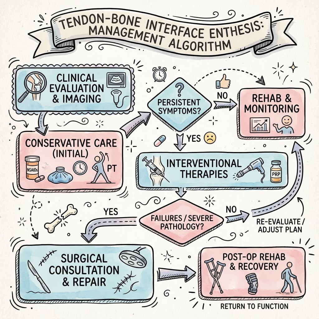
Management of Enthesopathy
Conservative Management
- Relative rest from aggravating activities
- Cross-training with low-impact alternatives
- Gradual return to activity when symptoms improve
- Eccentric loading programs (Achilles, patellar tendon)
- Stretching and flexibility
- Strengthening proximal and distal to insertion
- Biomechanical correction (orthotics, footwear)
- NSAIDs for pain and inflammation
- Corticosteroid injection (caution near weight-bearing tendons)
- PRP injection (emerging evidence)
- Indication
- Achilles, patellar tendinopathy
- Evidence
- Strong evidence
- Indication
- Chronic enthesopathy
- Evidence
- Moderate evidence
- Indication
- Refractory cases
- Evidence
- Emerging evidence
- Indication
- Acute inflammation
- Evidence
- Risk of tendon weakening
Surgical Principles for Tendon-Bone Repair
Tendon-to-Bone Fixation Techniques
- Tunnels through bone for suture passage
- Good bone-tendon apposition
- Lower cost than anchors
- Anchors placed in bone at insertion footprint
- Single-row or double-row techniques
- Suture bridge constructs maximize footprint coverage
- Medial row anchors at articular margin
- Lateral row anchors for suture bridge compression
- Improved footprint contact and initial fixation
- Advantages
- Low cost, good healing
- Considerations
- Technically demanding
- Advantages
- Simpler, faster
- Considerations
- Smaller footprint contact
- Advantages
- Maximum footprint
- Considerations
- Higher cost, more implants
Postoperative Rehabilitation
Rehabilitation Phases
- Immobilization or protected motion
- Passive ROM only
- Avoid active contraction of repaired muscle
- Protect healing interface
- Active-assisted motion begins
- Light loading permitted
- Progressive ROM exercises
- Avoid heavy resistance
- Timeline
- 0-6 weeks
- Activities
- Passive ROM only
- Timeline
- 6-12 weeks
- Activities
- Active-assisted, light loading
- Timeline
- 12-16 weeks
- Activities
- Progressive resistance
- Timeline
- 4-6 months
- Activities
- Sport-specific training
Outcomes of Enthesis Healing
Healing Biology Outcomes
- Healing forms scar tissue at interface
- Fibrocartilage zones do not regenerate
- Collagen organization remains disorganized
- Biomechanical strength 60-80% of native
- Pain relief: Generally good (80-90%)
- Functional improvement: Good
- Structural healing: Variable (60-80% intact at 2 years for rotator cuff)
- Rate
- 80-90%
- Comments
- Good even with re-tear
- Rate
- 60-80%
- Comments
- Varies by tear size
- Rate
- 70-85%
- Comments
- Depends on sport demands
Clinical Relevance
Enthesopathies
Enthesopathies are diseases targeting the enthesis, most commonly in spondyloarthropathies (ankylosing spondylitis, psoriatic arthritis, reactive arthritis).
- Immune-mediated inflammation at enthesis
- IL-23/IL-17 pathway activation
- Erosive changes initially, then new bone formation
- Can lead to ossification and ankylosis
- Achilles insertion to calcaneus
- Plantar fascia insertion
- Patellar tendon insertion (Osgood-Schlatter in juveniles)
- Spinal ligament entheses
Overuse Injuries
Repetitive loading can cause enthesopathy even without systemic disease.
- Rotator cuff tendinopathy (supraspinatus insertion)
- Lateral epicondylitis (common extensor origin)
- Achilles tendinopathy (calcaneal insertion)
Repeated microtrauma exceeds healing capacity, leading to degenerative changes, fibrocartilage calcification, and eventual failure.
Guidelines, Registries & Global Practice
Global Epidemiology and Guideline Comparison
Enthesis-related disorders are among the commonest musculoskeletal presentations worldwide. Full-thickness rotator cuff tear prevalence rises steeply with age (uncommon under 50, present in roughly a third of those over 60 on imaging, much of it asymptomatic). Achilles, patellar and lateral-epicondyle enthesopathies dominate sports and occupational practice. Inflammatory enthesitis is the defining lesion of spondyloarthropathy (population prevalence of the spectrum approximately 0.5 to 1.5 percent).
- Stance on first-line care
- Exercise-based therapy first; limited evidence for steroid, modest for surgery in chronic tears
- Surgery / key point
- Repair reasonable for symptomatic full-thickness tears failing non-op care
- Stance on first-line care
- Structured physiotherapy first-line; corticosteroid sparingly
- Surgery / key point
- Earlier repair favoured for acute traumatic and younger patients
- Stance on first-line care
- Conservative, graded loading; image-guided injection selectively
- Surgery / key point
- Refer for surgery if function-limiting after structured rehab
- Stance on first-line care
- Eccentric / heavy-slow loading for tendinopathy
- Surgery / key point
- Footprint-restoring repair; biologic augmentation still investigational
- Recommendation
- NSAIDs first-line; escalate to TNF or IL-17 inhibitors for persistent enthesitis
- Note
- Local steroid only with caution near load-bearing tendons
- Recommendation
- TNF inhibitor preferred over IL-17 in axial disease; either for peripheral enthesitis
- Note
- Conventional DMARDs ineffective for axial enthesitis
Controversies and Areas of Uncertainty
Clinical dogma favours early motion to prevent stiffness, yet controlled rat studies show immobilisation can improve repair-site mechanics (Gimbel 2007) while total unloading is harmful (Galatz 2009). The unresolved question is the optimal balance: protect the healing insertion without inducing shoulder stiffness. Human RCTs comparing early vs delayed rehab show broadly similar healing with possibly faster early ROM recovery, leaving protocols surgeon-dependent.
Double-row / suture-bridge constructs restore more footprint and have higher biomechanical strength, but higher-level clinical evidence shows only modest, often non-significant differences in functional scores, with some reduction in re-tear for larger tears. Cost and implant burden remain real trade-offs.
PRP, bone-marrow stimulation, scaffolds and growth factors (e.g. TGF-beta3) aim to regenerate the graded interface. Pre-clinical signals are promising but no biologic has reliably re-created the four-zone enthesis in humans; clinical benefit remains unproven and variable.
Whether spondyloarthropathy enthesitis is primarily biomechanical micro-damage at stressed entheses driving an IL-23/IL-17 response, or a primary autoimmune process, is debated. The 'enthesis organ' and mechano-inflammation models attempt to reconcile the two; it underpins why entheses at high-stress sites are preferentially affected.
Key Exam Points and MCQ Practice
Q: What are the four zones of a fibrocartilaginous enthesis from tendon to bone? A: Tendon → Uncalcified fibrocartilage → Calcified fibrocartilage → Bone. Separated by tidemark between zones 2 and 3. Sharpey fibers insert from zone 3 into zone 4.
Q: At what angle do Sharpey fibers insert into bone at the enthesis? A: 10-15 degrees from the tendon axis. This angled insertion distributes stress and provides superior pull-out resistance compared to perpendicular insertion.
Q: What does the tidemark represent in a fibrocartilaginous enthesis? A: The boundary between uncalcified (zone 2) and calcified (zone 3) fibrocartilage, representing the mineralization front. Similar to tidemark in articular cartilage.
Q: Does rotator cuff repair healing restore native four-zone enthesis architecture? A: No - Healing forms scar tissue predominantly collagen III, not organized enthesis. Fibrocartilage zones do not regenerate. Strength plateaus at 60-80% of native.
Q: Why is uncalcified fibrocartilage softer than tendon? A: Acts as compliant layer to absorb deformation and prevent stress concentration at the stiffer bone interface. Contains collagen II and aggrecan which are more compliant than parallel collagen I.
MCQ Practice Points
Q: What are the four zones of a fibrocartilaginous enthesis?
A: Fibrocartilaginous entheses (e.g., rotator cuff, Achilles, patellar tendon) have four distinct zones: Zone 1: Pure tendon (Type I collagen, tenocytes); Zone 2: Uncalcified fibrocartilage (Types I, II, III collagen, fibrocartilage cells); Zone 3: Calcified fibrocartilage (Type II collagen, hypertrophic chondrocytes); Zone 4: Bone (Type I collagen, osteocytes). The tidemark separates zones 2 and 3 (calcified from uncalcified). This gradual transition from soft tissue to bone dissipates stress concentration. These zones are not regenerated after surgical repair - heals with fibrous scar (Zone 1 directly to bone).
Q: What is the difference between fibrocartilaginous and fibrous entheses?
A: Fibrocartilaginous enthesis: Found where tendons attach at acute angles and experience compression as well as tension (rotator cuff, Achilles, patellar tendon, ACL). Has four zones with fibrocartilage transition. Fibrous enthesis: Found where tendons attach at obtuse angles (periosteal attachments like deltoid to humerus). Direct insertion of Sharpey fibers (collagen) into bone without fibrocartilage transition - only 2 zones (tendon and bone). Fibrous entheses are less prone to degeneration but also heal with fibrous scar after injury.
Q: Why is enthesis healing after surgical repair inferior to native tissue?
A: Native enthesis has four-zone graduated structure developed during skeletal maturation through endochondral ossification. After surgical repair: 1) Healing occurs through scar formation (fibrovascular tissue directly to bone) without fibrocartilage zones; 2) Collagen is disorganized (not aligned with direction of pull); 3) Higher stress concentration at repair site; 4) Lower ultimate tensile strength (50-70% of native). Strategies to improve healing include: biological augmentation (PRP, growth factors, stem cells), mechanical stimulation, optimizing surgical technique (footprint preparation, compression at interface).
Q: What is enthesopathy and what conditions affect the enthesis?
A: Enthesopathy refers to pathology at the tendon-bone interface. Degenerative enthesopathy: Chronic overload leads to microdamage, failed healing response, calcification within tendon (calcific tendinitis); common at rotator cuff, Achilles, lateral epicondyle. Inflammatory enthesopathy: Hallmark of seronegative spondyloarthropathies (ankylosing spondylitis, psoriatic arthritis, reactive arthritis) - inflammation at enthesis with eventual ossification; affects Achilles, plantar fascia, SI joints. Enthesophytes: Bony spurs at enthesis from chronic traction or inflammation. MRI shows bone marrow edema adjacent to enthesis in active enthesitis.
Q: What factors influence tendon-to-bone healing after surgical repair?
A: Biological factors: Growth factors (TGF-β, BMP, FGF promote healing), stem cells, vascularity at repair site, patient age (younger heals better), diabetes/smoking (impair healing). Mechanical factors: Tension at interface (some load beneficial for healing, excessive detrimental), motion (controlled early motion may improve healing), compression at footprint. Surgical factors: Footprint preparation (abrade to bleeding bone), fixation strength, contact area, number of anchor points. Time: Peak weakness at 3-6 weeks (inflammatory phase ending, remodeling not complete). Rehabilitation protocols balance protection with early motion.
At a Glance
The enthesis is the specialized fibrocartilaginous junction where tendon inserts into bone, featuring a four-zone architecture that creates a graded transition in mechanical properties: tendon (Zone 1) → uncalcified fibrocartilage (Zone 2) → calcified fibrocartilage (Zone 3) → bone (Zone 4). This ~1mm transition zone minimizes stress concentration between soft tissue (modulus 0.5-1 GPa) and rigid bone (10-20 GPa). Sharpey fibers anchor collagen bundles into bone at 10-15 degrees. The tidemark separates calcified from uncalcified fibrocartilage. Critically, enthesis healing never restores native four-zone architecture, forming mechanically inferior scar tissue instead—explaining high re-tear rates (20-40%) after rotator cuff repair and the 12-16 week minimum protection period.
TUCBFour Zones of Fibrocartilaginous Enthesis
Hook:TUCB - the Tough Universal Connection to Bone!
GRADSEnthesis Mechanical Properties
Hook:The enthesis GRADS from soft to hard tissue!
Exam Viva Scenarios
Practise clinical reasoning and management decisions out loud
“Examiner asks: Describe the anatomy of a fibrocartilaginous enthesis and explain its functional significance.”
“You repair a full-thickness rotator cuff tear. Three months postop the patient asks when the tendon will be 'back to normal'. Explain the healing process and why it never fully restores native tissue.”
“A 28-year-old man presents with painful heels, an episode of knee pain, and a swollen finger. Examination shows tenderness at both Achilles insertions and the plantar fascia origin. How do you approach this, and how does enthesis biology explain the picture?”
Four-Zone Structure
- Zone 1: Tendon (collagen I, tenocytes)
- Zone 2: Uncalcified FC (collagen I+II, aggrecan, chondrocytes)
- Zone 3: Calcified FC (mineralized, tidemark boundary)
- Zone 4: Bone (Sharpey fibers insert at 10-15 degrees)
Biomechanics
- Graded modulus: tendon 0.5-1 GPa → bone 10-20 GPa (100x)
- Uncalcified FC softer than tendon (compliant layer)
- Sharpey fibers: 10-15 degree angle, 200-400 μm insertion
- Stress distribution prevents concentration at interface
Types of Enthesis
- Fibrocartilaginous: High compression sites (rotator cuff, Achilles)
- Fibrous (direct): No compression (flexor tendons)
- Fibrocartilaginous has four zones and tidemark
- Fibrous is direct collagen I insertion to bone
Healing Timeline
- Weeks 0-2: Inflammation (weak, protection essential)
- Weeks 2-6: Proliferation (collagen III scar)
- Weeks 6-12: Remodeling (30-50% strength)
- Weeks 12-24+: Late remodeling (plateaus at 60-80% strength)
Healing Limitations
- Scar tissue forms, NOT four-zone enthesis
- Fibrocartilage zones do NOT regenerate
- Collagen III predominates over collagen I (inferior to native)
- Re-tear rate 20-40% (rotator cuff)
- Never regains native biomechanical properties
Evidence Base
Enthesis Organ Concept and Structure-Function Relationship
- Definitive review of where tendons and ligaments meet bone (entheses)
- Fibrocartilaginous entheses are sites of stress concentration prone to overuse enthesopathy
- Introduces the 'enthesis organ' concept: adjacent tissues (fibrocartilage, fat pad, bursa) jointly dissipate stress
- Most sports enthesopathies are degenerative rather than inflammatory
Insertion-Site Development Produces the Four Zones (Not Recapitulated in Adult Healing)
- Murine supraspinatus insertion does not form four mature zones until ~21 days postnatally
- Type II collagen (chondrocytes) appears from day 7; type X (mineralised fibrocartilage) from day 14
- Ordered fibrocartilaginous transition depends on a growth-plate-like signalling cascade
- Adult healing produces disorganised scar without this fibrocartilage formation
Tendon-to-Bone Healing Forms Scar, Not Organised Enthesis
- Rat supraspinatus repair followed to 56 days
- Early type I and III collagen rise then partially recede; repair never reorganises
- TGF-beta1 (not TGF-beta3) peaks at day 10 with cell proliferation
- Repair tissue stays histologically disorganised and biomechanically inferior to the uninjured insertion at the longest time point
Complete Removal of Load Is Detrimental to Tendon-to-Bone Healing
- Rat supraspinatus repair with botulinum-toxin muscle paralysis to remove load
- Paralysed shoulders had less scar volume and weaker structural properties
- Combining paralysis with immobilisation was most detrimental
- Free range of motion gave only modest improvement and did not overcome the effect of paralysis
Prolonged Immobilisation Can Improve Repair-Site Mechanics
- Rat supraspinatus repair compared immobilisation, cage activity and exercise at 4 and 16 weeks
- At 16 weeks, immobilised insertions had superior elastic properties (immobilisation greater than cage equals exercise)
- Decreased activity improved collagen organisation early and mechanics over time
- Activity level had no effect on elastic properties at 4 weeks
Functionally Graded Interface and Tissue-Engineering Strategies
- Enthesis is a functionally graded material grading soft tendon to mineralised bone
- Graded transition minimises damaging interfacial stress concentration
- Transition region is lost after injury and is not regenerated by repair or natural healing
- Reviews scaffold and graded-mineral strategies aiming to restore the gradient
Re-tear Rate and Predictors After Massive Cuff Repair
- 66 arthroscopic suture-bridge repairs of massive cuff tears, MRI at minimum 1 year
- Re-tear rate 42.4% despite generally satisfactory function
- Fatty infiltration of infraspinatus and extent of retraction were the strongest predictors of re-tear
- Healed shoulders had significantly better pain, UCLA and Constant scores
Enthesitis as the Primary Lesion in Spondyloarthropathy
- Enthesitis is a characteristic, possibly primary, lesion of spondyloarthropathy
- MRI shows enthesitis is widespread, often adjacent to synovial joints
- Entheseal inflammation may drive secondary synovitis in these diseases
- Transgenic TNF-alpha and BMP-6 over-expression models reproduce entheseal-based arthropathy