Complex Deformity Correction | Pedicle Subtraction | Vertebral Column Resection
- PSO (Grade 3) achieves 30-40° correction at single level through posterior closing wedge
- VCR (Grade 4-5) allows multiplanar correction for severe rigid deformities
- Neuromonitoring mandatory - wake-up test if signal changes occur
- Blood loss can exceed 2000mL - cell saver and preoperative autologous donation essential
- Biomechanical hinge is anterior column - protect at all costs during PSO
- “PSO apex is at posterior cortex - cutting too anterior risks anterior column failure
- “VCR requires anterior AND posterior support with mesh cage and rod construct
- “Schwab Grade 3 (PSO) is most common three-column osteotomy for sagittal imbalance
- “Complication rate higher with VCR (30%) versus PSO (20%) but greater correction achieved
PSO closes posteriorly, hinges anteriorly. Anterior column is tension band - must remain intact. Greenstick fracture of anterior cortex allows controlled closure. Breaking anterior column risks catastrophic failure requiring anterior reconstruction.
Highest risk: VCR at thoracic levels (1-10% deficit). Cord ischemia from canal compromise or vascular injury. PSO safer (1-2% risk) but still requires SSEP/MEP monitoring. Immediate wake-up test if signal loss.
Expect 1500-3000mL blood loss. Epidural venous plexus bleeding during posterior decompression. Cell saver mandatory. Tranexamic acid reduces loss by 30%. Type and cross 4-6 units preoperatively.
Sagittal Vertical Axis (SVA) drives indication. SVA greater than 5cm indicates sagittal imbalance. Pelvic incidence minus lumbar lordosis (PI-LL) mismatch greater than 10° requires correction. PSO at L2-L3 most effective for global sagittal balance.
Overview and Epidemiology
Three-column osteotomies represent the most powerful tools for correction of complex spinal deformity. These procedures involve resection of all three spinal columns (anterior, middle, posterior) to achieve significant sagittal and coronal plane realignment. The two main types are pedicle subtraction osteotomy (PSO, Schwab Grade 3) and vertebral column resection (VCR, Schwab Grade 4-6).
Adult spinal deformity with sagittal imbalance causes progressive disability and pain. Sagittal Vertical Axis (SVA) greater than 5cm and PI-LL mismatch greater than 10° predict poor quality of life. Three-column osteotomies restore sagittal balance when less invasive procedures cannot achieve adequate correction. PSO is the workhorse for fixed sagittal deformity, while VCR is reserved for the most severe rigid curves or tumor resection.
- Fixed sagittal imbalance with SVA greater than 5cm
- Pelvic incidence minus lumbar lordosis (PI-LL) mismatch greater than 10°
- Ankylosing spondylitis with severe kyphosis
- Post-traumatic kyphosis
- Flatback syndrome after prior fusion
- Severe rigid deformity greater than 70° unresponsive to PSO
- Congenital hemivertebra
- Sharp angular kyphosis requiring multiplanar correction
- Spinal tumor requiring en bloc resection
- Revision with junctional kyphosis
Pathophysiology
The anterior longitudinal ligament and anterior vertebral cortex form the biomechanical hinge during PSO closure. Greenstick fracture of the anterior cortex allows controlled closure of the posterior wedge. Complete fracture through the anterior column creates instability requiring anterior column reconstruction with cage support. Fluoroscopic monitoring during closure is mandatory to detect anterior column failure.
- Anatomical Structures
- Anterior longitudinal ligament, anterior vertebral body, anterior annulus
- Resection in PSO
- PRESERVED as hinge (greenstick fracture)
- Resection in VCR
- Complete resection, replaced with mesh cage
- Anatomical Structures
- Posterior vertebral body, posterior annulus, posterior longitudinal ligament
- Resection in PSO
- Complete resection within wedge
- Resection in VCR
- Complete resection
- Anatomical Structures
- Pedicles, facets, lamina, ligamentum flavum, interspinous ligaments
- Resection in PSO
- Complete bilateral resection
- Resection in VCR
- Complete resection
Vascular Anatomy at Risk
Segmental arteries arise from aorta at posterior vertebral body. At risk during anterior cortex perforation or VCR. Injury causes massive hemorrhage. Preoperative CT angiography identifies aberrant vasculature. Vascular surgery on standby for VCR cases.
Valveless venous network within spinal canal. Major source of bleeding during decompression. Hemostasis with bipolar cautery, thrombin-soaked Gelfoam, and bone wax on bleeding bone surfaces. Cell saver recovers 30-50% of blood loss.
Differential Diagnosis: Causes of Fixed Sagittal Imbalance
Before committing to a three-column osteotomy, the underlying driver of positive sagittal balance must be identified, because some causes are correctable with lower-grade osteotomies or address a different anatomical segment.
- Distinguishing Features
- Prior lumbar fusion in kyphosis, loss of lumbar lordosis, no remaining mobile segments
- Typical Apex / Segment
- Lower lumbar
- Preferred Osteotomy Strategy
- PSO (Grade 3) at L3 within or below old fusion
- Distinguishing Features
- Bamboo spine, raised inflammatory markers, fused rigid segments, poor horizontal gaze
- Typical Apex / Segment
- Thoracolumbar
- Preferred Osteotomy Strategy
- PSO at L2-L3; caution re osteoporosis and fragility
- Distinguishing Features
- Older patient, disc collapse, often partly flexible on supine/extension films
- Typical Apex / Segment
- Multilevel lumbar
- Preferred Osteotomy Strategy
- Posterior column (Ponte/SPO) if flexible; PSO only if rigid
- Distinguishing Features
- Prior vertebral fracture, focal angular deformity
- Typical Apex / Segment
- Single segment (often TL junction)
- Preferred Osteotomy Strategy
- PSO or VCR if vertebral body destroyed
- Distinguishing Features
- Sharp angular curve, anomalous segmentation on CT
- Typical Apex / Segment
- Focal, often thoracic
- Preferred Osteotomy Strategy
- VCR (Grade 4-6) / hemivertebra resection
- Distinguishing Features
- Long sweeping collapsing curve, pelvic obliquity, underlying disorder
- Typical Apex / Segment
- Thoracolumbar, pelvis
- Preferred Osteotomy Strategy
- Long fusion to pelvis; osteotomy only for fixed segments
Supine, fulcrum-bending, or traction radiographs distinguish a flexible deformity (correctable with posterior column osteotomies or positioning) from a truly fixed one that mandates a three-column osteotomy. Performing a PSO/VCR on a deformity that would correct with lower-grade techniques exposes the patient to unnecessary neurological and haemorrhagic risk.
Closing-Wedge (PSO) versus Opening-Wedge (SPO): the Column-Shortening Principle
The topic contrasts the Smith-Petersen / Ponte osteotomies (Grades 1-2) with the PSO (Grade 3) and stresses the intact anterior hinge, but never explains the single principle that makes the PSO the safer and more powerful osteotomy for a fixed (fused) spine: it shortens the column rather than lengthening it.
- Opening-wedge (Smith-Petersen) osteotomy. Resects the posterior elements and hinges open on the anterior column, lengthening the anterior column and opening an anterior gap at the disc/osteotomy. In a mobile disc space this is well tolerated, but in a rigidly fused (ankylosed) spine it forcibly stretches the anterior longitudinal ligament, aorta and viscera across a fixed anterior column - risking aortic rupture, mesenteric/visceral vascular injury and cord stretch - and each level yields only modest correction (roughly 10 degrees), leaving a non-apposed anterior gap that must fill with bone.
- Closing-wedge (pedicle subtraction) osteotomy. Resects a posterior V-shaped wedge and closes it around an intact anterior cortex/ALL hinge, shortening the posterior column while the anterior column pivots rather than lengthens. Because the column is shortened, not stretched, the cord and great vessels are not placed under tension, bone appose to bone (no anterior gap, so better fusion), and a single level delivers 30-40 degrees.
- Why it matters. This is exactly why the PSO replaced the opening-wedge Smith-Petersen as the operation of choice for fixed sagittal deformity, especially the ankylosed AS spine: closing-wedge column shortening is the safe way to generate a large correction. An over-aggressive closure that snaps the anterior hinge converts the shortening osteotomy into an unstable anterior gap - hence the emphasis on a controlled greenstick rather than a complete anterior fracture.
Q: Why is a closing-wedge PSO preferred over an opening-wedge Smith-Petersen osteotomy for a fixed ankylosing-spondylitis kyphosis? A: A PSO shortens the posterior column around an intact anterior hinge - the cord and aorta are not stretched, bone apposes to bone (no anterior gap), and a single level gives 30-40 degrees. An opening-wedge SPO lengthens the anterior column, which in a fused spine risks aortic rupture, visceral vascular injury and cord stretch and yields only ~10 degrees with a non-apposed anterior gap. Column shortening is the safe way to make large corrections.
Chin-Brow Vertical Angle: Planning Horizontal Gaze
The Scenario-1 viva turns on a "chin-brow vertical angle of 30 degrees" and the patient's inability to look horizontally, but the body never defines this parameter - the specific tool for planning correction of chin-on-chest (ankylosing spondylitis) kyphosis.
- What it is. The chin-brow vertical angle (CBVA) is the angle between a line drawn from the brow to the chin and the vertical, measured with the patient standing, the neck in its fixed/fused position, and the hips and knees extended. It quantifies the direction of the patient's gaze.
- What it means. A large positive CBVA means the gaze is directed downward toward the floor (chin-on-chest) - the disabling deformity of AS that prevents forward vision, eating and safe walking. The osteotomy correction is planned to bring the CBVA into a functional range (roughly -10 to +10 degrees, i.e. near-horizontal gaze, often aiming slightly positive so the patient can still see the ground for walking and reading).
- How it drives the plan. CBVA (for horizontal gaze) is planned alongside the global spinopelvic targets (SVA, PI-LL - developed in the sagittal-balance-parameters and spinopelvic-parameters topics): a lumbar PSO restores truncal balance, but the wedge angle needed in a chin-on-chest AS deformity is set to normalise the CBVA. Over-correcting the CBVA leaves the patient staring at the ceiling, so the target is a slightly downward, functional gaze rather than a strict zero.
Q: How do you quantify and plan correction of horizontal gaze in an ankylosing-spondylitis chin-on-chest deformity? A: The chin-brow vertical angle (CBVA) - the angle between the brow-to-chin line and the vertical, taken standing with the neck fixed and hips/knees extended. A large positive CBVA means a downward (floor-directed) gaze; the osteotomy is planned to restore a near-horizontal, functional gaze (about -10 to +10 degrees), aiming slightly positive so the patient can see the ground. It is planned alongside the global spinopelvic targets, and over-correction leaves the patient looking at the ceiling.
Classification
Schwab Anatomical Osteotomy Classification
The Schwab/SRS comprehensive anatomical classification (validated by Schwab et al, Neurosurgery 2014, Fleiss kappa 0.96) grades osteotomies 1-6 by extent of bony resection and destabilising potential, with an approach modifier (posterior, or combined anterior-posterior). Grades 3-6 are the three-column osteotomies.
- Osteotomy Type
- Partial facet resection (SPO)
- Columns Involved
- Posterior only
- Expected Correction
- 5-10°
- Osteotomy Type
- Complete facet resection (Ponte)
- Columns Involved
- Posterior only
- Expected Correction
- 10-15°
- Osteotomy Type
- Pedicle subtraction osteotomy (PSO)
- Columns Involved
- All three
- Expected Correction
- 30-40°
- Osteotomy Type
- Posterior VCR with cage
- Columns Involved
- All three
- Expected Correction
- 40-60°
- Osteotomy Type
- Complete VCR (anterior + posterior)
- Columns Involved
- All three
- Expected Correction
- 60-90°
- Osteotomy Type
- Multiple VCR
- Columns Involved
- All three at multiple levels
- Expected Correction
- Greater than 90°
Clinical Presentation
Preoperative Workup
Standing scoliosis radiographs (36-inch cassette) to measure global sagittal and coronal alignment. Measure Sagittal Vertical Axis (SVA), Pelvic Incidence (PI), Lumbar Lordosis (LL), and PI-LL mismatch. CT spine for bone quality assessment and pedicle anatomy. MRI if neurological symptoms or to rule out stenosis.
Cardiopulmonary clearance for patients over 60 years or with comorbidities. Autologous blood donation (2-4 units) if time permits. Nutritional optimization - albumin greater than 3.5g/dL. Smoking cessation mandatory for fusion. Osteoporosis treatment if DEXA T-score less than -2.5.
Determine osteotomy level - L2 or L3 most effective for global sagittal balance correction. Calculate correction needed - each PSO provides 30-40° lordosis. Plan instrumentation - extend 3-4 levels above and below osteotomy. Arrange neuromonitoring and wake-up test protocol.
- PSO (Schwab Grade 3)
- 30-40° sagittal
- VCR (Schwab Grade 4-5)
- 60-90° multiplanar
- PSO (Schwab Grade 3)
- 4-6 hours
- VCR (Schwab Grade 4-5)
- 6-10 hours
- PSO (Schwab Grade 3)
- 1500-2000mL
- VCR (Schwab Grade 4-5)
- 2000-4000mL
- PSO (Schwab Grade 3)
- 1-2%
- VCR (Schwab Grade 4-5)
- 5-10%
- PSO (Schwab Grade 3)
- Posterior only
- VCR (Schwab Grade 4-5)
- Anterior and posterior or posterior only
- PSO (Schwab Grade 3)
- Fixed sagittal imbalance
- VCR (Schwab Grade 4-5)
- Severe rigid deformity, tumor, hemivertebra
Investigations and Surgical Planning
Patient Positioning
Position: Prone on radiolucent Jackson table or OSI frame Padding: All bony prominences, avoid abdominal compression (improves venous drainage) Arms: Tucked at sides or on arm boards at less than 90° abduction Neuromonitoring: SSEP and MEP electrodes placed before positioning C-arm: Position for AP and lateral lumbar imaging
Avoid abdominal compression to minimize epidural venous engorgement and bleeding. Ensure chest rolls or frame allow abdomen to hang free. Hip flexion reduces lumbar lordosis making posterior decompression easier. Confirm neuromonitoring baseline signals before prepping.
Exposure
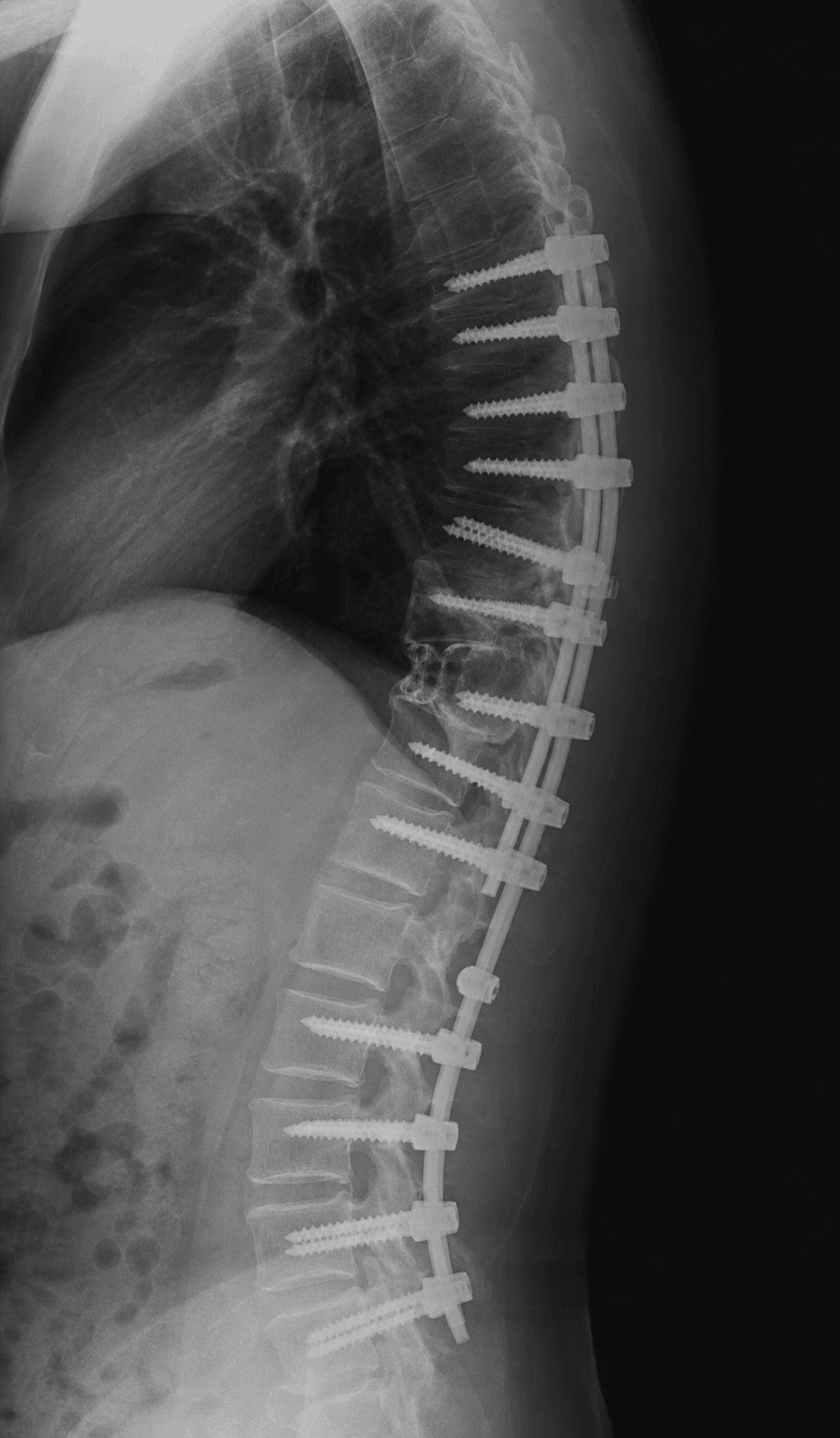
Incision: Midline from upper instrumented vertebra to lower instrumented vertebra Dissection: Subperiosteal exposure of posterior elements from lamina to tips of transverse processes Levels: Expose 3-4 levels above and 3-4 levels below planned osteotomy Landmarks: Identify osteotomy level with intraoperative fluoroscopy Instrumentation: Place pedicle screws at all levels except osteotomy level (screws placed after PSO closure)
Subperiosteal dissection protects paraspinal muscles and minimizes bleeding. Use bipolar cautery liberally for hemostasis.
Management: Vertebral Column Resection (VCR)
Vertebral Column Resection Principles
Definition: Complete resection of vertebral body, pedicles, and posterior elements (all three columns) at one or more levels
Schwab Grades:
- Grade 4: Posterior VCR with posterior cage support
- Grade 5: Complete VCR with anterior and posterior support
- Grade 6: Multiple VCR
Correction: 60-90° multiplanar correction possible with VCR
- Indication
- Sagittal and coronal deformity
- Advantages
- Single approach, shorter operative time
- Disadvantages
- More difficult anterior reconstruction
- Indication
- Severe rigid deformity, tumor
- Advantages
- Optimal anterior column reconstruction
- Disadvantages
- Two approaches, longer operative time, higher morbidity
- Indication
- Lumbar deformity
- Advantages
- Access to anterior column laterally
- Disadvantages
- Learning curve, lumbar plexus risk
VCR provides maximum correction but carries higher risk than PSO. Reserved for most severe deformities.
Management Algorithm

Complications
- Incidence
- 1-5%
- Risk Factors
- Thoracic level, cord ischemia, canal compromise during closure
- Management
- Immediate wake-up test, open osteotomy if deficit confirmed, MRI to rule out hematoma
- Incidence
- 10-20%
- Risk Factors
- VCR, epidural plexus injury, segmental vessel injury
- Management
- Cell saver, transfusion protocol, vascular surgery consultation if uncontrolled
- Incidence
- 5-10% of PSO
- Risk Factors
- Osteoporosis, excessive closure force, thin anterior cortex
- Management
- Anterior column reconstruction with cage, convert to VCR construct
- Incidence
- 10-15%
- Risk Factors
- Extensive decompression, adhesions, revision surgery
- Management
- Primary repair, fibrin glue, lumbar drain if large tear, bed rest 48 hours
- Incidence
- 2-8%
- Risk Factors
- Prolonged operative time, blood loss, diabetes, obesity
- Management
- Antibiotics, irrigation and debridement, hardware retention if stable
- Incidence
- 10-20%
- Risk Factors
- Smoking, osteoporosis, anterior column fracture, infection
- Management
- Revision fusion, bone graft, anterior column support if deficient
- Incidence
- 20-30%
- Risk Factors
- Osteoporosis, abrupt transition, inadequate proximal fixation
- Management
- Extend fusion proximally, prophylactic vertebroplasty at UIV, tethering
Postoperative neurological deficit requires emergent assessment. Obtain wake-up test in OR before emergence. If deficit present, obtain stat MRI to rule out epidural hematoma. If hematoma present, emergent decompression. If no hematoma, consider cord ischemia or intraoperative injury - supportive care and high-dose steroids (controversial). Document deficit and serial neurological exams.
Postoperative Care and Rehabilitation
Postoperative Protocol
ICU monitoring for first 24-48 hours. Hourly neurological checks for lower extremity motor and sensory function. Hemodynamic monitoring and transfusion as needed for anemia or hemodynamic instability. Drain output monitored, expect 200-500mL first 24 hours. Pain control with PCA narcotics and muscle relaxants.
Out of bed to chair on postoperative day 1 or 2 with brace. Physical therapy for ambulation with walker. TLSO brace for 3 months when out of bed. Drain removal when output less than 30mL per 8 hours. DVT prophylaxis with sequential compression devices and pharmacological prophylaxis.
Brace wear whenever out of bed. No bending, lifting, or twisting (BLT precautions). Gradual increase in ambulation distance. Wound check at 2 weeks, staples removed if healing well. Pain management transition from narcotics to non-narcotics.
Standing X-rays at 6 weeks to assess alignment and hardware position. Brace weaning if early fusion signs present. Physical therapy for core strengthening and flexibility. Return to sedentary work possible at 8-12 weeks.
Radiographs at 3, 6, and 12 months to assess fusion. Full activity after solid fusion confirmed, typically 9-12 months. Monitor for complications including proximal junctional kyphosis, pseudarthrosis, and hardware failure.
Outcomes and Prognosis
Patient-Reported Outcomes
Pain and function: Oswestry Disability Index (ODI) improves by 15-20 points on average after PSO for sagittal imbalance. Patients with severe preoperative disability benefit most. Pain scores (VAS) decrease from 7-8/10 to 3-4/10 on average.
Quality of life: SF-36 and SRS-22 scores improve significantly in physical function domains. Mental health scores less reliably improved. Patient satisfaction 70-80% at 2 years.
Radiographic correction: PSO achieves 30-40° sagittal correction and SVA improvement from 10-15cm to less than 5cm. VCR achieves 60-90° multiplanar correction. Correction maintained in 80-90% of patients without junctional failure.
- PSO
- 30-40° per level
- VCR
- 60-90° per level
- PSO
- 85-90%
- VCR
- 75-85%
- PSO
- 15-20%
- VCR
- 25-35%
- PSO
- 1-2%
- VCR
- 5-10%
- PSO
- 10-15%
- VCR
- 15-25%
Factors associated with worse outcomes include: smoking (doubles pseudarthrosis risk), osteoporosis (T-score less than -2.5), obesity (BMI greater than 35), diabetes, age over 70 years, proximal junctional kyphosis at upper instrumented vertebra, inadequate sagittal correction (residual SVA greater than 5cm), and anterior column fracture during PSO closure requiring conversion to VCR.
Guidelines, Registries & Global Practice
Global Epidemiology
Adult spinal deformity prevalence rises sharply with age, reported in 30-68% of adults over 60 years on screening radiographs, with sagittal malalignment the strongest driver of disability and the main indication for three-column osteotomy. Three-column osteotomy itself remains a low-volume, high-acuity procedure concentrated in tertiary deformity centres worldwide.
Side-by-Side Society Guidance
- Patient Selection
- Reserve for fixed/rigid sagittal or coronal deformity not correctable by Ponte/SPO
- Neuromonitoring
- SSEP plus transcranial MEP standard; M and M database reporting encouraged
- Service Model
- High-volume deformity surgeons; multidisciplinary M and M review
- Patient Selection
- Stratify by SRS-Schwab and age-adjusted alignment targets (PI-LL, SVA, PT)
- Neuromonitoring
- Multimodality monitoring with checklist response to alerts
- Service Model
- Knowledge Forum Deformity education and credentialing pathways
- Patient Selection
- Specialist spinal deformity MDT decision; concentrate in commissioned centres
- Neuromonitoring
- Intra-operative monitoring expected for corrective osteotomy
- Service Model
- NHS specialised commissioning to designated units
- Patient Selection
- Evidence-based selection emphasising sagittal targets and frailty assessment
- Neuromonitoring
- Monitoring with documented alert protocol
- Service Model
- High-volume centres; outcome benchmarking
Registry and Database Evidence
No joint-replacement-style implant registry captures osteotomies. Outcome and complication tracking relies on prospective deformity databases (SRS Morbidity and Mortality database, International Spine Study Group, European Spine Study Group), which underpin most multicentre complication estimates.
High-resource centres: routine cell salvage, high-dose TXA, multimodality monitoring, ICU and vascular surgery backup, staged or single-stage VCR. Limited-resource settings: monitoring and blood-bank capacity may constrain case selection, favouring lower-grade osteotomies or staged correction and careful patient triage to referral centres.
Informed consent must include:
- Neurological deficit risk (1-5% depending on procedure) with possibility of permanent paralysis
- Massive blood loss and transfusion requirements (multiple units likely)
- Infection risk (2-8%) with potential need for hardware removal
- Pseudarthrosis (10-20%) requiring revision surgery
- Proximal junctional kyphosis (20-30%) may require extension of fusion
- Medical complications: DVT/PE, cardiac events, prolonged recovery (3-6 months)
- Alternative treatments discussed including continued conservative management
Documentation requirements:
- Preoperative radiographic measurements (SVA, PI-LL mismatch) justifying surgery
- Medical optimization completed (smoking cessation, osteoporosis treatment, cardiac clearance)
- Neuromonitoring records with any signal changes documented and managed
- Operative report detailing osteotomy level, extent of resection, closure technique, any complications
- Postoperative neurological exam documented immediately in PACU and daily thereafter
Common litigation issues:
- Unrecognized neurological deficit in immediate postoperative period - ensure hourly neuro checks
- Inadequate informed consent regarding paralysis risk - document discussion of worst-case scenarios
- Anterior column fracture not recognized or inadequately managed - fluoroscopy before final closure mandatory
- Pseudarthrosis from inadequate fusion technique - ensure adequate bone graft and biologics used
Controversies and Areas of Uncertainty
Posterior-only VCR avoids a second approach and its pulmonary/vascular morbidity, but anterior column reconstruction is technically harder and the rate of intraoperative monitoring alerts is high. The optimal approach for the most rigid angular curves remains debated and surgeon/experience dependent.
L3 maximises lumbar lordosis restoration and global SVA correction, while L4 risks the L5 root and lumbosacral fixation strain. Whether to favour a more proximal or distal level for a given pelvic incidence is not standardised and is individualised to deformity geometry.
Classic fixed thresholds (SVA over 5cm, PI-LL over 10°) are being replaced by age-adjusted targets, since older patients tolerate (and may do better with) modest residual positive balance. Overcorrection in younger patients raises proximal junctional kyphosis risk.
Proximal junctional kyphosis/failure remains common (20-30%). Prophylactic strategies (UIV and UIV+1 cement augmentation, tethers, hook fixation at the top, transitional rods) show promise but none has been definitively proven superior in high-level trials.
Routine high-dose methylprednisolone for an intraoperative or postoperative deformity-related cord injury is not evidence-supported and carries real harms; it is no longer recommended as a reflex. Priority is to rule out and treat a compressive cause (haematoma, malpositioned hardware, residual canal compromise) and to optimise mean arterial pressure for cord perfusion.
MCQ Practice Points
Q: What is the expected sagittal plane correction achieved with a single-level pedicle subtraction osteotomy (PSO)? A: 30-40° - A single-level PSO typically provides 30-40° of sagittal plane correction. This is achieved through posterior column closing wedge with anterior column acting as hinge. Multiple PSOs (e.g., two-level) can achieve 60-80° correction but with increased morbidity.
Q: In the Schwab classification of spinal osteotomies, what defines a Grade 3 osteotomy? A: Pedicle subtraction osteotomy (PSO) - Schwab Grade 3 is defined as pedicle and partial vertebral body resection (pedicle subtraction osteotomy). Grade 1-2 are posterior column only. Grades 4-6 involve vertebral column resection with increasing complexity.
Q: What structure serves as the biomechanical hinge during PSO closure? A: Anterior longitudinal ligament and anterior vertebral cortex - The anterior column must remain intact during PSO to serve as hinge. Greenstick fracture allows controlled closure. Complete anterior column fracture requires cage placement and converts procedure to modified VCR.
Q: What is the approximate major complication rate for PSO versus VCR? A: PSO 15-20%, VCR 25-35% - Major complications include neurological deficit, massive hemorrhage, infection, and pseudarthrosis. VCR has higher complication rate due to more extensive resection and greater destabilization during procedure.
Q: What is the optimal level for PSO to correct global sagittal imbalance in adult spinal deformity? A: L2 or L3 - PSO at L2 or L3 provides maximum correction of sagittal vertical axis (SVA). More proximal osteotomies correct regional kyphosis but less effective for global SVA. L4 or L5 PSO increases risk of L5 nerve root injury.
Q: What is the expected blood loss for PSO and what strategies reduce bleeding? A: 1500-2500mL for PSO - Strategies to reduce blood loss include: tranexamic acid (1g bolus + 1g infusion reduces loss by 30%), cell saver autotransfusion, preoperative autologous donation, meticulous hemostasis with bipolar cautery, avoiding abdominal compression to reduce epidural venous engorgement, and hypotensive anesthesia (controversial).
At a Glance
Three-column osteotomies are powerful spinal deformity correction techniques classified by the Schwab system (Grades 1-6). Pedicle subtraction osteotomy (PSO, Grade 3) achieves 30-40° correction per level by creating a posterior closing wedge that hinges on the intact anterior column—this tension band must be preserved to avoid catastrophic failure. Vertebral column resection (VCR, Grade 4-6) enables 60-90° multiplanar correction for severe rigid deformities. Complication rates are significant (PSO 20%, VCR 30%) with neurological deficit risk of 1-5%; neuromonitoring (SSEP/MEP) is mandatory with immediate wake-up test if signals change. Blood loss typically exceeds 2000mL requiring cell saver and tranexamic acid.
PRECISEPSO Steps - Safe Execution
Hook:Execute PSO with PRECISION - every step critical to avoid catastrophic anterior column failure or neurological injury!
SHARPVCR Indications
Hook:SHARP deformities need SHARP corrections - VCR cuts through all three columns for maximum flexibility!
BLEEDSComplications of Three-Column Osteotomies
Hook:Three-column osteotomies BLEED and carry major risks - prepare patient and surgical team accordingly!
Exam Viva Scenarios
Practise clinical reasoning and management decisions out loud
“A 45-year-old man with ankylosing spondylitis presents with severe thoracolumbar kyphosis. He cannot look horizontally and has chronic back pain. Standing radiographs show 60° thoracolumbar kyphosis, chin-brow vertical angle of 30°, and SVA of 18cm. How would you assess and manage this patient?”
“You are performing a PSO at L3 for sagittal imbalance. You have completed the posterior column resection and are beginning to close the osteotomy. During closure, you hear a loud crack and feel sudden loss of resistance. Fluoroscopy shows discontinuity of the anterior vertebral cortex. What has happened and how do you manage it?”
“Postoperative day 0 after L2 PSO for sagittal imbalance. Patient underwent 6-hour procedure with 2500mL estimated blood loss, transfused 4 units PRBCs. In PACU, patient is somnolent but arousable. When you perform neurological exam, patient has 0/5 bilateral lower extremity motor function and sensory level at L1. Intraoperative neuromonitoring was stable throughout case. How do you assess and manage?”
Key Anatomy and Biomechanics
- Three columns: Anterior (ALL + anterior VB), Middle (posterior VB + PLL), Posterior (pedicles + facets + lamina)
- PSO hinge = anterior column (ALL + anterior cortex) - MUST preserve for stability
- Greenstick anterior cortex fracture allows controlled closure - complete fracture requires cage
- Epidural venous plexus is major bleeding source - valveless system drains to IVC
Classification and Correction
- Schwab Grade 3 = PSO = 30-40° correction per level (posterior only)
- Schwab Grade 4-5 = VCR = 60-90° correction (all three columns resected)
- L2 or L3 optimal level for PSO to correct global sagittal imbalance
- SVA greater than 5cm and PI-LL mismatch greater than 10° indicate need for three-column osteotomy
Surgical Technique Pearls
- PSO apex at posterior cortex - cutting too far anterior risks anterior column fracture
- Gradual closure over several minutes prevents sudden anterior column failure
- SSEP/MEP monitoring mandatory - wake-up test if signals lost during closure
- VCR requires temporary rod stabilization before vertebral body resection to prevent collapse
- Mesh cage with bone graft provides anterior column support in VCR
Major Complications
- Neurological deficit 1-5% (higher with VCR and thoracic levels) - cord ischemia or compression
- Massive hemorrhage 1500-3000mL - epidural plexus and segmental vessels at risk
- Anterior column fracture 5-10% of PSO - requires cage placement or conversion to VCR
- Pseudarthrosis 10-20% - smoking, osteoporosis, anterior fracture are risk factors
- Proximal junctional kyphosis 20-30% - extend fusion if UIV osteoporotic
Management Algorithms
- Ankylosing spondylitis with kyphosis greater than 60° → PSO at L2/L3 for correction
- Severe rigid deformity greater than 70° unresponsive to PSO → VCR for maximum correction
- Anterior column fracture during closure → STOP, check neuromonitoring, place cage, convert to VCR construct
- Postoperative neurological deficit → STAT MRI, evacuate hematoma if present emergently (within 6 hours)
Key Evidence and Outcomes
- Bridwell 2003 (n=27): PSO gives mean 34° lordosis gain, SVA improves ~13.5cm; thoracic pseudarthrosis the main failure
- Lenke 2009 (n=43): posterior-only VCR feasible but 18% intraoperative MEP alerts - monitoring mandatory
- Schwab 2014: anatomical osteotomy grades 1-6, almost-perfect reliability (kappa 0.96)
- Elwatidy 2008 RCT (n=64): high-dose TXA cuts blood loss 49% and transfusion 80%
- Cho 2012 (n=166 revisions): 34% major complication rate; three-column osteotomy an identified risk factor
Evidence Base and Key Trials
PSO for Fixed Sagittal Imbalance (Foundational Series)
- Consecutive series of 27 patients with fixed sagittal imbalance treated by lumbar PSO
- Average increase in lordosis 34.1°; C7 sagittal plumb line improved by a mean of 13.5cm
- One lumbar pseudarthrosis at the osteotomy site; six thoracic pseudarthroses elsewhere
- Significant improvement in Oswestry (p less than 0.0001) and pain scores (p = 0.0002) at minimum 2 years
Lumbar PSO: Minimum 5-Year Follow-up
- 35 patients (mean age 53; osteotomies 1 at L1, 13 at L2, 20 at L3, 1 at L4), mean follow-up 5.8 years
- Ten pseudarthroses (29%) in 8 patients, all away from the osteotomy site (thoracolumbar/lumbosacral junctions)
- No significant degradation in regional alignment or SRS/ODI scores between 2 years and final follow-up
- Maintaining sagittal vertical axis under 8cm predicted better SRS outcome scores (p = 0.038)
Posterior-Only Vertebral Column Resection for Severe Deformity
- 43 patients undergoing single-stage posterior-only VCR with pedicle screws and anterior cages
- 7 patients (18%) lost intraoperative neurogenic motor evoked potentials during correction
- All recovered to baseline or improved after prompt intervention; no patient was neurologically worse
- Two transient nerve-root palsies resolved by 2 weeks and 6 months
The Comprehensive Anatomical Spinal Osteotomy Classification
- Proposes 6 anatomic resection grades (1-6) of increasing destabilising potential plus an approach modifier
- Grade 3 = PSO, Grades 4-6 escalate toward complete and multiple vertebral column resection
- Inter-observer reliability Fleiss kappa 0.96 for resection type and 0.88 for approach modifier
- Intra-observer reliability averaged 0.96, rated 'almost perfect'
Prophylactic High-Dose Tranexamic Acid in Spine Surgery (RCT)
- Double-blind RCT of 64 patients undergoing spinal surgery, randomised to TXA versus placebo
- TXA loading 2g then 100mg/h infusion intra-operatively and for 5 hours postoperatively (adult dosing)
- 49% reduction in total blood loss (p less than 0.007) and 80% less transfusion (p less than 0.008)
- No TXA-related complications observed
Major Complications and the Cost of Three-Column Osteotomy in Revision Deformity Surgery
- 166 patients undergoing multilevel revision adult deformity surgery, mean follow-up 3.5 years
- 34.3% developed a major complication (19.3% perioperative, 18.7% during follow-up)
- Performance of a three-column (pedicle subtraction) osteotomy was an identified risk factor for complications
- Follow-up (delayed) major complications, not perioperative ones, drove worse final outcome scores
AOSpine / SRS Deformity Principles (Reference Guidance)
- Three-column osteotomy reserved for fixed/rigid deformity not correctable by posterior column (Ponte/SPO) osteotomies
- Multimodality intraoperative neuromonitoring (SSEP plus transcranial MEP) is considered standard of care
- Sagittal alignment targets guided by age-adjusted PI-LL, SVA, and pelvic tilt rather than fixed numeric cut-offs
- High-volume deformity centres with blood-management and critical-care support recommended