A1 Pulley | Stenosing Tenosynovitis | Injection | Release
- A1 Pulley: Location of stenosis at MCP level. Release A1, NOT A2.
- Ring and Middle Fingers: Most commonly affected digits.
- Steroid Injection: 70% success rate. Lower in diabetics (50%).
- A2 Pulley: CRITICAL - never release. Causes bowstringing if released.
- Diabetics: 10% prevalence, multiple digits, lower injection success.
- “Palpable nodule at A1 (MCP level)
- “Worse in morning (tissue edema)
- “Diabetics: 10% prevalence, multiple fingers
- “Preserve A2 = fundamental principle
Stenosis occurs at A1 pulley (MCP level). Nodule in flexor tendon catches at pulley during motion. Release A1, never A2.
A2 is critical for tendon function. Releasing A2 causes bowstringing and loss of grip. A2 and A4 are the critical pulleys.
10% prevalence in diabetics. Multiple digits common. Injection success only 50%. May need surgery first-line.
Inject into tendon SHEATH, not tendon. At A1 pulley level. 70% success. May repeat once. Avoid tendon injection.
- Presentation
- Pain only, no catching
- Treatment
- Conservative, splint
- Key Action
- Trial of splint and activity modification
- Presentation
- Catching, active extension OK
- Treatment
- Injection first
- Key Action
- 70% success with steroid injection
- Presentation
- Locking, passive unlock needed
- Treatment
- Injection or surgery
- Key Action
- Consider surgery if injection fails
- Presentation
- Fixed locked
- Treatment
- Surgery
- Key Action
- A1 pulley release indicated
A1-A2-A3-A4-A5Annular Pulley System
Hook:A2 and A4 are the CRITICAL pulleys - never release them! A1, A3, A5 are minor and can be released safely.
DRAGTrigger Finger Risk Factors
Hook:DRAG = Diabetes, RA, Age 50-60, Gripping - know the risk factors for trigger finger!
Overview and Epidemiology
Trigger finger is extremely common in the exam. Key points: A1 pulley location, preserve A2, injection 70% success, diabetics have higher prevalence and lower injection response.
Trigger Finger (stenosing tenosynovitis) is catching or locking of a digit due to stenosis at the A1 pulley.
- Prevalence: 2-3% general population
- Gender: Female greater than male (6:1)
- Age: Peak 50-60 years
- Digits: Ring greater than middle greater than thumb greater than index greater than small
Middle-aged women are the classic demographic.
- Diabetes: 10% prevalence (vs 2% general)
- Rheumatoid arthritis: Inflammatory tenosynovitis
- Gout: Crystal-induced
- Repetitive gripping: Occupational
- Carpal tunnel: Associated conditions
Screen diabetics for trigger finger.
Pathophysiology and Mechanisms
There are 5 annular pulleys (A1-A5) and 3 cruciate pulleys (C1-C3). A2 and A4 are CRITICAL for tendon function - never release them. A1, A3, A5 are minor and can be safely released.
- A1 Pulley: At MCP joint level - where trigger finger occurs
- A2 Pulley: Over proximal phalanx - CRITICAL, do not release
- A3 Pulley: At PIP joint level - minor
- A4 Pulley: Over middle phalanx - CRITICAL, do not release
- A5 Pulley: At DIP joint level - minor
- Repetitive friction causes tendon sheath thickening at A1
- Nodule forms in flexor tendon (FDS or FDP)
- Nodule catches at narrowed A1 pulley
- Results in catching, triggering, or locking
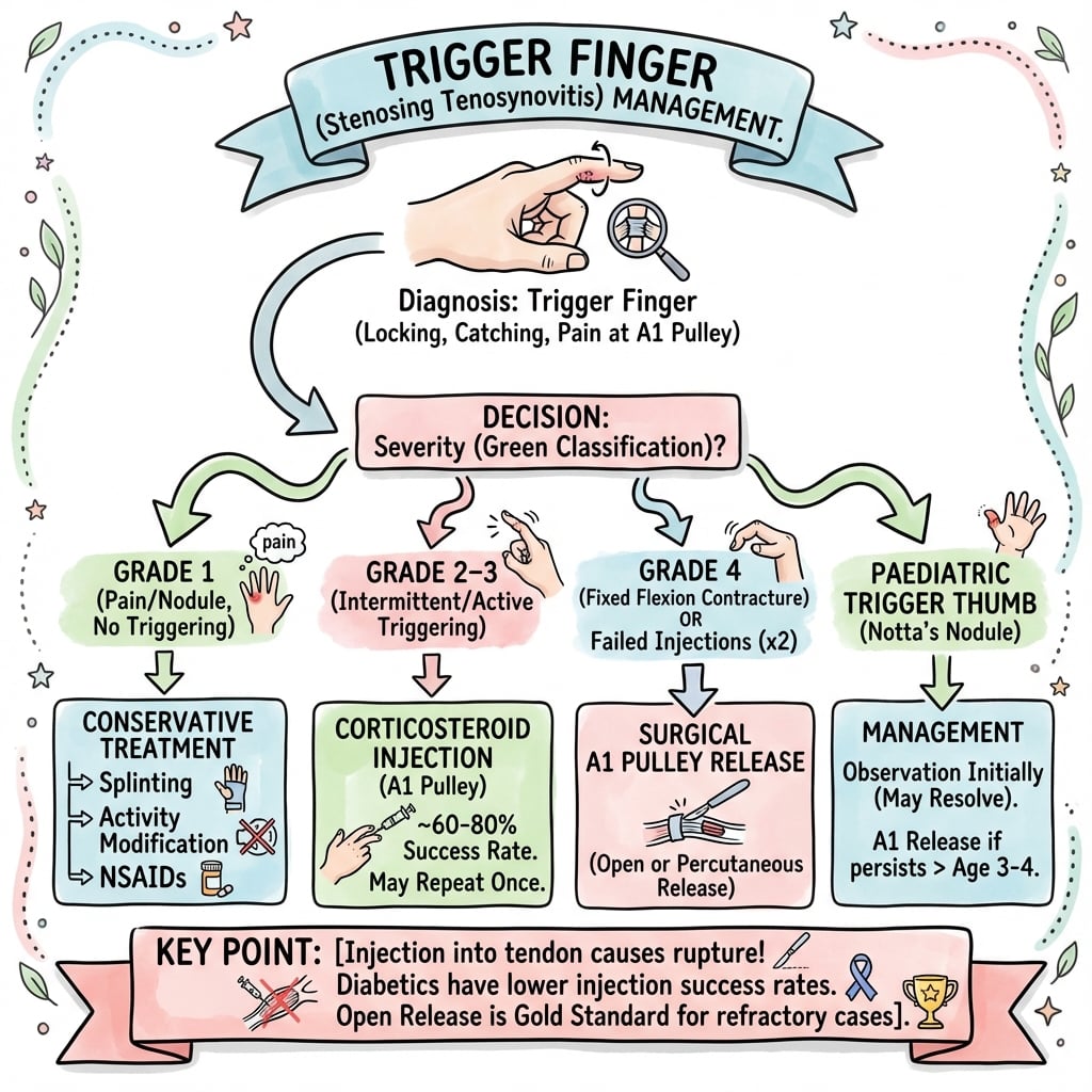
Classification Systems
Green Severity Grading
- Description
- Pain, palpable nodule
- Clinical Features
- History of catching, no demonstrable catching
- Treatment
- Splinting, activity modification
- Description
- Catching
- Clinical Features
- Demonstrable catching, can actively extend
- Treatment
- Steroid injection (70% success)
- Description
- Locking
- Clinical Features
- Requires passive extension to unlock
- Treatment
- Injection or surgery
- Description
- Fixed locked
- Clinical Features
- Unable to passively extend the digit
- Treatment
- Surgical release indicated
Higher grades progressively more likely to need surgery.
Clinical Assessment
- Catching or clicking: During flexion/extension
- Locking: May need passive unlock
- Worse in morning: Tissue edema after rest
- Pain at palm: Over MCP crease
- Progressive: Gets worse over time
Ask about diabetes and occupation.
- Palpable nodule: At A1 pulley (MCP crease)
- Tenderness: Over A1 pulley
- Triggering: Demonstrable with flexion/extension
- Grade: Can actively extend, needs passive, or fixed?
- Other fingers: Check all digits
Always palpate at the MCP crease, not PIP.
Differential Diagnosis
- Key feature
- Palpable nodule at A1 (MCP crease) with catching/locking on flexion
- Discriminator from trigger finger
- Triggering reproduced; nodule moves with tendon
- Key feature
- Palpable cord/nodule in palmar fascia, fixed flexion contracture
- Discriminator from trigger finger
- Cord is superficial and fixed; no triggering, no active unlocking
- Key feature
- Mechanical locking with joint-line tenderness, crepitus
- Discriminator from trigger finger
- Locking is at the joint, not the pulley; radiographs abnormal
- Key feature
- Extensor tendon subluxation at MCP, snapping on extension
- Discriminator from trigger finger
- Dorsal not volar; snapping on extension over the knuckle
- Key feature
- Diffuse sheath swelling, Kanavel signs if septic
- Discriminator from trigger finger
- No discrete nodule; systemic/inflammatory features, pain on passive extension
- Key feature
- Mechanical catching without nodule, often post-injury
- Discriminator from trigger finger
- History of trauma; no A1 nodule; may need imaging
Investigations
Investigation Protocol
Clinical diagnosis in most cases. Palpable nodule at A1 with triggering is pathognomonic. No imaging needed routinely.
HbA1c or fasting glucose if not known diabetic. Diabetics have 10% trigger finger prevalence and need careful counseling about lower injection success.
Rarely required. Can show tendon sheath thickening, A1 pulley thickening, tendon nodule. Useful if diagnosis uncertain.
No imaging is required for typical trigger finger with clear clinical findings.

Management Algorithm
Non-Operative Management
Conservative Options
Reduce gripping activities. Especially repetitive gripping. Modify work tasks if occupational.
Night splint keeping MCP in extension. Prevents tendon nodule catching overnight. Variable success.
Short-term anti-inflammatory. May reduce symptoms but won't resolve stenosis.
Conservative treatment has limited long-term success as sole treatment.
Surgical Technique

Open A1 Pulley Release
Surgical Steps
Local anesthesia. Wide-awake local anesthesia no tourniquet (WALANT) preferred. Hand on table.
Transverse incision at MCP crease. 1-1.5cm. Follow skin crease for cosmesis.
Blunt spread to expose A1 pulley. Identify and protect digital nerves (lateral).
Incise A1 pulley longitudinally. Divide completely. STOP at A2.
Test tendon excursion. Ask patient to flex/extend. Confirm no triggering.
Skin only. Interrupted sutures or steri-strips. Soft dressing.
Simple, quick procedure (less than 10 minutes). LA suitable.
Complications and Consent for the Steroid Injection
The injection is the first-line treatment and the topic stresses "into the sheath, not the tendon" — but it never says what actually goes wrong, and these are exactly the points to consent for.
- Subcutaneous fat atrophy and skin hypopigmentation at the injection site. Steroid that tracks into the dermis/subcutaneous fat causes a depressed, pale patch — cosmetically distressing, often slow to recover or permanent, and more conspicuous in darker skin. Warn patients, and keep the injection deep to the dermis in the sheath.
- Transient hyperglycaemia in diabetics. A single corticosteroid injection can raise blood glucose for several days — relevant both to consent and to the "inject-or-operate-first in diabetes" debate, given injection already succeeds in only roughly a third to a half of diabetics.
- Flexor tendon attenuation or rupture. Rare, but the reason the steroid goes into the sheath and not the tendon, and the reason to limit the number of injections — repeated intratendinous steroid weakens the tendon.
- Steroid flare, infection and depigmentation. A short-lived increase in pain in the first 24-48 hours is common; infection is rare with aseptic technique.
How many injections? Because recurrence reaches around half of patients at one year (Rozental), repeated injections of diminishing benefit carry cumulative cost and complication risk — most clinicians proceed to release after one or two failed injections, sooner in high-risk patients (insulin-dependent diabetes, multiple digits, young age).
Q: What specific complications must you mention when consenting a patient for a trigger-finger steroid injection? A: Subcutaneous fat atrophy and skin hypopigmentation (worse in darker skin, often permanent), transient hyperglycaemia in diabetics, the rare flexor tendon attenuation/rupture (why you inject the sheath not the tendon and limit injections), a short post-injection flare, and infection. Recurrence is common (~56% at a year), so plan to move to surgery after one or two failed injections.
SHEATHInjection Technique
Hook:SHEATH = where you inject! Into the tendon sheath, not the tendon itself.
Complications
- Incidence
- 5%
- Prevention/Management
- Ensure complete A1 division, check glide
- Incidence
- Less than 1%
- Prevention/Management
- Direct vision, stay central, protect nerves
- Incidence
- Rare
- Prevention/Management
- NEVER release A2 - critical pulley
- Incidence
- Less than 1%
- Prevention/Management
- Sterile technique
- Incidence
- 5%
- Prevention/Management
- Early motion, hand therapy if needed
- Incidence
- Less than 3%
- Prevention/Management
- Complete release, address underlying disease
A2 pulley injury causing bowstringing is a serious complication. Prevention is key - know anatomy.
When Triggering Persists After A1 Release
The surgical steps end with "check glide" and "may need to break up adhesions", and a viva asks "what if the finger is still stiff after release?" — but the topic never explains what to do when the digit is not cured. The first move is to separate two different problems, because they have different solutions.
- What it is
- Still catching/locking despite a confirmed complete A1 release
- What to do
- Look at the flexor digitorum superficialis (FDS) - a thickened nodule or catching at the FDS decussation (Camper's chiasm); resect ONE slip (usually ulnar) of FDS to debulk and free the glide. NEVER release A2.
- What it is
- A fixed PIP flexion contracture, no triggering - common after a long-standing locked digit
- What to do
- Early active motion and hand therapy, not more surgery; a contracture may need serial splinting
- What it is
- Triggering returns after initial cure (~3% after surgery)
- What to do
- Usually incomplete A1 release or unaddressed underlying disease (diabetes, RA); revise and confirm a complete release
Why WALANT matters here: operating wide awake lets the patient actively flex and extend on the table, so residual triggering from an FDS nodule is caught and dealt with at the index operation rather than discovered at the post-operative review. And the cardinal rule still holds — the answer to persistent triggering is to address the A1 release and the FDS, never to divide the critical A2 pulley.
Q: A digit still triggers despite a complete A1 release on the table — what is the cause and the fix? A: A flexor digitorum superficialis problem — a thickened FDS nodule or catching at the FDS decussation. Resect one slip (usually the ulnar) of FDS to debulk the tendon; do not release A2 (bowstringing). Distinguish this from residual stiffness (a PIP contracture), which is treated with early motion and hand therapy, not more surgery.
Postoperative Care
Postoperative Protocol
Soft dressing. Immediate finger ROM encouraged. No splint needed.
Active and passive ROM. Full motion as tolerated. Keep wound dry.
Remove sutures. Most patients fully functional by this point. Scar massage.
Return to all activities. Including gripping and lifting. Scar tenderness may persist.
Return to work: Light duties immediately. Full duties 2-4 weeks.
Outcomes and Prognosis
Success Rates:
- Conservative (splint): Variable (30-40%)
- Injection: 70% (lower in diabetics - 50%)
- Surgery: 95-100%
Prognostic Factors:
- Better Outcome
- Short (under 6 months)
- Worse Outcome
- Long (over 1 year), fixed locked
- Better Outcome
- Non-diabetic
- Worse Outcome
- Diabetic (50% injection success)
- Better Outcome
- Lower grade
- Worse Outcome
- Higher grade (4 = surgery)
- Better Outcome
- Single digit
- Worse Outcome
- Multiple digits
Most patients do very well with injection or surgery.
Guidelines, Registries & Global Practice
Global epidemiology: Lifetime prevalence is approximately 2-3% in the general population, rising to 5-20% in people with diabetes. Peak onset is in the fifth-to-sixth decade, with a female predominance (roughly 2-6:1 across series). Trigger finger is one of the commonest reasons for elective hand surgery worldwide and is a recognised marker of impaired glucose tolerance.
Side-by-side guidance:
- First-line
- Corticosteroid injection for adult primary trigger finger
- Surgery
- Reserved for failed injection or fixed locking
- Notable position
- Injection is the default initial intervention
- First-line
- Single corticosteroid injection; up to two before surgery
- Surgery
- Open A1 release if injections fail
- Notable position
- Emphasises informed consent re digital nerve and recurrence
- First-line
- Injection, with splinting as adjunct
- Surgery
- Open or ultrasound-guided percutaneous release
- Notable position
- Percutaneous release accepted where expertise exists
- First-line
- Stepwise: activity modification then injection
- Surgery
- Definitive A1 release
- Notable position
- Diabetics counselled toward earlier surgery
Trigger finger is not tracked by arthroplasty registries (no implant), so the evidence base is observational and trial-derived rather than registry-derived. Pooled trial and cohort data consistently show injection short-term success of roughly 55-70% (markedly lower in diabetes, ~30-50%), recurrence in around half of patients by one year, and open surgical release success exceeding 95% with complication rates under 3%.
- High-resource settings: Office-based injection, WALANT release, and increasing use of ultrasound-guided percutaneous release; readily available hand therapy.
- Limited-resource settings: Greater reliance on a single definitive open release to avoid repeat visits; injection still first-line where steroid is accessible. Ultrasound guidance is less available, so blind injection and open release predominate.
Document grade and prior treatment, and consent specifically for digital nerve injury, incomplete release/recurrence, infection, and the principle of A1 release with A2 preservation. Record diabetes status and the counselling given about lower injection success.
Controversies and Areas of Uncertainty
Most surgeons offer one or two injections before recommending release, but the optimal ceiling is debated. With recurrence around 56% at one year (Rozental 2008), some advocate proceeding to surgery sooner, particularly in high-risk patients, rather than repeating injections of diminishing benefit.
Randomised data show equivalent clinical outcomes for open and percutaneous (including ultrasound-guided) release. Percutaneous is faster and incisionless but carries a higher theoretical risk of digital nerve injury and incomplete release, especially in the thumb. Choice is largely operator-expertise dependent.
Ultrasound-guided injection improves accuracy and may speed early recovery, but several trials show no difference in 3-6 month outcomes versus a blind landmark-based injection. Routine use is not yet standard and depends on availability.
Injection success is markedly lower in diabetes (≈30-50%) and type 1 patients respond poorly. Whether to attempt injection at all in insulin-dependent diabetics, given transient glycaemic disturbance and low success, remains a point of practice variation.
Paediatric trigger thumb (Notta's node) is a distinct entity, not adult stenosing tenosynovitis. Many resolve spontaneously in the first years of life; surgery is considered for persistent fixed flexion, and injection has little role. Do not extrapolate adult management to children.
MCQ Practice Points
Q: At what level does trigger finger occur? A: A1 pulley at the MCP joint level. This is where the stenosis and nodule catching occurs.
Q: Which pulley must be preserved during trigger finger release? A: A2 pulley. A2 and A4 are critical for tendon function. Releasing A2 causes bowstringing.
Q: What is the success rate of steroid injection for trigger finger? A: 70% in general population. Only 50% in diabetics.
Q: What is the prevalence of trigger finger in diabetics? A: 10% (vs 2-3% general population). Multiple digits common. Lower injection success.
Q: Which digit is most commonly affected in trigger finger? A: Ring finger (30%), followed by middle (25%), thumb (20%), index (15%), small (10%).
Q: What is WALANT and why is it ideal for trigger finger release? A: Wide Awake Local Anesthesia No Tourniquet. Allows patient to actively flex/extend to confirm complete release and resolution of triggering.
Exam Viva Scenarios
Practise clinical reasoning and management decisions out loud
“A 55-year-old woman has catching in her ring finger for 2 months. She feels a click at the palm and sometimes needs to straighten the finger with her other hand. There is a tender nodule at the MCP crease. What is your management?”
“A 60-year-old diabetic man has triggering in his ring, middle, and index fingers. He has had one injection to each finger with only partial improvement. What is your approach?”
“A 70-year-old woman presents with her middle finger locked in flexion for 2 weeks. She cannot straighten it actively or passively. Her MCP is tender. What is your management?”
Pathology
- Stenosing tenosynovitis at A1 pulley
- Ring greater than middle most common
- Nodule catches at narrowed pulley
- Diabetics: 10% prevalence
Clinical
- Catching/locking with flexion
- Worse in morning
- Palpable nodule at MCP crease
- Tender at A1 pulley
Green Classification
- 1: Pain, no catching
- 2: Catching, active extension
- 3: Locking, passive unlock
- 4: Fixed locked = surgery
Treatment
- Injection: 70% success (50% diabetics)
- Surgery: 95%+ success
- Grade 4 = surgery first-line
- Can repeat injection once if partial response
Surgical Principle
- Release A1 pulley completely
- PRESERVE A2 pulley (critical)
- Check tendon glide
- Protect digital nerves (lateral)
Key Points
- A2 and A4 = critical pulleys
- Diabetics: 10% prevalence
- Ring finger most common
- Multiple digits in diabetics
Evidence Base
- Two RCTs, 63 participants pooled
- Corticosteroid plus lidocaine superior to lidocaine alone at 4 weeks (RR 3.15, 95% CI 1.34 to 7.40)
- Number needed to treat to benefit = 3
- Benefit lasted up to 4 months in one trial; no adverse events reported
- Double-blind RCT, 50 adults in general practice
- Triamcinolone vs saline: satisfactory immediate response 16/25 vs 5/25 (p less than 0.001)
- Greater reduction in pain and perceived improvement with steroid
- Beneficial effect maintained across 12 months of follow-up
- 118 trigger digits (92 nondiabetic, 26 diabetic)
- Injection success: nondiabetics 57% vs diabetics 32% (p = 0.04)
- All 5 type 1 diabetic digits failed injection and required surgery
- Surgical A1 release successful in 71/72 (99%), no excess complications in diabetics
- 124 digits prospectively followed for 1 year after injection
- 56% had symptom recurrence (median 5.6 months); freedom from recurrence 70% at 6 months, 45% at 12 months
- Insulin-dependent diabetes, younger age, multiple digits and other upper-limb tendinopathies predicted failure
- Duration and severity of symptoms did NOT predict outcome
- 62 resistant trigger digits randomised to open vs percutaneous A1 release
- Ultrasound-measured bowstringing increased at 12 weeks in both groups, greater after open release
- Bowstringing resolved by 24 weeks with no inter-group difference
- No association between bowstringing and any clinical outcome
- 72 grade 2+ trigger digits randomised to open surgery vs ultrasound-guided needle release
- Both groups improved significantly in VAS and Quinnell grade at 7 and 30 days
- No significant difference between techniques at any timepoint up to 180 days
- Level II therapeutic evidence