Adult Acquired Flatfoot Deformity (PTTD)
Weight-bearing radiographs and clinical photographs are provided.
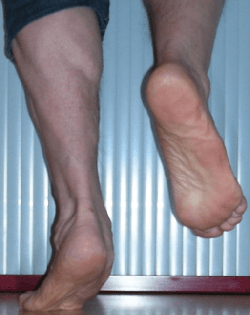
Clinical image for Adult Acquired Flatfoot Deformity (PTTD)
Image source: Open Access medical literature (NIH/PubMed Central) • CC-BY License
Questions
What is the anatomy and function of the posterior tibial tendon? Why is it susceptible to degeneration?
Describe the clinical staging of posterior tibial tendon dysfunction (PTTD). What clinical and radiographic features differentiate each stage?
What is your assessment approach for this patient? What radiographic parameters should be measured?
Outline the non-operative management options for PTTD. When is surgery indicated?
For a Stage II PTTD with flexible deformity, describe the surgical options and your preferred approach.
When and how would you perform a subtalar/triple arthrodesis? What are the goals and outcomes?
Exam Tips
- →3-6 months conservative trial
- →NWB 6-8 weeks after reconstruction
- →Union rate 90-95% for fusion
- →Adjacent joint OA 10-20% at 10 years