Ankle Fractures (Weber/AO Classification)
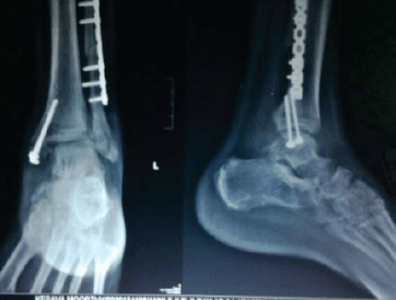
AP and lateral radiographs of the ankle demonstrating a Weber B bimalleolar ankle fracture. The fibular fracture is at the level of the syndesmosis (trans-syndesmotic). There is an associated transverse medial malleolar fracture. The mortise view shows lateral talar shift with increased medial clear space (>4mm) indicating deltoid ligament injury or medial malleolar fracture. The posterior malleolus is intact on lateral view.
Image source: Open Access medical literature (NIH/PubMed Central) • CC-BY License
Questions
Describe the classification systems for ankle fractures.
How do you assess stability of an ankle fracture?
Describe the surgical approach and fixation principles.
When do you address the syndesmosis and posterior malleolus?
What are the complications of ankle fractures?
Discuss outcomes and special circumstances.
Must Mention
- •Weber: A (below), B (at), C (above) syndesmosis
- •Medial clear space >4mm = unstable
- •SER most common mechanism
- •Syndesmosis: fix Weber C, stress test Weber B
- •Posterior malleolus: fix if >25% or unstable
- •Fix fibula first (restores length/rotation)
Common Pitfalls
- •Missing Maisonneuve
- •Not stress testing Weber B
- •Fixing medial before fibula
- •Wrong ankle position for syndesmosis
- •Missing posterior malleolus
- •Not checking mortise alignment