Carpal Tunnel Syndrome
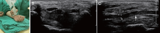
Cross-sectional anatomy of the carpal tunnel at the level of the hook of hamate. The median nerve lies superficially, just deep to the transverse carpal ligament (flexor retinaculum). The nine flexor tendons (4 FDP, 4 FDS, FPL) fill the tunnel. The median nerve is flattened and enlarged proximally. The thenar branch exits radially. Release involves dividing the transverse carpal ligament while protecting the nerve and palmar cutaneous branch.
Image source: Open Access medical literature (NIH/PubMed Central) • CC-BY License
Questions
Describe the anatomy of the carpal tunnel and median nerve.
What are the clinical features and examination findings?
Describe the diagnostic workup including nerve conduction studies.
What is the treatment algorithm including surgical technique?
What are the complications and outcomes of surgery?
What are the differential diagnoses for hand numbness?
Must Mention
- •10 structures in tunnel (median nerve, 9 tendons)
- •Palmar cutaneous branches before tunnel (spared)
- •Thenar branch variations (Lanz)
- •NCS: motor >4.2ms, sensory >3.5ms
- •Splint → injection → surgery algorithm
- •90%+ success with surgery
Common Pitfalls
- •Wrong tunnel contents
- •Missing palmar cutaneous
- •Wrong NCS values
- •No thenar branch mention
- •Missing pronator syndrome
- •No differential diagnosis