Allman / Neer Classification of Clavicle Fractures
- Confusing Neer IIA and IIB. IIA = fracture medial to BOTH CC ligaments (both stay on the distal fragment, proximal end untethered); IIB = conoid torn / between the ligaments (only trapezoid holds the distal fragment). Both are unstable, both 20-30% nonunion, both operative — but examiners want the precise distinction.
- Treating "displaced" as an automatic operate. For Allman Group I (midshaft) the classification identifies who benefits — fixation is a displacement + demand decision (shortening over 20 mm, no cortical contact, comminuted/vertical, high-demand), not a mandate to plate every clavicle.
- Missing the medial-third danger. Group III is rare but sits over the subclavian vessels, brachial plexus and mediastinum — assess them (and get CT) before calling it 'just a clavicle'.
- Forgetting the absolute indications. Open, neurovascular injury, floating shoulder, scapulothoracic dissociation, skin tenting, polytrauma → fix regardless of the numbers.
- Mislabelling the paediatric injury. In children the lateral 'Neer II equivalent' is usually a physeal separation with an intact periosteal sleeve that remodels — don't over-operate.
The Allman classification
The Allman classification (Allman, 1967) divides clavicle fractures into three groups by the location of the fracture line along the bone. It is the starting point for every clavicle fracture assessment.
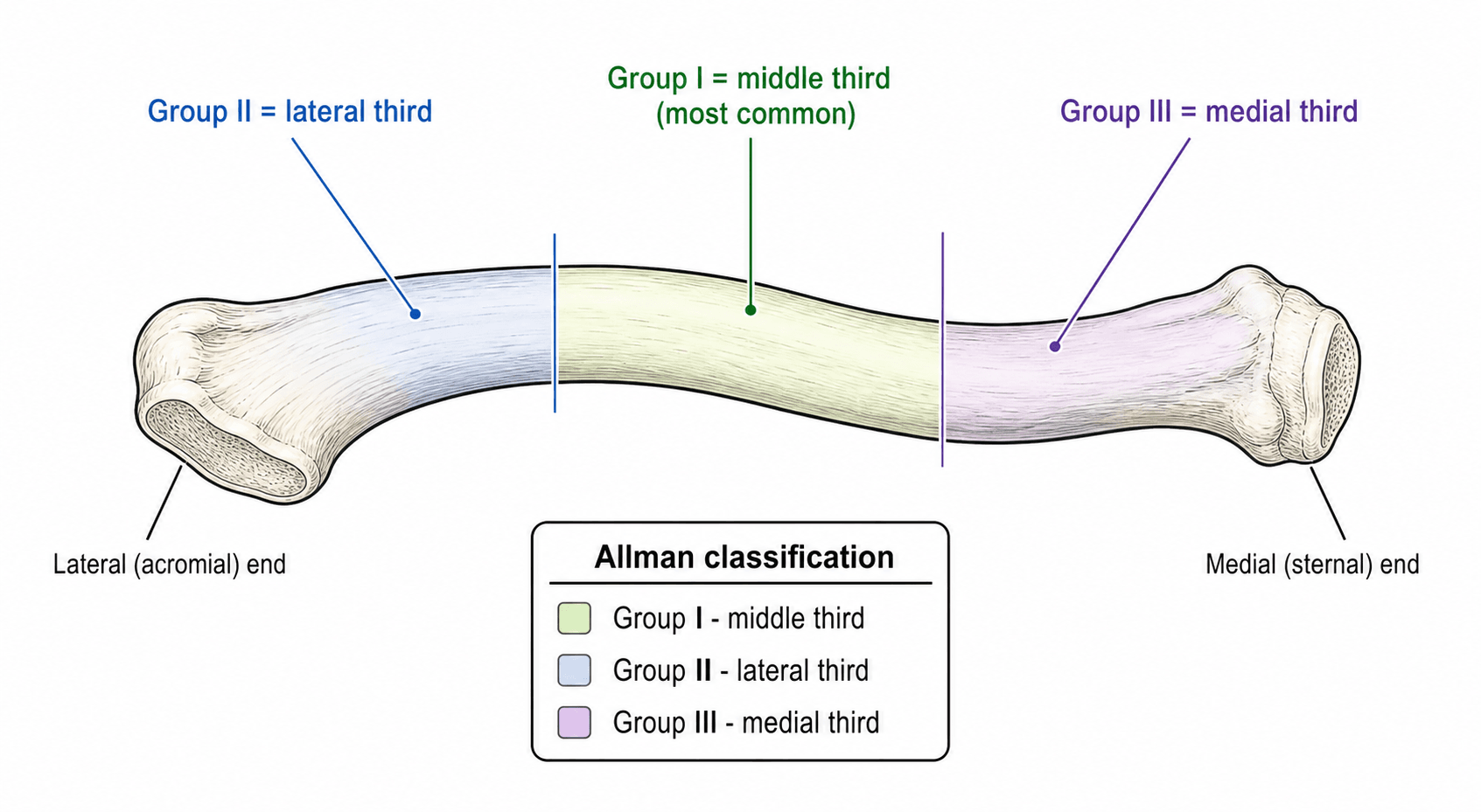
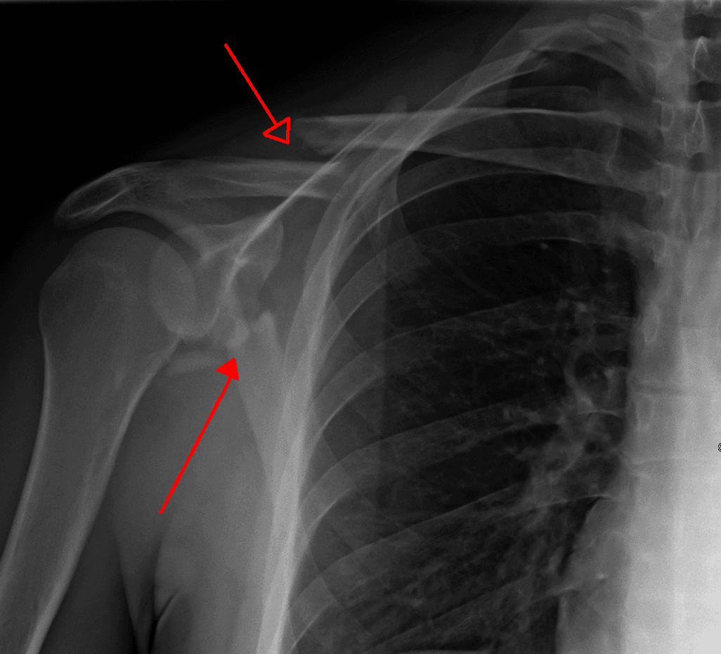
- Location
- Middle third (midshaft)
- Prevalence
- About 80 percent
- Typical mechanism
- Direct blow to shoulder or fall onto point of shoulder
- Key consideration
- Most heal with a sling; displacement and pattern decide whether to fix
- Location
- Lateral (distal) third
- Prevalence
- About 15 percent
- Typical mechanism
- Direct lateral impact or fall onto acromion
- Key consideration
- Neer subtypes critical: Type II is high-risk for nonunion
- Location
- Medial (sternal) third
- Prevalence
- About 5 percent
- Typical mechanism
- Direct blow to sternum or high-energy medial force
- Key consideration
- Assess subclavian vessels, brachial plexus, pneumothorax, mediastinal injury
MLMPrevalence by Allman group
Hook:MLM — Most are Middle, Less are Lateral, Minimal are Medial.
The middle third is the weakest segment because it is the transitional zone between the robust medial sternal articulation and the lateral coracoclavicular ligament complex. It has the thinnest soft-tissue envelope and no strong ligamentous tether — this is why it fractures first and why displacement is common in higher-energy injuries.
Classification is only step two — the OSCE wants the assessment that comes first:
- Presentation: pain with the arm supported across the trunk, a visible/palpable deformity or prominence, and skin tenting over a displaced or comminuted spike (a warning of imminent conversion to an open fracture).
- Examination (never skip): a full neurovascular exam — the brachial plexus (especially the medial cord) and the subclavian/axillary artery (limb perfusion, distal pulses, an expanding haematoma) — and a respiratory exam for pneumothorax (particularly with a medial-third or high-energy injury); inspect for an open wound.
- Imaging: an AP clavicle plus a cephalic-tilt view (about 15 to 30°) to judge displacement; the Zanca view (about 10 to 15° cephalic tilt, reduced exposure) best profiles the AC joint / lateral third; bilateral views to measure shortening; and CT for the medial third / sternoclavicular joint and to characterise comminution.
Neer subtypes of lateral third fractures
Neer (1968) subclassified Allman Group II (lateral third) fractures by the relationship of the fracture line to the coracoclavicular (CC) ligaments and by the degree of displacement. This sub-classification is clinically essential because it predicts union and guides treatment.
The CC ligaments have two components: the trapezoid (lateral, attaching to the trapezoid ridge of the coracoid and the undersurface of the clavicle) and the conoid (medial, attaching to the base of the coracoid and the conoid tubercle of the clavicle). Their integrity relative to the fracture line determines displacement and stability.
- Fracture position
- Between or lateral to the CC ligaments (interligamentous)
- Ligament status
- Both trapezoid and conoid intact; CC ligaments hold the medial fragment
- Displacement
- Minimal
- Nonunion risk
- Low
- Treatment
- Sling for 3 to 6 weeks; most unite without issue
- Fracture position
- Medial to BOTH CC ligaments
- Ligament status
- Both conoid and trapezoid remain on the distal fragment; the proximal (medial) fragment has no ligamentous tether
- Displacement
- Significant — proximal fragment displaces superiorly under SCM/trapezius pull
- Nonunion risk
- High (up to ~30 percent)
- Treatment
- ORIF: hook plate, CC fixation, or plate-and-suture-button construct
- Fracture position
- Between the conoid and trapezoid, OR with the conoid torn
- Ligament status
- Trapezoid stays on the distal fragment; conoid disrupted (or left on the proximal fragment)
- Displacement
- Significant — proximal fragment displaces superiorly
- Nonunion risk
- High (up to ~30 percent)
- Treatment
- ORIF: hook plate, CC screw, or dual small-fragment plating
- Fracture position
- Intra-articular, extending into the AC joint
- Ligament status
- CC ligaments intact; AC capsule involved
- Displacement
- Variable (minimal bony displacement)
- Nonunion risk
- Low for bony union; risk is late AC arthrosis
- Treatment
- Symptomatic relief; if late AC arthrosis develops, distal clavicle excision
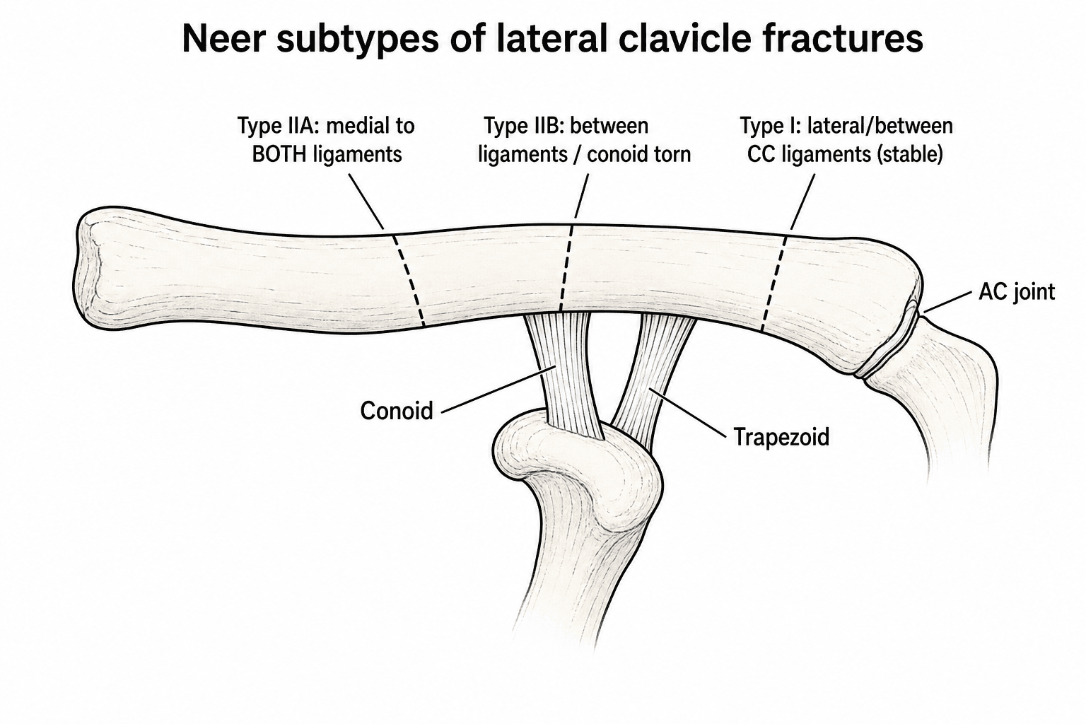
The defining error to avoid: do not confuse Neer Type IIA and IIB. Type IIA is the fracture medial to both CC ligaments — both conoid and trapezoid stay on the distal fragment, leaving the proximal fragment completely untethered. Type IIB is the fracture between the ligaments or with the conoid torn — only the trapezoid holds the distal fragment. Both are unstable Type II patterns with a 20 to 30 percent nonunion rate non-operatively, because the proximal fragment retracts superiorly under sternocleidomastoid and trapezius traction while the distal fragment is tethered down — the muscle forces hold the fracture surfaces apart. Both are operative candidates.
SIDENeer Type II displacement mechanism
Hook:SIDE — SCM pulls Up, Intra-articular enters the joint, Distal stays Down, Evaluate the ligaments to call IIA vs IIB.
In children and adolescents, the equivalent of a Neer Type II lateral third fracture is usually a physeal separation (Salter-Harris equivalent) rather than a true ligament disruption: the periosteal sleeve stays intact and the bone remodels. Treat non-operatively in the young patient unless there is gross displacement or neurovascular compromise.
The medial third (Allman Group III) — the dangerous rarity
Group III is only about 5 percent of clavicle fractures, but it is high-stakes because of what lies immediately behind it.
The medial clavicular physis is the last in the body to ossify (around age 18 to 20) and fuse (around age 23 to 25). So in a patient under about 25, what looks like a sternoclavicular dislocation is usually a physeal (Salter-Harris) fracture-separation through this growth plate — which has a far better remodelling potential than a true dislocation.
Anterior medial/sternoclavicular displacement is usually benign (often reduced and accepted even when it re-displaces). POSTERIOR (retrosternal) displacement is a surgical emergency: the medial fragment can compress the trachea, oesophagus and great vessels (brachiocephalic/subclavian vein, aortic arch), producing dyspnoea, stridor, dysphagia, venous congestion or diminished pulses. CT with contrast (CT angiogram) is the investigation of choice (plain films, including the serendipity view, are unreliable). Manage posterior displacement with urgent closed reduction under general anaesthesia (a scapular bump, abduction-traction, and a sterile towel clip to grasp the medial clavicle) with cardiothoracic surgery on standby, proceeding to open reduction if closed reduction fails.
Displacement & the decision to operate
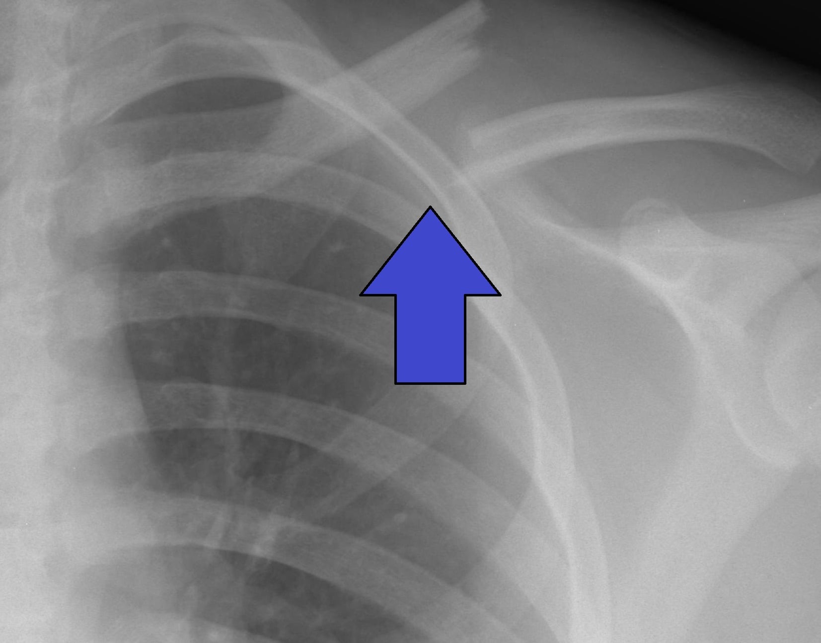
For Allman Group I (midshaft) fractures the classification alone does not dictate treatment — the surgeon must assess displacement, shortening and fracture pattern to choose between a sling and plate fixation.
- 1Is there an absolute indication?Open fracture, neurovascular injury, floating shoulder, scapulothoracic dissociation, skin tenting with imminent open conversion, or polytrauma needing early upper-limb use → fix.
- 2Measure displacement & patternShortening over 20 mm, no cortical contact between main fragments, a vertical/short-oblique pattern, or comminution with multiple fragments are relative indications for plate fixation.
- 3Weigh the patient's demandA young, active or high-demand patient with a displaced fracture benefits most from anatomical restoration and early mobilisation; an elderly, low-demand patient with a malunion is often asymptomatic.
- 4Choose the constructSuperior anatomically precontoured plate with interfragmentary compression where the pattern allows. Intramedullary fixation is an option for simple patterns but plates carry stronger evidence.
- Indication
- Open fracture
- Rationale
- Wound management and infection control; debridement and fixation
- Indication
- Neurovascular injury (brachial plexus, subclavian vessels)
- Rationale
- Urgent exploration/decompression; fixation stabilises the reduction
- Indication
- Floating shoulder (ipsilateral scapular neck fracture)
- Rationale
- Superior shoulder suspensory complex disrupted; fixation restores stability
- Indication
- Scapulothoracic dissociation
- Rationale
- Lethal if missed; vascular repair and clavicle reconstruction required
- Indication
- Skin tenting with imminent open conversion
- Rationale
- Fix to protect and close the soft-tissue envelope
- Indication
- Polytrauma needing upper-limb mobilisation
- Rationale
- Early fixation permits rehabilitation and care of other injuries
- Indication
- Shortening greater than 20 mm
- Rationale
- Alters shoulder biomechanics; correlates with weakness and poorer outcome in active patients
- Indication
- No cortical contact between main fragments
- Rationale
- Predicts nonunion and symptomatic malunion non-operatively
- Indication
- Vertical or short-oblique pattern
- Rationale
- Mechanically unstable; tends to shorten further under the weight of the arm
- Indication
- Comminution (multiple cortical fragments)
- Rationale
- Marks high-energy injury; shortening progresses during non-operative care
- Indication
- Young, active or high-demand patient
- Rationale
- Functional demands justify anatomical restoration and faster return to sport/work
SIXORIF decision for midshaft fractures
Hook:SIX — Shortening, Interfragmentary gap, eXtremes: if any are present, consider fixation.
Allman vs the Robinson (Edinburgh) classification
A common viva escalation is "what are the limitations of Allman, and what do you use instead?" Allman gives a simple anatomical framework, but it does not capture displacement or comminution — the very features that drive the nonunion risk and the fixation decision. The Robinson (Edinburgh) classification (1998) was designed to fix exactly that, which is why modern trauma units quote it.
- Allman (1967)
- Anatomical third only (middle / lateral / medial)
- Robinson / Edinburgh (1998)
- Anatomical fifth PLUS displacement and comminution/articular extension
- Allman (1967)
- 3 groups (I middle, II lateral, III medial)
- Robinson / Edinburgh (1998)
- Type 1 medial fifth, Type 2 diaphyseal, Type 3 lateral fifth - each split A (undisplaced/cortical contact) vs B (displaced), and further sub-grouped
- Allman (1967)
- No - location alone
- Robinson / Edinburgh (1998)
- Yes - displaced 2B (diaphyseal) and 3B (lateral) carry the highest nonunion; comminution adds risk
- Allman (1967)
- Simple but coarse
- Robinson / Edinburgh (1998)
- Satisfactory inter- and intra-observer reliability (validated on 1000 fractures)
- Allman (1967)
- Quick communication / the classic exam answer
- Robinson / Edinburgh (1998)
- Stratifying who is at risk and who benefits from fixation
Name Allman for the quick anatomical answer, then say you would stratify with Robinson because it incorporates displacement and comminution — the features that actually predict nonunion (displaced 2B/3B) and therefore drive the decision to fix. For lateral-third fractures, add the Neer/Craig subtypes to decide stability.
Guidelines, registries & global practice
- Displaced midshaft fractures: the Canadian Orthopaedic Trauma Society (COTS) multicentre RCT showed plate fixation gives better Constant and DASH scores and lower nonunion than non-operative care at one year — but subsequent meta-analyses confirm most patients still unite and many do well without surgery, so reserve fixation for the displaced, high-demand subgroup.
- AAOS / BOA practice: non-operative care for undisplaced fractures; operative fixation for absolute indications and for displaced fractures in active patients after shared decision-making. There is no global consensus to operate on every displaced clavicle.
- Lateral third (Neer II): higher nonunion risk than shaft fractures and less predictable operative outcomes — fixation is generally recommended in active patients, but a symptomatic nonunion in the elderly may be acceptable.
- Construct trends: superior anatomical plating predominates; hook plates remain a recognised option for very lateral fragments but carry subacromial impingement and require routine removal; suture-button CC reconstruction is increasingly used to avoid a second operation.
Viva practice
Exam viva
Practise clinical reasoning and management decisions out loud
“A 28-year-old professional rugby player falls directly onto the point of his left shoulder during a match. Radiographs show a displaced midshaft fracture of the left clavicle with 25 mm of shortening and no cortical apposition between the main fragments. The fracture is a short oblique configuration with a butterfly fragment. How would you classify this, and what would you recommend?”
“A 45-year-old builder presents after a fall onto his right shoulder. Radiographs show a fracture of the distal third of the right clavicle with the distal fragment sitting in anatomical position and the proximal fragment displaced 18 mm superiorly. What is the diagnosis, how would you classify it, and what would you do?”
Exam cheat sheet
- Allman by third: I middle (~80%), II lateral (~15%), III medial (~5%). Middle third is the weakest, transitional segment.
- Neer subtypes the lateral third by the CC ligaments. Type I interligamentous/stable; Type IIA medial to both ligaments (both on distal fragment); Type IIB conoid torn / between the ligaments. Both Type II patterns: 20 to 30 percent nonunion non-operatively → ORIF.
- Group I fixation is a displacement decision: over 20 mm shortening, no cortical contact, vertical/comminuted pattern, or a high-demand patient.
- Absolute indications: open, neurovascular injury, floating shoulder, scapulothoracic dissociation, skin tenting, polytrauma.
- Group III is rare but dangerous: assess subclavian vessels, brachial plexus, pneumothorax, mediastinum.
- In children, the lateral injury is usually physeal with an intact periosteal sleeve — it remodels.
Allman three-group classification
- Group I: middle third, about 80 percent; most heal with a sling, fix if displaced or high-demand
- Group II: lateral third, about 15 percent; use Neer subtypes to decide management
- Group III: medial third, about 5 percent; rare but assess subclavian vessels, brachial plexus, pneumothorax, mediastinum
Neer subtypes for lateral third fractures
- Type I: between/lateral to CC ligaments, minimal displacement, sling
- Type IIA: fracture medial to BOTH CC ligaments — both ligaments on the distal fragment, proximal fragment untethered, high nonunion, ORIF
- Type IIB: conoid torn or fracture between the ligaments — only trapezoid on the distal fragment, high nonunion, ORIF
- Type III: intra-articular into the AC joint; if late arthrosis develops, distal clavicle excision
When to fix a midshaft clavicle fracture
- Absolute: open, neurovascular injury, floating shoulder, scapulothoracic dissociation, skin tenting, polytrauma
- Relative: shortening greater than 20 mm, no cortical contact, vertical/short-oblique pattern, comminution, young active patient
- Construct: superior anatomical plate with interfragmentary compression; intramedullary fixation is an option but plates have stronger evidence
- Complications: infection, hardware irritation, nonunion (rare after fixation), malunion, supraclavicular nerve injury, refracture after plate removal
Evidence Base
Fractures of the clavicle in the adult. Epidemiology and classification
- 1000 consecutive adult clavicle fractures (Edinburgh); a bimodal age-sex incidence pattern was defined.
- Introduced the Edinburgh classification by anatomical site, displacement, comminution and articular extension, with satisfactory inter- and intra-observer reliability.
- Displaced diaphyseal (type 2B) and displaced outer-fifth (type 3B) fractures had the highest nonunion risk; comminution in 2B added further risk.
Fractures of the clavicle
- Undisplaced shaft and lateral fractures unite well non-operatively with good function.
- Displaced shaft fractures carry a higher nonunion/functional-deficit rate than older series suggested, but outcomes remain hard to predict.
- Displaced lateral-end fractures have a higher nonunion risk than shaft fractures and operative results there are less predictable.
According to PubMed, the epidemiology and the displacement-based nonunion stratification come from Robinson 1998 (DOI) and the synthesis of operative-versus-non-operative outcomes from Khan et al. 2009 (DOI). The Allman three-group system, the Neer/Craig lateral-third subtypes, and the COTS fixation evidence are standard, well-established clavicle-trauma teaching.