ALPSA Lesions
Overview
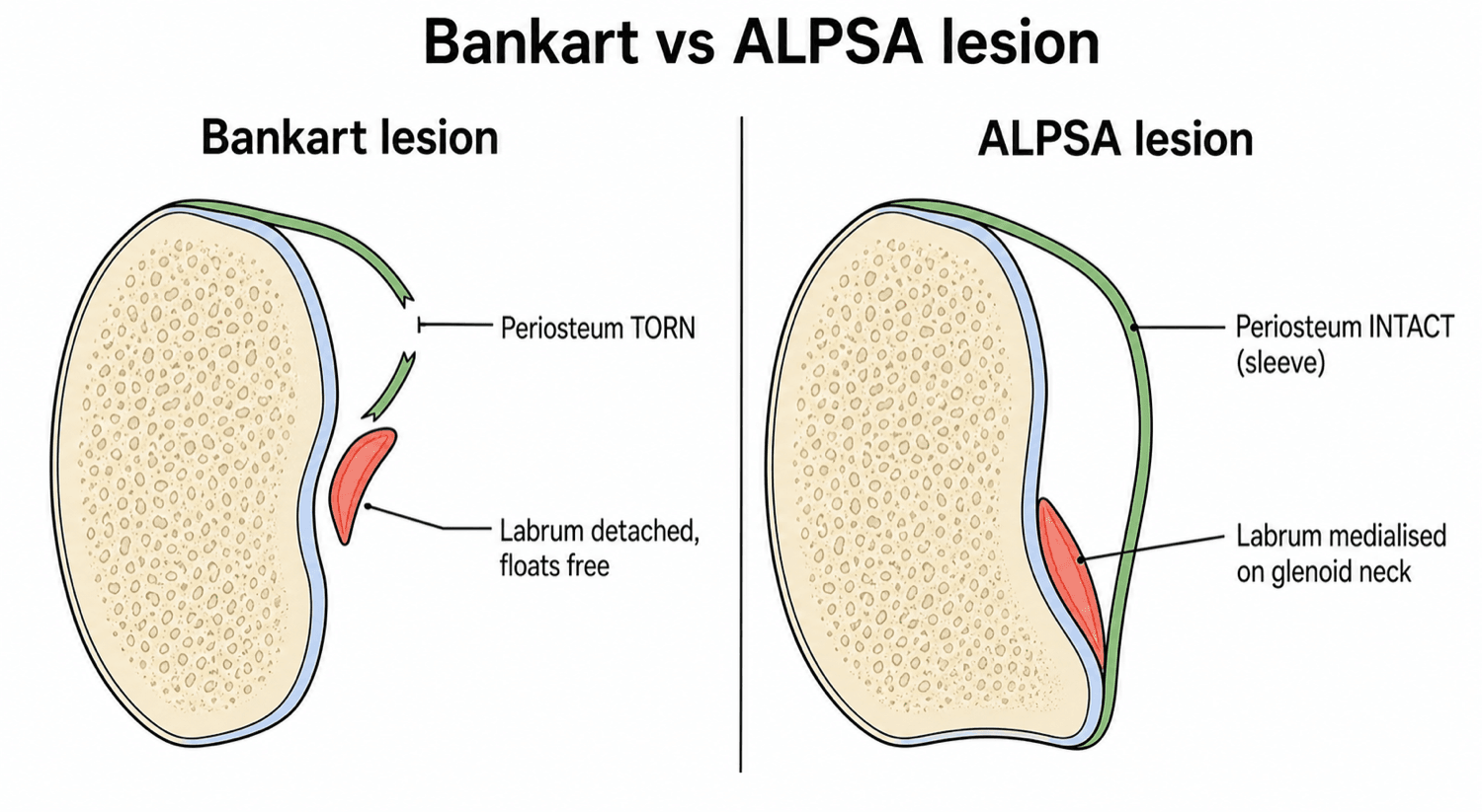
What it is. An ALPSA lesion (Anterior Labroligamentous Periosteal Sleeve Avulsion) is an avulsion of the anterior labrum and IGHL from the glenoid rim in which the anterior scapular periosteum remains intact but stripped. Because the periosteum holds, the tissue behaves as a sleeve: the labrum slides medially down the glenoid neck and heals there, inferiorly on the neck, scarred into a non-functional position.
How it differs from a Bankart. In a Bankart lesion the periosteum tears, creating a distinct separation: the detached labroligamentous structures float free anterior to the rim and the lesion does not heal, leaving a gap. In an ALPSA the tissue is still present but medialised, and that "medialisation" eliminates the glenoid depth, predisposing the joint to recurrent anterior instability even with minor trauma.
Pathophysiology and Mechanisms
The labroligamentous complex. The structures involved are the labrum, the inferior glenohumeral ligament and the periosteum:
- Labrum: a fibrocartilaginous bumper that increases glenoid depth by 50%
- Inferior glenohumeral ligament (IGHL): the main static restraint
- Periosteum: the thick anterior scapular periosteum
The mechanism. A forceful anterior dislocation strips the periosteum without rupturing it. The stripped periosteum forms a sleeve, and the labrum and ligament slide medially within it.
The healing trap. Because the tissue remains vascularised through the periosteum and stays in contact with bone, it heals. It heals on the glenoid neck, medial to the rim, so the glenoid concavity is lost (the "flat glenoid") and the chock-block effect is gone. This is what explains the high recurrence rates in untreated ALPSA.
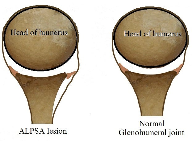
Biomechanical consequences. Loss of labral height reduces the effective glenoid concavity by up to 50%. Medialised healing elongates the IGHL complex, leaving it effectively lax and reducing its tension; force distribution across the glenohumeral joint is disrupted, and the force required to cause a dislocation is significantly reduced.
The compression-concavity mechanism. The labrum normally creates a chock block that resists humeral head translation. When the labrum is medialised that effect is lost even though the tissue is present, and the joint becomes functionally equivalent to a glenoid with bone loss. This is why a repair without mobilisation fails: the bumper never returns.
Classification Systems
Neviaser classification. Neviaser staged the lesion by its age and the state of the tissue, and the distinction is crucial for surgical planning:
- Duration
- Less than 3 months
- Tissue
- Mobile
- Surgical implication
- Can be reduced easily
- Duration
- Greater than 3 months
- Tissue
- Scarred and fixed
- Surgical implication
- Requires sharp dissection and release
Chronic lesions often require a capsular release, anterior and inferior, to gain excursion.
Clinical Presentation
History. The patient reports recurrent instability, often described as a "loose" shoulder, and often gives a history of multiple dislocations. Clicking or catching can occur. The provoking position is abduction and external rotation, the ABER position, and the lesion is common in young patients under 25.
What the history can tell you. The history is identical to Bankart instability. Chronic symptoms suggest an ALPSA rather than an acute Bankart.
Red flags. These warrant urgent imaging and specialist referral:
- First-time dislocation over age 40 (rotator cuff tear risk)
- Neurological symptoms (axillary nerve injury)
- Unable to reduce (locked posterior dislocation)
Examination
Findings. The tests and what they show:
- Finding
- Positive
- Finding
- Positive: relief with a posterior force
- Finding
- Increased translation (grade 2-3)
- Finding
- May be present (multidirectional component)
- Finding
- Assesses inferior capsular laxity
The limit of the examination. You cannot clinically distinguish an ALPSA from a Bankart. Imaging is the only differentiator.
Imaging
Why MR arthrography. The lesion is defined on imaging. Direct MR arthrography (MRA), with intra-articular gadolinium, is substantially more sensitive and specific for labroligamentous lesions than conventional non-contrast MRI, because the contrast distends the joint and tracks into the lesion and under the stripped periosteal sleeve, outlining the medialised labrum. A healed, scarred chronic ALPSA and a re-apposed Perthes can look near-normal on non-contrast MRI and even on neutral-position MRA, and are easily missed.
The ABER view. Abduction-external rotation is the key manoeuvre: it tensions the anteroinferior IGHL-labral complex and pulls contrast into the lesion, unmasking non-displaced or healed anteroinferior labral lesions that the neutral view misses. A suspected instability lesion therefore warrants MR arthrography, ideally including ABER images; CT arthrography is the alternative when bone loss must be quantified precisely or MRI is contraindicated.
What to look for. On axial T2 or PD fat-saturated images, find the labrum, the "black triangle". Normally it sits on the rim. In an ALPSA it is a rounded, scarred bundle sitting medial to the rim on the glenoid neck, and the periosteum may be visualised as a low-signal line connecting the labrum to the scapula: the sleeve. Look also for the "cul-de-sac" deep to the capsule.
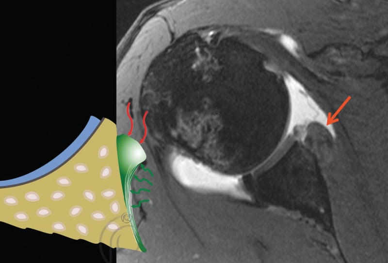
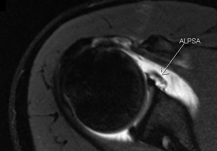
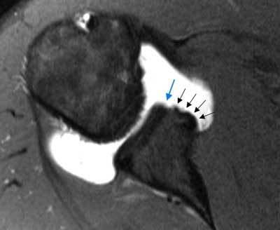
The MRI signs. Medialisation is the key sign. The others to report:
- Synovial stripping: contrast tracking under the sleeve
- Bone loss: assess for a concomitant bony Bankart, often remodelled in chronic cases
- Hill-Sachs: usually present because of the recurrence
CT arthrography. CTA is particularly useful for precise quantification of glenoid bone loss, for chronic cases in which scarring makes MRI interpretation difficult, and for preoperative planning with 3D reconstructions. It is superior to MRI for detecting subtle bone loss of less than 10%. In an ALPSA the contrast tracks medially with the labrum, the intact periosteal sleeve is visible as a soft-tissue connection to the scapula, and the glenoid morphology is assessed for erosive bone loss versus an inverted-pear glenoid.
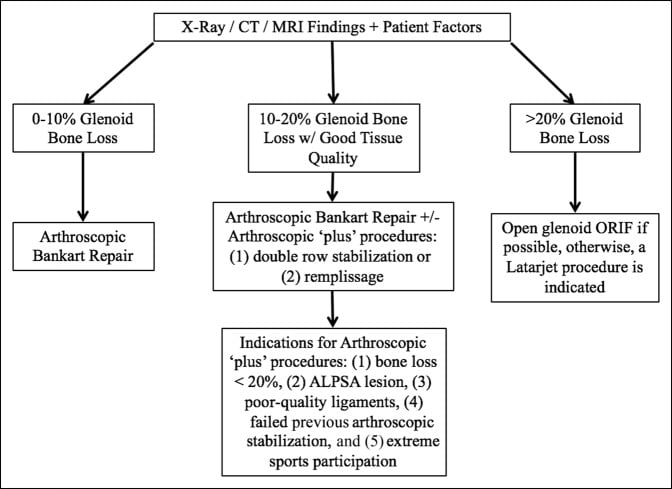
Glenoid bone loss. Quantifying bone loss is critical for surgical planning, because significant bone loss may require a Latarjet. The measurement methods:
- Best-fit circle: measure the defect as a percentage of the inferior glenoid circle
- Glenoid index: the ratio of anterior to posterior glenoid width
- En-face view: 3D CT reconstruction for direct visualisation
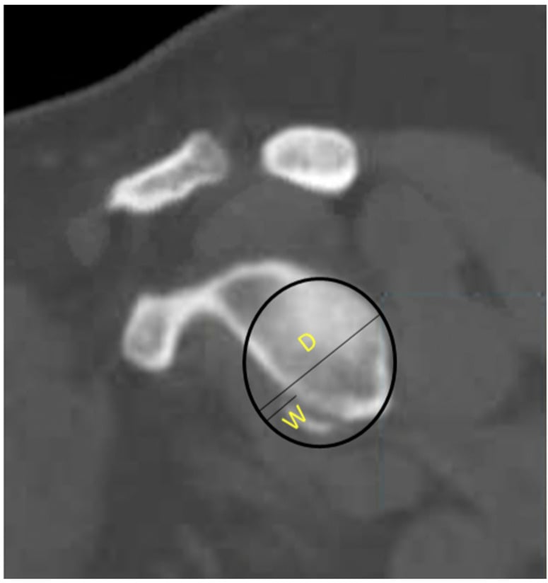
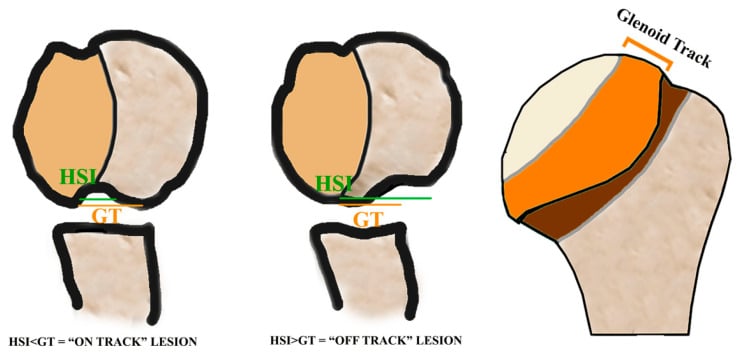
Thresholds. The percentage of bone loss guides the surgical decision:
- Less than 15%: soft-tissue repair adequate
- 15-25%: consider augmentation (remplissage, bone graft)
- Greater than 25%: Latarjet typically required
Differential Diagnosis
The anterior lesion family. A Bankart lesion is a true labral detachment with a floating labrum. A Perthes lesion is a non-displaced periosteal sleeve avulsion. A GLAD lesion is a glenolabral articular disruption, with the focus on the cartilage. Beyond these, consider a HAGL lesion (humeral avulsion of the glenohumeral ligament), a bony Bankart (labrum with an attached bone fragment) and multidirectional instability (global capsular laxity, atraumatic).
- Periosteum
- Torn
- Labrum position
- Floating/detached
- Stability
- Unstable
- Periosteum
- Intact
- Labrum position
- Medialised
- Stability
- Unstable
- Periosteum
- Stripped
- Labrum position
- In situ
- Stability
- Unstable
- Periosteum
- Intact
- Labrum position
- Intact
- Stability
- Stable
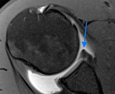
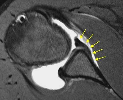
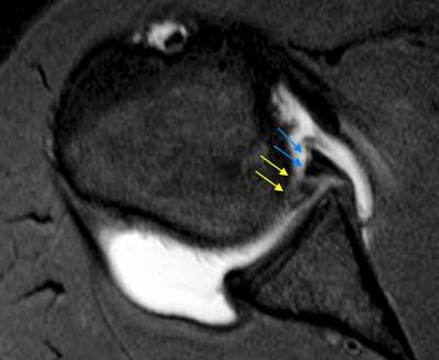
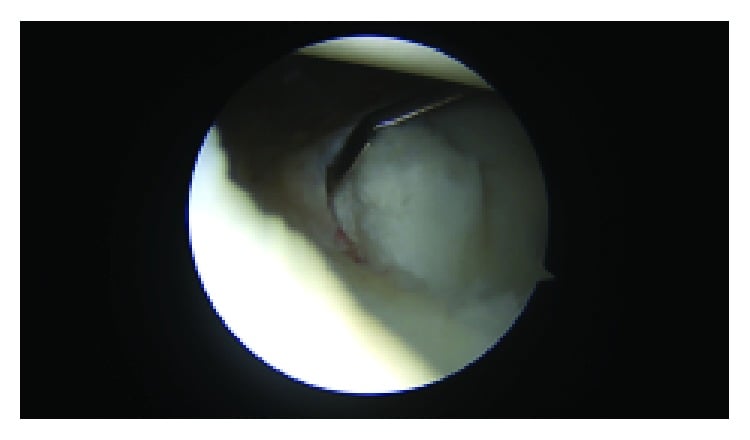
The posterior counterpart. Everything above is anterior. The posterior analogue is the POLPSA (Posterior Labrocapsular Periosteal Sleeve Avulsion), the direct mirror of the ALPSA: in posterior instability the posterior labrum and capsule are stripped with an intact periosteal sleeve and displace and medialise on the posterior glenoid neck, and the same principle applies, mobilise the medialised tissue before fixation. Posterior instability is more often atraumatic or from repetitive loading (linemen, bench-pressing) than a single dislocation, but the labroligamentous-sleeve concept is identical. The related posterior lesions:
- Reverse Bankart: a discrete postero-inferior labral detachment, the posterior equivalent of the Bankart
- Kim lesion: an incomplete, "concealed" avulsion of the postero-inferior labrum (a marginal crack with an intact superficial surface) that must be completed and then repaired
- Reverse Hill-Sachs (McLaughlin) lesion: the anteromedial humeral-head impaction of a posterior dislocation, classically after a seizure or electrocution
Management Algorithm
The decision. Non-operative treatment has a high failure rate in young patients and is not recommended for athletes. Operative treatment is indicated for recurrent instability. Arthroscopic repair is the gold standard; open repair is rarely needed unless there is massive bone loss. The key is the technical execution of the repair.
Timing. Early repair of an acute lesion is easier because the tissue is not scarred. Chronic repair is equally successful if the lesion is adequately mobilised. Do not delay once the diagnosis is confirmed.
Surgical Considerations
Arthroscopic repair, step by step.
- Diagnostic arthroscopy. Confirm the diagnosis. Probe the labrum: it may feel "healed" but it lacks bumper function.
- Mobilisation, the critical step. Use an elevator or shaver to detach the scarred labrum from the glenoid neck, and release anteriorly and inferiorly, around the corner. "Float the labrum": it must rise to the rim effortlessly.
- Preparation. Decorticate the glenoid rim to create a bleeding bed.
- Fixation. Suture anchors on the face, at the articular margin.
- Shift. Tension the tissue superiorly and laterally to restore the bumper.
Failure to mobilise turns this into a non-anatomical plication.
Because the ALPSA lesion has healed, it can look like a normal but small labrum. If you simply put anchors in and stitch it where it lies, you validate the malposition: the bumper has not been restored and recurrence is guaranteed. The healed tissue must be incised and the labrum elevated off the glenoid neck to the face before any anchor goes in.
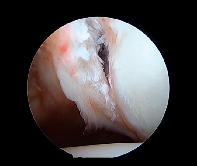
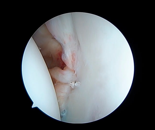
Pearls. The 5 o'clock portal is essential for the inferior release. Switch to the anterosuperior portal to view the reduction of the drive-through sign. Use 3 or more anchors for robust fixation, and if the tissue is poor, incorporate the capsule (Bankart repair plus capsular shift).
Complications
Recurrence is the most common complication, and is often due to failure to mobilise the lesion fully. Stiffness follows excessive tightening (overtensioning). The axillary nerve is at risk during the inferior dissection. Infection is rare.
Hardware and chondrolysis. Anchors can loosen. Placing anchors on the articular face is necessary for an ALPSA repair, but they must not be prominent: metal or hard PEEK anchors that sit proud can destroy the humeral head cartilage (chondrolysis). Use soft anchors, or ensure that knotless anchors are countersunk.
Rehabilitation
The protocol. Rehabilitation is identical to that after a Bankart repair:
- Content
- Sling immobilisation; pendulums
- Restriction or milestone
- Protect external rotation (usually restricted to 0 or 30 degrees)
- Content
- Regain range of motion: active assisted to active
- Restriction or milestone
- Scapular control
- Content
- Strengthening; proprioception
- Restriction or milestone
- Return to sport at 6 months (contact sports)
Sling removal criteria.
- No pain at rest
- Control of the scapula
- Ability to perform activities of daily living at waist level
Contact progression.
- 4 months: non-contact drills (passing, catching)
- 5 months: controlled contact (pad work)
- 6 months: unrestricted contact (tackling)
Return to sport criteria.
- Full, pain-free range of motion
- Symmetrical strength (external rotation, internal rotation, abduction)
- Psychological readiness (ACL-RSI or a similar scale)
- Completion of sport-specific training drills
- No apprehension in the apprehension position
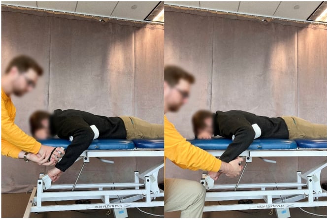
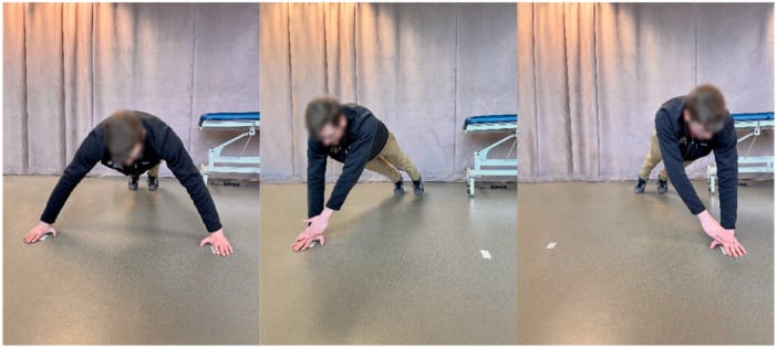
Prognosis
Recurrence. The recurrence rate is higher than for a discrete Bankart even after arthroscopic repair: 19.2% (5/26) versus 7.4% (5/67) at a mean 47 months (P = .05, Level IV). Proper mobilisation of the medialised lesion is necessary but does not, on the available evidence, bring the failure rate down to that of a discrete Bankart. Without mobilisation, recurrence rates are significantly higher still.
Why. ALPSA patients arrive after significantly more instability events, a mean of 12.3 dislocations or subluxations versus 4.9, so the lesion is a marker of a more damaged shoulder as well as a harder repair. That is the honest reading: the medialisation is part of the problem rather than the whole of it.
Other outcomes. Return to sport is high (greater than 85%). A long-term risk of arthritis exists because of the initial cartilage damage, and a GLAD component often co-exists.
Prognostic factors.
- Age: younger patients have a higher recurrence risk
- Bone loss: significant glenoid bone loss requires a Latarjet, by the thresholds in Imaging (typically over 25%)
- Hyperlaxity: a Beighton score greater than 5 increases the failure risk
- Sport: collision athletes have a higher recurrence
Keep the ALPSA in proportion: bone loss outranks it, and by a wide margin. Across 194 arthroscopic Bankart repairs, significant bone defects, an inverted-pear glenoid or an engaging Hill-Sachs, raised recurrence to 67% versus 4% without, and to 89% in contact athletes with bone loss. The authors identified those bony defects, not the soft-tissue pattern, as the dominant driver of arthroscopic repair failure, and recommended bony reconstruction where glenoid loss is significant.
The practical consequence for a medialised chronic lesion is that recognising and mobilising the ALPSA is necessary but is the smaller half of the assessment. Quantify the glenoid bone loss and characterise the Hill-Sachs before committing to a soft-tissue repair: a beautifully mobilised ALPSA repaired over an inverted-pear glenoid still fails most of the time. The evidence is a retrospective case series that predates the glenoid-track concept, so use it for the magnitude of the effect rather than as a modern decision rule.
Guidelines, Registries & Global Practice
- No ALPSA-specific society guideline exists — it is a lesion subtype rather than a stand-alone diagnosis, so management follows the broader anterior shoulder instability literature (ISAKOS, AAOS, BOA-BESS and shoulder-society instability working groups) on labral repair and bone-loss thresholds.
- Epidemiology: ALPSA is over-represented in chronic, recurrent instability and in collision/contact athletes worldwide (rugby, American football, AFL, wrestling, ice hockey), reflecting the repeated dislocations that drive the medialised, scarred lesion.
- Standard of care: Recognition of the medialised lesion and mobilisation (takedown and float to the rim) before fixation is a universally expected technical standard; an "in-situ" repair that fixes the labrum on the neck is regarded internationally as a technical error and a cause of revision.
- Bone-loss integration: Across guidelines the decision is increasingly driven by quantified glenoid bone loss and the glenoid track (subcritical/critical thresholds, off-track Hill-Sachs), so a chronic ALPSA with significant bone loss may need a bony procedure (Latarjet) rather than soft-tissue repair alone.
- Practice variation reflects access to MR arthrography/CT arthrography for diagnosis and to bony-augmentation options, not disagreement on the mobilisation principle.
MCQ Practice Points
Q: What structure remains intact in an ALPSA lesion that is torn in a Bankart? A: The anterior scapular periosteum.
Q: What does ALPSA stand for? A: Anterior Labroligamentous Periosteal Sleeve Avulsion.
Q: What is the most critical step in ALPSA repair? A: Mobilization (Takedown) of the labrum.
Q: A chronic, fixed ALPSA lesion is classified as: A: Neviaser Type II.
Q: What MRI finding differentiates ALPSA from Bankart lesion? A: Medially displaced labrum with intact periosteal sleeve (labrum lies against glenoid neck rather than at rim).
Q: Why do ALPSA lesions have higher recurrence rates if not properly mobilized? A: The medialized labrum heals in a non-anatomic position, failing to restore the labral bumper effect and capsular tension.
Viva Scenarios
Practise clinical reasoning and management decisions out loud
“You are shown an MRI of a 19-year-old rugby player. There is a dark lump of tissue sitting on the anterior glenoid neck, medial to the rim. What is this and how does it differ from a Bankart?”
“You are scoping a 'Bankart'. You enter the joint and the labrum looks 'healed' but the shoulder is loose (Drive-through sign positive). Probe shows it is firmly attached but medial. What do you do?”
“A patient had a stabilisation elsewhere and re-dislocated. MRI shows the anchors are in the glenoid neck, not the face. The labrum is still medial. Why did it fail?”
“You are shown a CT arthrography of a 26-year-old footballer with chronic anterior instability. Contrast tracks medially along the glenoid neck with the labrum. What is the diagnosis and how does this affect your surgical planning?”
Diagnosis
- History: Recurrent Instability
- MRI: Medialized Labrum
- MRI: Intact Periosteal Sleeve
- Sign: Drive-through positive
Management
- Arthroscopic Repair is Gold Standard
- CRITICAL: Mobilize/Takedown lesion
- Fix to Glenoid Face (Not neck)
- Restore Bumper Effect
Key Differentiators
- Bankart: Torn Periosteum, Floating
- ALPSA: Intact Periosteum, Medialized
- GLAD: Cartilage defect
- Perthes: Undisplaced sleeve
Evidence Base
Neviaser's 1993 paper is the original description; Ozbaydar's 2008 comparative series is the key outcome data — and it shows ALPSA has a HIGHER recurrence than a discrete Bankart after repair, not an equivalent one. Bigliani's glenoid-rim-lesion classification and Burkhart & De Beer's bone-loss paper frame the associated bony pathology that worsens instability outcomes.
The anterior labroligamentous periosteal sleeve avulsion lesion (original description)
- Original description of the ALPSA lesion, found in 4 of 8 acute primary anterior dislocations
- Unlike a Bankart, the anterior scapular periosteum does NOT rupture, allowing the labroligamentous structures to displace medially and rotate inferiorly on the scapular neck
- The lesions heal in this medialised position and cause recurrent dislocation through subsequent incompetence of the anterior inferior glenohumeral ligament; the described arthroscopic technique converts the ALPSA back into a Bankart before repair
Results of arthroscopic capsulolabral repair: Bankart lesion versus ALPSA lesion
- 93 shoulders undergoing arthroscopic suture-anchor repair: 67 (72%) discrete Bankart, 26 (28%) ALPSA
- Recurrence at a mean 47 months was HIGHER in the ALPSA group (5/26, 19.2%) than the Bankart group (5/67, 7.4%) (P = .05)
- ALPSA patients had significantly more pre-operative dislocations/subluxations (mean 12.3 vs 4.9)
Glenoid rim lesions associated with recurrent anterior dislocation of the shoulder
- Classified anterior glenoid rim lesions in 25 unstable shoulders into Type I (displaced avulsion fracture with capsule), Type II (a medially displaced fragment malunited to the rim — analogous to the medialised ALPSA concept), and Type III (rim erosion, IIIA under 25% / IIIB over 25%)
- Detected on plain radiographs and CT-arthrograms; treatment reattached the fragment and/or capsule to the rim and addressed capsular laxity
- 88% (22/25) had satisfactory results without recurrence; 12% redislocated