The Other Side of Instability
- HAGL stands for Humeral Avulsion of the Glenohumeral Ligament.
- It is a cause of recurrent instability often missed on standard MRI.
- The 'J Sign' on coronal oblique MRI is pathognomonic.
- It is frequently associated with subscapularis tears (Open repair addresses both).
- Failure to recognise HAGL leads to failed Bankart repairs.
- “In a patient with instability but NO Bankart lesion, look for the HAGL.
- “Arthroscopic repair is technically demanding (70-degree scope helps).
- “Open repair is the historical gold standard.
Overview
What it is. A HAGL lesion (humeral avulsion of the glenohumeral ligament) is a traumatic detachment of the inferior glenohumeral ligament (IGHL) complex from its insertion on the anatomical neck of the humerus. The classic Bankart lesion is failure of the same complex on the glenoid side; the HAGL is the humeral side of the capsular failure spectrum. It is a potent cause of anterior instability because the hammock of the IGHL is disabled.
Why it was overlooked. The lesion was once considered rare, a "lesion of exclusion". Modern MRI techniques and arthroscopic vigilance have revealed it to be a significant contributor to recurrent instability, particularly in contact athletes. In anterior instability a Bankart lesion is found in 80-90% and a HAGL in 2-9%.
The spectrum of capsular failure. Identifying the specific failure pattern directs the surgical management, so the continuum has to be in mind from the start:
- Bankart: glenoid-side labral avulsion
- ALPSA: medialised glenoid-side failure with intact periosteum
- HAGL: humeral-side avulsion of the ligament proper
- Floating IGHL: bipolar failure, both ends detached, the most severe
Anatomy and Stabilisers
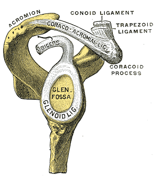
How the shoulder stays in. Glenohumeral stability is a balance, and it explains why a HAGL matters yet mid-range stability can be normal. The static (passive) stabilisers are the bony glenoid, which is shallow and contributes little alone; the labrum, which deepens the socket by roughly 50 percent and forms a chock-block and suction seal; the negative intra-articular pressure with the adhesion-cohesion of the synovial fluid; and the capsuloligamentous complex (SGHL, MGHL, IGHL), acting mainly as end-range check-reins. The dynamic (active) stabilisers are the rotator cuff producing concavity-compression, pressing the convex head into the concave glenoid, the long head of biceps, and scapular positioning by the periscapular muscles, which keeps the glenoid under the head.
Mid range and end range. In the mid range the dynamic cuff centres the head; at end range, in abduction and external rotation (ABER), the static IGHL takes over. A HAGL is a failure of that end-range static restraint, so the shoulder feels stable in the mid range but apprehensive in ABER: the end-range anterior check-rein has been lost.
The glenohumeral ligament complex. The IGHL that avulses in a HAGL is one of three discrete capsular thickenings, and the dominant restraint changes with arm position:
- SGHL: primary restraint to inferior translation in adduction (arm by the side) and a contributor to anterior stability; part of the rotator interval with the coracohumeral ligament
- MGHL: main restraint to anterior translation in the mid range (around 45 degrees of abduction) with external rotation, and the most anatomically variable ligament. The Buford complex, a cord-like MGHL with an absent antero-superior labrum, is a normal variant not to be repaired
- IGHL: the anterior band is the primary restraint to anterior translation at 90 degrees of abduction with external rotation, the position of instability, and is the part that must be restored; the posterior band restrains posterior translation in flexion and internal rotation; the axillary pouch between them forms the hammock
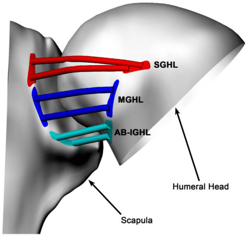
The humeral insertion. The IGHL inserts onto the anatomical neck just below the articular surface, approximately 1-2 cm from the articular margin, and the insertion spans 2-3 cm along the neck. Its blood supply is from branches of the anterior circumflex humeral artery. This insertion is the weak link in a HAGL, and anchors must be placed at the anatomical footprint to restore it.
Pathophysiology and Mechanism
Mechanism. Hyper-abduction and external rotation, the same mechanism as a Bankart, but the failure point is humeral. Studies suggest a HAGL may occur when the rotator cuff is intact (a stiff humerus) or with a concurrent subscapularis strain; the strain is a frequent companion because the two share anterior structures.
What is lost. The IGHL functions as a hammock supporting the humeral head in ABER, and a HAGL removes that support. The anterior check-rein is gone, so the head translates anteriorly without resistance, and the axillary pouch loses its tension and fails to cradle the head in abduction. The force required to cause dislocation is reduced by up to 50%.
Associated injuries. Subscapularis tears are often associated with the anterior HAGL, Hill-Sachs lesions are common, and glenoid bone loss occurs but is less common than with a Bankart. A HAGL can also coexist with a Bankart, dual pathology with both sides affected.
Classification
West Point classification (Bui-Mansfield). Types A and B name the band that has avulsed, Type C is the bipolar lesion, and a bone fragment can accompany any of them:
- Type A, anterior HAGL (93%): avulsion of the anterior band from the humeral neck; the most common
- Type B, posterior HAGL (7%): avulsion of the posterior band; rare, and associated with posterior instability
- Type C, floating AIGHL: complete detachment from both glenoid and humerus, a devastating loss of all inferior restraints
- Bony HAGL (BHAGL, 20%): any of the above with a bone fragment from the humeral neck, and the only variant visible on radiographs
Prognostic implications. Healing potential and repair difficulty follow the type.
- Healing potential
- Moderate
- Repair difficulty
- Standard
- Healing potential
- Good (bone to bone, if fixed)
- Repair difficulty
- May be easier
- Healing potential
- Poor
- Repair difficulty
- Most complex
- Healing potential
- Variable
- Repair difficulty
- Dual repair needed
Clinical Presentation and Examination
History. The event is a traumatic dislocation, classically high-energy: contact sport, a fall, a motor accident. In Bokor's series 94% had a violent first dislocation. Patients tend to be slightly older than those with isolated Bankart instability, though the lesion is also seen in adolescents as an "atypical" lesion. Afterwards they report instability or apprehension with overhead or ABER activity, and may recall a pop or tearing sensation deep in the axilla.
The failed repair. The other presentation is persistent instability after a "successful" Bankart repair that "never felt tight". Always question the diagnosis in a failed instability repair and in any traumatic dislocation without a labral tear.
Examination. Apprehension is positive in ABER and relocation is positive, with relief of pain or apprehension. Check the subscapularis with lift-off and belly-press, because a tear is often associated, and always verify axillary nerve sensation and deltoid function. Without imaging the lesion is clinically indistinguishable from Bankart instability: apprehension is sensitive but not specific for the type of lesion.
Imaging
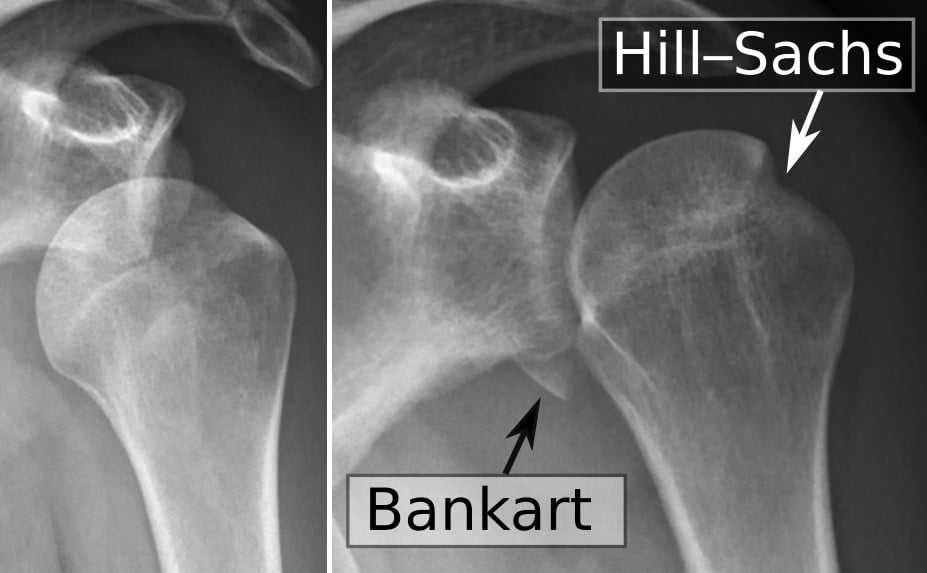
Radiographs. The film shows the bony footprints of an anterior dislocation, a bony Bankart or a Hill-Sachs, but a soft-tissue HAGL is radiographically occult, and it is the lesion to suspect when a traumatic dislocation leaves no Bankart. Only a bony HAGL, a fleck off the humeral neck, shows on plain film.
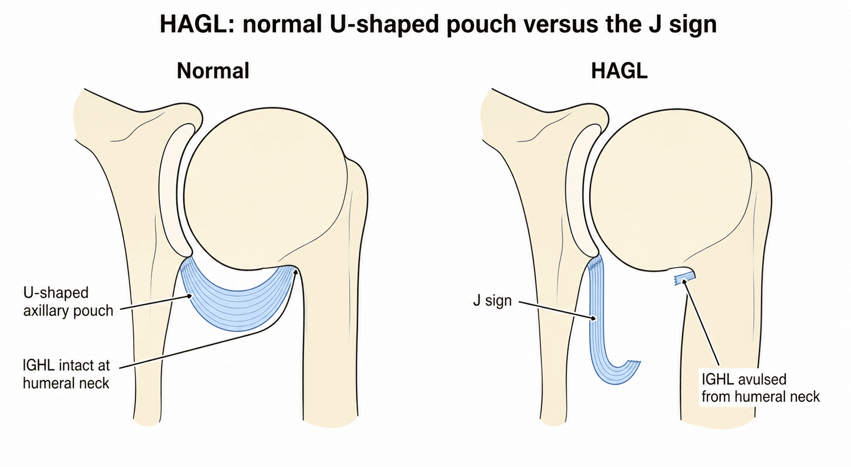
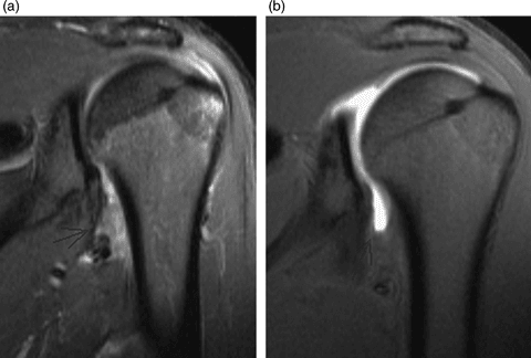
The J sign. On coronal oblique T2 images the axillary pouch normally forms a "U" under the humeral head, slung between the inferior glenoid and the medial humeral neck. In a HAGL the lateral (humeral) attachment is gone, and the IGHL falls inferiorly against the chest wall and glenoid, forming a deep "J".
Other MRI signs. Always evaluate the integrity of the capsule on the humeral side, not just the labrum. Beyond the J sign, look for:
- Double axillary pouch: fluid collects both within the joint and in the disrupted capsule, creating the appearance of two pouches
- Extravasation on MR arthrography: contrast escapes through the humeral-side capsular defect and tracks inferiorly along the humeral neck and shaft, in contrast to the glenoid-side leak typical of a capsular Bankart variant
- Oedema: bone marrow oedema at the humeral neck in acute lesions
The "fake" HAGL. The IGHL is lax in adduction, and folds can look like tears. Look for fluid extending below the teres minor, or frank detachment, and remember that MR arthrography significantly increases sensitivity. A high index of suspicion is required to differentiate artefact from pathology, and HAGL is missed on roughly one-third of preoperative MRIs even in specialist centres.
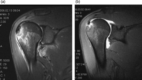
Timing of the scan. A later study can appear normal. In both cases below the lesion is present on the initial STIR image and the oblique coronal T1 fat-saturated MR arthrogram a few weeks later shows full recovery.
Differential Diagnosis
The clinical picture of "traumatic anterior instability" is shared by several lesions. The discriminator is almost always imaging and arthroscopy, not the bedside exam. Knowing how each entity differs determines where you place your anchors.
- Site of Failure
- Antero-inferior labrum / glenoid
- Key Discriminator
- Labral detachment with intact periosteum
- Implication
- Standard anterior arthroscopic Bankart repair
- Site of Failure
- Glenoid side, medialised
- Key Discriminator
- Labrum healed medially on the scapular neck
- Implication
- Mobilise and re-tension labrum back to glenoid rim
- Site of Failure
- Glenoid side, non-displaced
- Key Discriminator
- Labrum detached but held by intact periosteum, lies anatomically (often MRI-occult; ABER view helps)
- Implication
- Mobilise the labroperiosteal sleeve and re-fix to the rim
- Site of Failure
- Humeral insertion of IGHL
- Key Discriminator
- J sign on MRI, capsule stripped off humeral neck
- Implication
- Repair to humeral neck (open or 5 o'clock scope)
- Site of Failure
- Antero-inferior glenoid bone
- Key Discriminator
- Glenoid fragment, bone loss on CT
- Implication
- Address bone loss (fragment fixation or Latarjet)
- Site of Failure
- Global capsular redundancy
- Key Discriminator
- Sulcus sign, bilateral, atraumatic, no discrete lesion
- Implication
- Rehabilitation first; capsular plication if it fails
A patient with recurrent anterior instability after a genuinely traumatic, unidirectional dislocation, whose MRI report says "no labral tear", is not MDI and is not "functional". Reclassify the imaging as inadequate and review it specifically for the J sign at the humeral capsular insertion: the IGHL may be stripped from the humerus. A Bankart repair in that patient tightens the wrong side of the hammock, and the instability persists.
Management
The decision. Non-operative treatment is reserved for low-demand or elderly patients. Unlike a Bankart, a HAGL has limited spontaneous healing, because the avulsed capsule retracts and the synovial environment limits it, so bracing has little role. Operative repair is for young patients, athletes and recurrent instability.
Open or arthroscopic. Open repair is the gold standard: the lesion is easier to find and fix, and an associated subscapularis tear is addressed in the same exposure. Arthroscopic repair is technically difficult and needs a 70-degree scope and accessory portals (5 o'clock). The choice depends largely on surgeon experience and the presence of subscapularis pathology.
Timing. Acute repair, within 3 weeks, allows mobilisation of the tissue. A chronic HAGL may be scarred and retracted, making arthroscopic repair impossible, and chronic cases often require open release of the capsule to mobilise it.
Surgical Technique
Open repair. Deltopectoral approach. The subscapularis is taken down with an L-shaped tenotomy, or its lower half is taken down, to reach the deep capsule; the L-shaped tenotomy gives excellent exposure of the inferior pouch. External rotation of the humerus reveals the avulsed ligament, which is fixed with double-loaded suture anchors in the humeral neck, and the subscapularis is repaired robustly at closure. The approach buys visualisation and protection of the axillary nerve at the cost of subscapularis morbidity.
Arthroscopic repair. Standard posterior and anterior portals, with an accessory low anterior (5 o'clock) or trans-subscapularis portal. A standard anterior portal is too high to reach the inferior pouch, so the 5 o'clock portal is established low, with localised cutaneous nerve protection. Visualisation is key, so switch portals frequently.
- View from the anterior portal (or a high anterolateral portal)
- Prepare the bone bed on the humerus; the angle is difficult
- Pass sutures through the ligament first (outside-in or with a penetrator)
- Fix to the humerus
The axillary nerve. It is at risk in both open and arthroscopic repair. It runs at the 6 o'clock position, approximately 1-2 cm inferior to the glenoid and to the inferior capsule at the surgical neck, which puts it dangerously close to the 5 o'clock portal. The free edge of the lesion can lie far closer: Bokor measured only about 4.8 mm at 6 o'clock intra-operatively, and MRI overestimated the distance in 52% of cases.
- Open: identify and protect the nerve before the capsular repair; bluntly dissect and visualise it if unsure
- Arthroscopic: keep portals and instruments high, above the equator, when possible, or work within safe zones; establish the 5 o'clock portal with care, using a blunt trocar; never pass needles blindly in the inferior pouch
Complications
Intraoperative
Axillary nerve injury is the most feared complication; its course and protection are described above. The others:
- Subscapularis damage: aggressive retraction or inadequate repair during the open approach leads to subscapularis insufficiency
- Anchor malposition: anchors too high (in cartilage) or too low (in the shaft) fail to restore the anatomy
- Inadequate mobilisation: failure to release scarred tissue in a chronic case leaves an insufficient repair
Early postoperative
- Recurrent instability if the repair fails or an associated Bankart or bone loss was missed
- Stiffness, specifically loss of external rotation from capsular tightening
- Infection, the standard surgical risk
- Haematoma, the risk increased by subscapularis dissection
Late
- Chronic instability, often from missed pathology or an inadequate repair
- Subscapularis failure: weakness of internal rotation and on lift-off testing
- Glenohumeral arthritis, the long-term sequel of recurrent instability
- Anchor migration: osteolysis around bioabsorbable anchors or loosening of metal ones
Rehabilitation
External rotation threatens both the subscapularis repair and the HAGL repair, so the programme limits it for the first six weeks. Rehabilitation is often slower than after a Bankart because of the subscapularis precautions after an open repair.
- Sling immobilisation
- External rotation limited, usually to less than 30 degrees
- Pendulums
- Active range of motion
- Progress external rotation
- Strengthening
- Return to sport from 6 months
Prognosis
By repair type. Open repair restores stability in greater than 90%. Arthroscopic results are variable, 75-90%, and highly surgeon and case dependent. BHAGL repair has excellent outcomes because of bone-to-bone healing; the floating IGHL is the most challenging and may require a combined open approach.
Function. 85-95% return to their pre-injury level of activity. Sport is permitted from 6 months (above) and the return typically takes 6-9 months, longer than after an isolated Bankart. External rotation may be slightly reduced, by 5-10 degrees, and full strength is expected if the subscapularis has healed properly.
Prognostic factors.
- Positive
- Acute (less than 3 weeks)
- Negative
- Chronic (greater than 6 months)
- Positive
- BHAGL
- Negative
- Floating IGHL
- Positive
- Isolated
- Negative
- Combined Bankart/bone loss
- Positive
- Younger patient
- Negative
- Older with tissue degeneration
Long term. A missed HAGL means the Bankart repair fails: a 100% failure rate when a HAGL is present. Ongoing instability leads to accelerated osteoarthritis. Re-dislocation is low, less than 5%, if the lesion is properly repaired and rehabilitated.
Guidelines, Registries & Global Practice
Global Epidemiology
- HAGL accounts for roughly 2-9% of operatively treated anterior instability depending on the population studied (≈2.3% in the multicentre MOON cohort, ≈9% in early arthroscopic series).
- In the selected subgroup of a violent first dislocation without a Bankart lesion and no multidirectional laxity, incidence rises to ~39% (Bokor series).
- Predominantly affects young to middle-aged males injured in high-energy contact sport (rugby codes, football, wrestling) or trauma; also described as an "atypical" lesion in adolescents.
Society Guidance (side by side)
There is no dedicated international guideline for HAGL specifically; recommendations are extrapolated from anterior instability guidance and expert consensus.
- Relevant Position
- Anterior instability guidance emphasises identifying all capsulolabral pathology and bone loss before stabilisation; no HAGL-specific recommendation
- Relevant Position
- Shoulder instability pathways stress thorough MRI/MRA assessment and surgeon experience for non-Bankart lesions
- Relevant Position
- Recognise HAGL as a distinct cause of failure; recommend dedicated imaging review and repair to the humeral footprint
- Relevant Position
- Highlights axillary nerve protection during anterior-inferior capsular surgery
Registry Notes
- Joint registries (NJR, AJRR, AOANJRR, Swedish, Norwegian) capture arthroplasty, not soft-tissue instability repair, so there is no large-scale registry survivorship data for HAGL repair. Evidence remains limited to case series and multicentre cohorts such as MOON.
High- vs Limited-Resource Practice Variation
- Well-resourced centres: ready access to MR arthrography, ABER-positioned MRI, 70-degree arthroscopes and accessory portals enables attempted arthroscopic repair.
- Limited-resource settings: reliance on plain radiographs and standard MRI increases the miss rate; open deltopectoral repair is often the pragmatic, reliable default and addresses any associated subscapularis tear in the same exposure.
Controversies and Areas of Uncertainty
The evidence base for HAGL is built almost entirely on small case series, cadaveric work and a handful of multicentre cohorts. There is no randomised trial, so several questions remain genuinely open.
Open versus arthroscopic repair. There is no high-level comparative trial. Open deltopectoral repair remains the historical reference standard and is favoured when a subscapularis tear coexists or the lesion is chronic and retracted; arthroscopic repair is increasingly reported with good results in expert hands but is technically demanding. The MOON cohort showed HAGL is strongly associated with open surgery in current practice.
Anchor position. Cadaveric data support placing anchors in the humeral neck rather than juxtachondrally to restore the anterior restraint, but the clinical magnitude of this difference is small and unproven in vivo.
True incidence. Reported figures range from about 2% (MOON multicentre) to about 9% (early arthroscopic series), and up to 39% in the highly selected subgroup of a violent first dislocation without a Bankart. The denominator drives the number; quote the population, not a single figure.
MRI versus MR arthrography. Whether routine MR arthrography or ABER positioning meaningfully closes the miss rate described under Imaging is not settled.
Posterior (reverse) HAGL. A rare entity, usually linked to posterior instability, but recent data show it can accompany recurrent anterior dislocation and respond to arthroscopic repair. Its optimal management is still being defined.
Role of non-operative treatment. Conservative management is generally reserved for low-demand patients, but the threshold is opinion-based rather than evidence-based.
MCQ Practice Points
Q: The 'J Sign' on coronal oblique MRI indicates: A: HAGL Lesion (Humeral Avulsion of Glenohumeral Ligament).
Q: What percentage of anterior instability cases involve a HAGL lesion? A: Approximately 9%.
Q: Which tendon tear is most commonly associated with an anterior HAGL? A: Subscapularis.
Q: A floating glenohumeral ligament (bipolar avulsion) corresponds to which West Point type? A: Type C.
Q: What is the most likely cause of a failed Bankart repair in a patient with persistent instability and no evidence of bone loss? A: Missed HAGL lesion - performing a glenoid-side repair when the lesion is on the humeral side leads to 100% failure.
Viva Scenarios
Practise clinical reasoning and management decisions out loud
“A 22-year-old rugby player returns 1 year after 'successful' Bankart repair with recurrent instability. He says it never felt tight. What are your thoughts?”
“A 30-year-old male dislocates his shoulder. Reduced in ED. Post-reduction X-ray shows a small fleck of bone lateral to the humeral head. What is this?”
“You perform an open Latarjet. During exposure, you notice the subscapularis tendon is partially torn inferiorly. What should you look for?”
Diagnosis
- History: Traumatic Dislocation
- Sign: Apprehension Positive
- MRI: J Sign (Inferior droop)
- MRI: Double Axillary Pouch
- X-ray: BHAGL Fleck
Classification (West Point)
- Type A: Anterior (93%)
- Type B: Posterior (7%)
- Type C: Floating (Bipolar)
- BHAGL: Bony Avulsion
Management
- Non-op: Elderly/Low demand
- Open: Gold Standard (Subscap approach)
- Scope: Technically demanding (5 o'clock portal)
- Rehab: Protect ER for 6 weeks
Evidence Base
The Original HAGL Description
- Prospective arthroscopic evaluation of 64 shoulders with anterior instability
- 6 shoulders (9.3%) had a HAGL lesion; 73.5% had a Bankart lesion
- Coined the term HAGL and described its pathological anatomy
- In instability without a primary Bankart lesion, HAGL must be ruled out
West Point Nomenclature
- Systematic review combining 6 institutional cases with a MEDLINE search
- Developed the West Point nomenclature for HAGL variants and associated injuries
- Standardised terminology because different variants require different surgical approaches
Imaging Features of HAGL
- Retrospective review of 6 HAGL lesions with radiographic and MR correlation
- Only 50% were detected on imaging (radiograph or MRI) prior to arthroscopy
- 68% had associated injuries (rotator cuff, Bankart, Hill-Sachs, osteochondral)
- Cited incidence of 7.5-9.4% across two large series