Systemic Disease | Central Erosion | Cuff Status Critical
- Confirm the inflammatory diagnosis and distinguish infection, crystal disease, cuff-tear arthropathy and primary OA
- Cuff, glenoid, deltoid and humeral bone are all decision variables; cuff status alone does not select one arthroplasty
- Medical control is foundational, but a persistently painful destroyed joint may still require surgery
- Perioperative medication plans are drug- and patient-specific and should be agreed with rheumatology
- Cervical symptoms or risk factors require assessment; do not order flexion-extension radiographs universally without indication
- “Assess neck symptoms, neurology and airway/positioning risk rather than applying a blanket cervical-radiograph rule
- “Anatomic and reverse arthroplasty selection depends on cuff function, glenoid/humeral bone, deltoid, deformity and patient goals
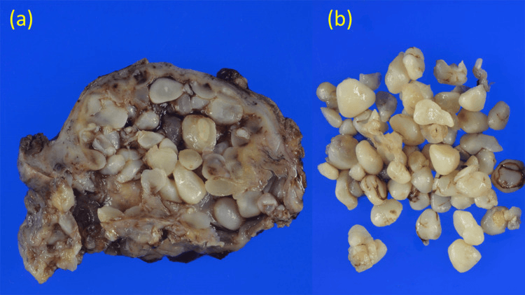
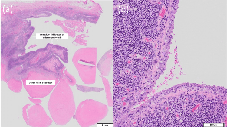
- “Rice bodies suggest chronic synovial inflammation but are not specific to rheumatoid arthritis
- “Obtain cultures or biopsy when infection, crystal disease or proliferative synovial disorder remains possible
Overview and Epidemiology
Definition. Inflammatory arthritis of the shoulder includes rheumatoid arthritis and seronegative, crystal and other inflammatory disorders affecting synovium, cartilage, bone, tendon and bursa. The imaging patterns overlap, so systemic context and exclusion of infection are essential.
Epidemiology. Shoulder symptoms are common in established inflammatory arthritis, but the historical prevalence estimates predate modern disease control. Modern DMARD and biologic therapy has changed the surgical case mix. Involvement is commonly bilateral, although asymmetry and alternative shoulder pathology are common too.
Anatomy and Biomechanics
The synovium. Normal synovium is thin, only a few cell layers deep. The bare area is the bone that lies within the capsule but is not covered by cartilage (the surgical neck and the glenoid neck, for example), and it is the site of the initial marginal erosions in RA.
The cuff. An intact rotator cuff allows concentric rotation of the humeral head. A massive tear leads to cuff tear arthropathy, with superior migration or escape of the head.
Pathophysiology
In RA the synovium is the primary site of pathology, in contrast to OA, where cartilage wear is the primary event. Inflammatory shoulder disease may be synovium-driven, but the phenotype varies.
Synovitis. The rheumatoid synovium undergoes massive hypertrophy and angiogenesis and forms pannus. Hypertrophic, vascular synovium may also produce an effusion and rice bodies.
Erosion and cartilage loss. Inflammatory mediators damage cartilage and marginal and subchondral bone.
Tendon and bursa. The pannus releases enzymes (collagenase, metalloproteinases) that attack the rotator cuff tendons from the undersurface. Supraspinatus is the tendon most commonly affected, the biceps is often inflamed (tenosynovitis) or ruptured, and the subacromial-subdeltoid bursa may be involved.
How often the cuff is torn. Full-thickness tears are common, conventionally reported in roughly a quarter to a half of RA shoulders. The range is wide because the figure depends on disease duration, imaging modality and whether the cohort predates biologic control. What matters operatively is not the population rate but this patient's cuff, because cuff function is one of the variables that selects anatomic against reverse arthroplasty.
The end stage. Central migration, deformity or cuff insufficiency may develop, but none is universal or disease-specific.
Mimics. Infection, CPPD, hydroxyapatite disease and proliferative synovial disorders can look inflammatory.
Beyond Rheumatoid: The Seronegative Spondyloarthropathy Shoulder
The seronegative spondyloarthropathies (psoriatic arthritis, ankylosing spondylitis, reactive and enteropathic arthritis) behave differently from RA in ways examiners probe.
Serology and the primary lesion. They are RF- and anti-CCP-negative and HLA-B27-associated. The primary lesion is enthesitis, inflammation where tendon, ligament or capsule inserts into bone, rather than the synovial pannus that drives RA, although a secondary synovitis also occurs.
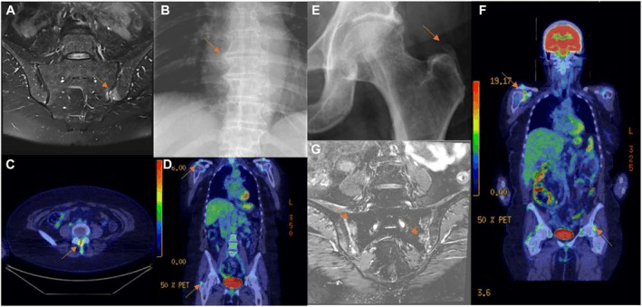
Radiology. The signature is mixed erosive and proliferative, not purely erosive: alongside the erosions there is new bone formation (enthesophytes, periostitis) and a tendency to ankylosis (bony fusion). Psoriatic arthritis classically produces the pencil-in-cup deformity and can swing between an erosive-mutilating form (arthritis mutilans) and a bone-forming one. Ankylosing spondylitis tends toward a stiff, ankylosed glenohumeral joint, often with supero-lateral humeral-head erosion (the "hatchet" head), and is accompanied by sacroiliitis and spinal disease.
- Rheumatoid arthritis
- RF / anti-CCP positive
- Seronegative (PsA / AS)
- RF / anti-CCP negative; HLA-B27 associated
- Rheumatoid arthritis
- Synovial pannus
- Seronegative (PsA / AS)
- Enthesitis (with secondary synovitis)
- Rheumatoid arthritis
- Purely erosive, osteopenic, central wear
- Seronegative (PsA / AS)
- Mixed erosive AND proliferative (new bone, enthesophytes)
- Rheumatoid arthritis
- Central erosion / instability
- Seronegative (PsA / AS)
- Tendency to ankylosis / fusion (especially AS)
- Rheumatoid arthritis
- Symmetric small-joint disease, nodules
- Seronegative (PsA / AS)
- Skin/nail psoriasis, dactylitis, sacroiliitis, uveitis, IBD
Surgical implications. The arthroplasty decision still hinges on cuff integrity and glenoid bone stock, as in RA, and the perioperative principles are shared: infection risk, osteopenia, holding biologics. Ankylosing spondylitis adds its own hazards:
- A stiff, ankylosed shoulder that is harder to mobilise
- A rigid kyphotic cervical spine that is brittle and difficult to intubate, with a high fracture risk on positioning
- A higher rate of heterotopic ossification after arthroplasty
Medical control. TNF and IL-17 inhibitors are central for axial spondyloarthritis and psoriatic disease, whereas methotrexate, a mainstay in RA, is less effective for axial AS.
Classification Systems
Neer's classic description of the rheumatoid stages guides the decision between soft-tissue procedures (synovectomy) and reconstruction. Larsen grading quantifies radiographic joint destruction.
- Name
- Wet
- Features
- Active synovitis, granulations, marginal erosions, osteopenia
- Treatment
- Medical / Synovectomy
- Name
- Dry
- Features
- Burned out: sclerosis, cysts, stiffness, loss of joint space
- Treatment
- Arthroplasty (TSA)
- Name
- Resorptive
- Features
- Severe bone loss, pencil-in-cup deformity
- Treatment
- Augmentation / Reverse
Clinical Assessment
History. Morning stiffness typically lasts over an hour. Ask about systemic symptoms (fatigue, malaise) and other joints, particularly the hands and feet. Nocturnal pain is common, and function is often poor because stiffness combines with weakness of the cuff.
Examination. Look for wasting of the spinati and a boggy swelling anteriorly, and feel for warmth, effusion and tenderness. Cuff strength is often reduced, from pain or from a tear; lag signs indicate a massive tear.
Always examine the neck. Cervical spine involvement is common in long-standing RA - reported prevalence varies enormously with the population and the era studied, and figures from historical cohorts predate modern DMARD control, so treat any single percentage with suspicion. Subluxation can cause radiculopathy mimicking shoulder pain, or myelopathy - and it matters before any anaesthetic, because intubation and positioning load an unstable atlantoaxial joint.
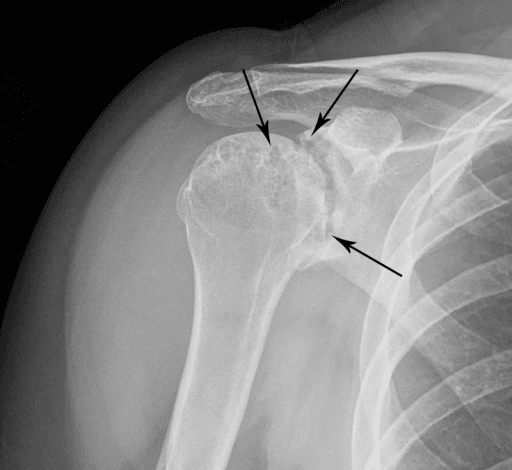
RA or OA? Rheumatoid disease may produce synovitis, periarticular osteopenia, marginal erosions, cartilage loss, cuff and biceps pathology, and central migration; the combination and severity vary with disease duration and control. Primary OA more often shows osteophytes, sclerosis and asymmetric glenoid wear, but posterior wear is not universal and cuff status must be assessed independently.
- Bone quality
- Osteopenic, erosions
- Wear pattern
- Central (acetabularisation)
- Rotator cuff
- Often thin/torn
- Discriminating clue
- Bilateral, symmetric, raised CRP/ESR, anti-CCP positive
- Bone quality
- Sclerotic, osteophytes
- Wear pattern
- Posterior (Walch B)
- Rotator cuff
- Usually intact
- Discriminating clue
- Goat's-beard osteophyte, normal inflammatory markers
- Bone quality
- Variable
- Wear pattern
- Superior (acetabularisation of acromion)
- Rotator cuff
- Massive deficiency
- Discriminating clue
- High-riding head, pseudoparalysis, often non-inflammatory
- Bone quality
- Rapid destruction
- Wear pattern
- Diffuse
- Rotator cuff
- Intact early
- Discriminating clue
- Acute, hot, systemic sepsis, synovial WBC very high
- Bone quality
- Erosive + new bone
- Wear pattern
- Mixed
- Rotator cuff
- Variable
- Discriminating clue
- Skin/nail disease, enthesitis, RF negative
- Bone quality
- Destructive
- Wear pattern
- Superior/central
- Rotator cuff
- Often massive tear
- Discriminating clue
- Chondrocalcinosis, bloody effusion with crystals
Investigations
Blood tests. The laboratory screen:
- ESR and CRP - markers of active inflammation
- RF and anti-CCP - specific for RA
- Full blood count - anaemia of chronic disease, or leukopenia (Felty's)
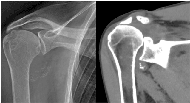
Radiographs. AP, Grashey and axillary lateral views. The rheumatoid findings:
- Periarticular osteopenia ("washed out"), which affects fixation and fracture risk
- Marginal erosions
- Symmetric joint space narrowing
- Central migration of the head (acetabularisation)
- A high-riding head, which indicates a massive cuff tear
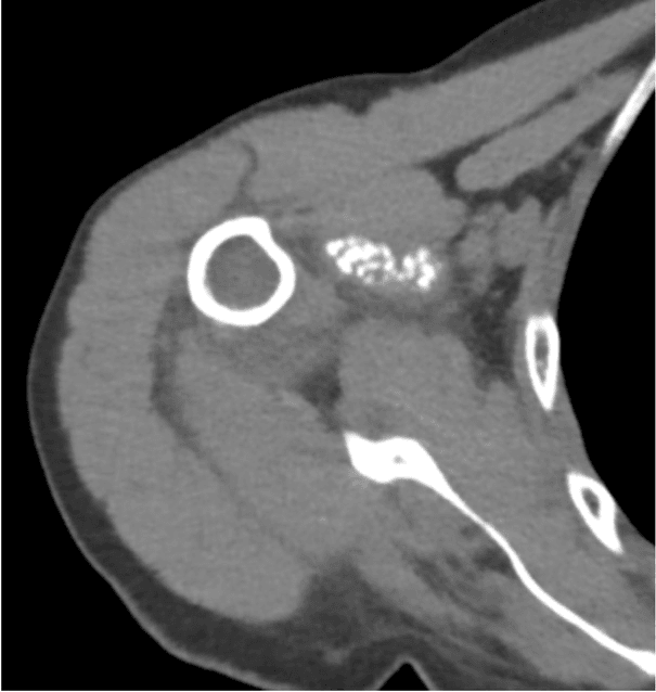
CT and MRI. Together they guide the choice between anatomic and reverse arthroplasty. CT is essential for glenoid version and bone stock, including central erosion; MRI is essential for rotator cuff integrity, which is critical for implant choice.
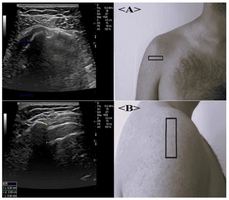
Synovial fluid. Aspirate when the diagnosis is unclear or infection (septic arthritis) is suspected. Rheumatoid fluid is inflammatory, with a white cell count of 2,000-50,000, and is cloudy, of low viscosity and low in glucose.
Before surgery. The preoperative work-up:
- Disease control and medication - review the DMARD schedule with the rheumatologist and coordinate systemic treatment
- Cervical spine - flexion-extension radiographs to rule out atlantoaxial instability when neck symptoms, neurology or risk factors indicate them; respiratory risk is likewise assessed as clinically indicated
- Cardiac - risk stratification
- Skin - inspect its integrity and healing risk
- Glenoid and humeral bone - plan fixation and reconstruction
- Sepsis - culture or biopsy when the diagnosis is uncertain
Rice Bodies in the Inflammatory Shoulder
Rice bodies are a marker of chronic inflammatory synovitis, and they carry an important "do not miss".
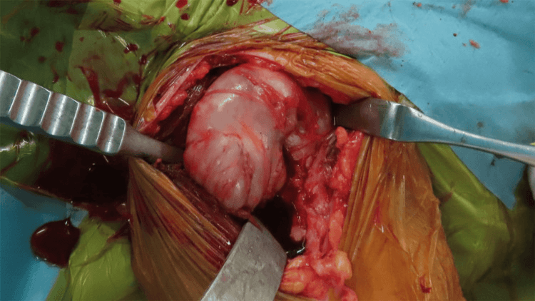
What they are. Small, smooth, white, ovoid intra-articular or intra-bursal loose bodies that resemble grains of polished rice, also called "melon-seed bodies". They are made of fibrin, collagen and degenerated or infarcted synovial fragments, formed when chronically inflamed hypertrophic synovium sheds and the fragments become encased in fibrin. They can fill the glenohumeral joint and, characteristically at the shoulder, the subacromial-subdeltoid bursa.
The tuberculosis caveat. The common associations are rheumatoid arthritis and juvenile idiopathic arthritis, but rice bodies are classically described in tuberculous and atypical-mycobacterial synovitis and tenosynovitis. Finding them, especially in a monoarticular or atypical presentation, should always prompt exclusion of tuberculosis.
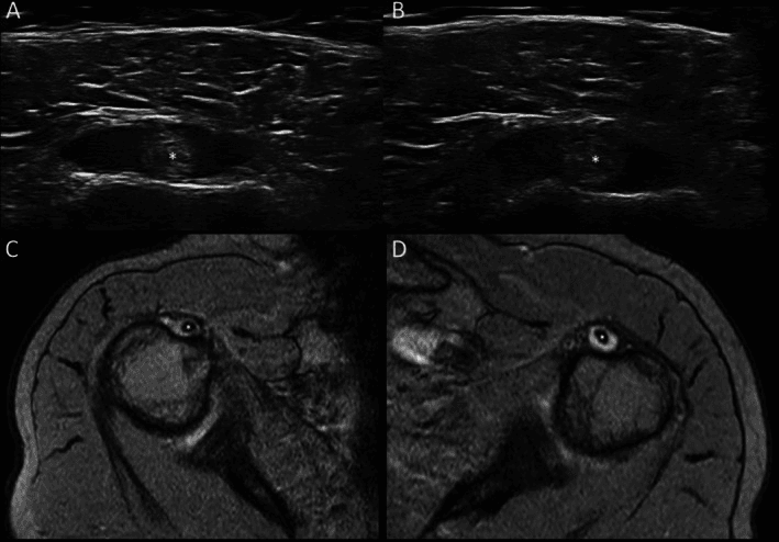
Imaging. Rice bodies are largely radiolucent and poorly seen on plain radiographs, being isodense to fluid, so a plain film mostly shows the background arthropathy. They are best demonstrated on MRI, as myriad small bodies of low-to-intermediate T1 and low T2 signal within a distended joint or bursa, or on ultrasound.
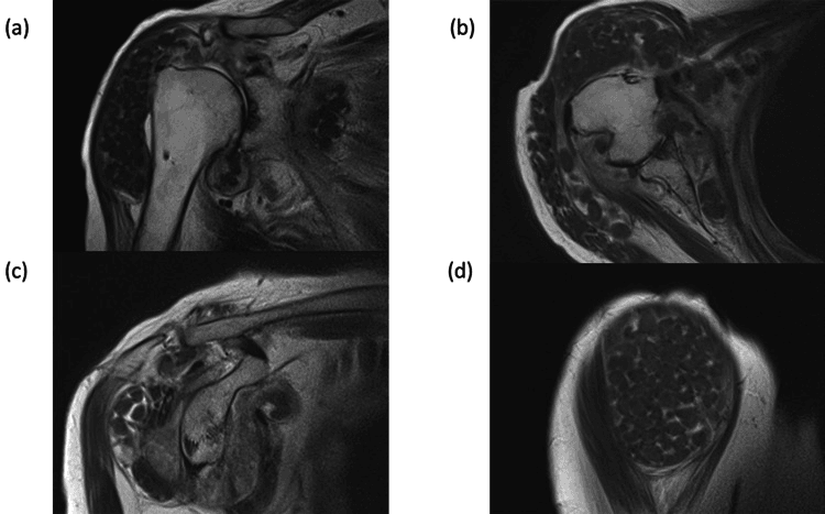
Management. Rice bodies are removed at synovectomy, open or arthroscopic, together with the diseased synovium; recurrence tracks ongoing synovitis, so disease control matters. Always send the synovium and bodies, and the fluid, for acid-fast staining, mycobacterial culture and histology to exclude an infective (mycobacterial) cause before attributing them to RA alone.
Management Algorithm
Choosing the operation. Implant choice depends on the cuff and the bone, and on the deltoid, deformity and the patient's goals as well; cuff status alone does not select one arthroplasty.
- Cuff / deltoid
- Function assessed
- Bone and joint
- No advanced destruction
- Possible Strategy
- Systemic optimisation; selective injection or synovectomy
- Cuff / deltoid
- Functional cuff and deltoid
- Bone and joint
- Reconstructible glenoid/humerus
- Possible Strategy
- Anatomic arthroplasty may be considered
- Cuff / deltoid
- Deltoid must function
- Bone and joint
- Assess glenoid and humeral bone
- Possible Strategy
- Reverse arthroplasty may be considered
- Cuff / deltoid
- Variable
- Bone and joint
- Deficient fixation or active disease
- Possible Strategy
- Staged, augmented, limited or non-operative strategy
Medical management. NSAIDs, DMARDs (methotrexate) and biologics (TNF-alpha inhibitors), with regular monitoring. Physiotherapy is gentle range of motion, avoiding aggressive strengthening if the cuff is fragile. Corticosteroid injection has a limited role because of the risk of infection and cuff atrophy.
Methotrexate can typically be continued perioperatively. Biologics are usually held for 1 dosing cycle before surgery to reduce infection risk. Check local guidelines.
Arthroscopic synovectomy. Indicated in the wet stage (active synovitis) with a preserved joint space and an intact cuff. The goal is to reduce pain and slow enzymatic destruction; it does not stop long-term progression. Reported pain relief is good in the short term, but the published series are small and uncontrolled and no study on this page quantifies it, so counsel improvement rather than a percentage.
Hemiarthroplasty. Indicated for severe glenoid bone loss and cuff tear arthropathy (historical). Pain relief is less reliable, with metal articulating on eroded bone.
Anatomic total shoulder arthroplasty. Needs an intact rotator cuff and adequate glenoid bone stock, and gives superior pain relief to hemiarthroplasty.
Reverse total shoulder arthroplasty. Indicated for a rotator cuff tear, severe glenoid erosion or the elderly patient, and becoming standard for many RA shoulders.
Surgical Technique (Key Points)
Anatomic TSA. Use a deltopectoral approach and handle the fragile soft tissues gently. The subscapularis is often thin and friable and needs careful takedown and repair. Central wear may need eccentric reaming or an augmented glenoid component, and pegged ingrowth components are preferred over cemented ones for better bone preservation.
Reverse TSA. Also deltopectoral. If central erosion is severe, medialise the reaming or use bone graft, and use long-peg baseplates in osteopenic bone. A "stove-pipe" humerus often requires a cemented stem. Deltoid tension is key.
Complications
- Risk in RA
- High
- Reason
- Immunosuppression, skin fragility
- Management
- Debridement, antibiotics, explant
- Risk in RA
- Moderate
- Reason
- Osteopenia, eccentric reaming
- Management
- Revision to Reverse / Bone graft
- Risk in RA
- High
- Reason
- Cortical thinning (disuse/steroids)
- Management
- ORIF vs Stem revision
- Risk in RA
- High
- Reason
- Progressive disease
- Management
- Revision to Reverse
In anatomic TSA, if the rotator cuff is unbalanced or fails, the humeral head migrates superiorly and eccentrically loads the superior rim of the glenoid component. This cyclic "rocking" leads to early loosening.
Postoperative Care
Rehabilitation. The protocol runs in phases:
- 0-6 weeks - sling, passive range of motion only to protect the subscapularis repair, pendulums
- 6-12 weeks - active assisted range of motion, pulleys
- 3-6 months - active range of motion, gentle strengthening
- 6 months onwards - full activity
In RA. Soft-tissue repairs such as the subscapularis take longer to heal. Osteopenia makes aggressive passive stretching a fracture risk, and the skin needs care with dressings and tape because it tears.
Outcomes and Prognosis
Pain and function. Arthroplasty, anatomic or reverse, gives reliable and substantial pain relief in RA: the Mayo series recorded significant long-term relief with both anatomic TSA and hemiarthroplasty (p less than 0.0001). Function is less predictable than in OA, because of the muscles and cuff.
Reading the survivorship figures. Be careful here: the arthroplasty registries do not separate inflammatory arthritis cleanly, and the RA-specific series are small or historical.
Anatomic TSA and hemiarthroplasty. The Mayo cohort (Sperling, operated 1976-1991) shows durable pain relief with anatomic TSA and a lower revision risk than hemiarthroplasty when the cuff was intact. Radiographic glenoid lucency was nonetheless present in 72% of those TSAs, and glenoid erosion in 98% of hemiarthroplasties. Hemiarthroplasty brings lower satisfaction and ongoing glenoid-sided pain, with progressive erosion the predictable failure mode.
Reverse TSA. There are no long-term RA-specific survivorship data. The Young series is 18 shoulders at a mean 3.8 years with no revisions, which is far too short to quote a ten-year figure.
Overall. Surgery significantly improves quality of life, but complication rates (infection, fracture) are higher than in OA.
Guidelines, Registries & Global Practice
Global epidemiology:
- RA affects roughly 0.5–1% of adults worldwide; shoulder symptoms develop in the majority over the disease course and involvement is frequently bilateral.
- Female-to-male ratio approximately 3:1; peak onset in the fourth-to-sixth decades.
- Widespread early use of methotrexate and biologic DMARDs has reduced the incidence of end-stage erosive shoulder disease and of synovectomy/early arthroplasty compared with historical cohorts — a trend seen across high-income health systems.
- Conventional DMARDs (e.g. methotrexate)
- Continue through surgery
- Biologic DMARDs
- Withhold; operate at end of dosing cycle, resume after wound healing (~14 days)
- Glucocorticoids
- Continue usual daily dose; avoid supraphysiologic 'stress' dosing
- Conventional DMARDs (e.g. methotrexate)
- Continue low-dose methotrexate perioperatively
- Biologic DMARDs
- Time surgery to end of dosing interval
- Glucocorticoids
- Maintain baseline dose
- Conventional DMARDs (e.g. methotrexate)
- Continue csDMARDs perioperatively
- Biologic DMARDs
- Individualised hold based on infection risk
- Glucocorticoids
- Maintain; minimise long-term dose
- Major arthroplasty registries (AOANJRR, NJR for England/Wales, the Nordic registries) show reverse arthroplasty use rising steeply, including for inflammatory arthritis with cuff or glenoid compromise.
- Revision rates for arthroplasty in RA run slightly higher than in OA, driven chiefly by infection and soft-tissue (cuff/subscapularis) failure rather than by polyethylene wear.
- Where biologics and early rheumatology access are routine, surgeons increasingly see "burned-out" dry-stage disease rather than florid synovitis.
- In limited-resource settings, patients more often present with advanced erosive ("resorptive") disease, severe glenoid bone loss and massive cuff deficiency — shifting practice toward reverse arthroplasty or, where implants/expertise are unavailable, hemiarthroplasty or arthrodesis.
- Combined rheumatology–orthopaedic clinics improve perioperative optimisation and are an aspirational model regardless of setting.
Controversies & Areas of Uncertainty
These are legitimate debates, and examiners reward a balanced answer that acknowledges the uncertainty rather than a dogmatic one.
Anatomic TSA or reverse in the cuff-intact RA shoulder. With an intact cuff, anatomic TSA gives better rotation and preserves bone, but progressive cuff attrition is intrinsic to RA and may cause later glenoid loosening ("rocking horse"). Some surgeons favour primary reverse arthroplasty in older RA patients to pre-empt this, accepting reduced rotation and notching. No high-level trial resolves the trade-off.
Holding biologics. The optimal hold interval is extrapolated from low-grade, mostly retrospective data; recommendations are consensus (Guideline-level), and the magnitude of infection reduction is uncertain. Over-holding risks a disabling flare.
Continuing or stopping methotrexate. Continuing is now standard, but practice still varies in patients with renal impairment or concurrent high-dose steroids.
The role of synovectomy. Its value in the biologic era is debated, and it is increasingly limited to medically refractory synovitis with preserved cartilage.
The glenoid component in soft bone. Whether to implant a glenoid (better pain relief) or accept hemiarthroplasty or ream-and-run (lower loosening risk) in severely osteopenic, eroded glenoids remains individualised.
Cervical spine clearance. Routine flexion-extension films before every RA arthroplasty are traditional teaching, but selective imaging guided by symptoms and examination is increasingly advocated.
MCQ Practice Points
Q: What is the most common pattern of glenoid wear in Rheumatoid Arthritis? A. Posterior wear (Retroversion) B. Superior wear C. Central wear (Medialization) D. Anterior wear Answer: C. RA causes central erosion/acetabularization. OA causes posterior wear (B2 glenoid).
Q: Which medication is typically held for 1-2 dosing cycles prior to arthroplasty? A. Methotrexate B. TNF-alpha inhibitors (e.g., Adalimumab) C. Prednisone D. Sulfasalazine Answer: B. Biologics are held to reduce infection risk. Methotrexate is typically continued.
Q: In an RA patient with end-stage arthritis and a massive rotator cuff tear, the best surgical option is: A. Arthroscopic Debridement B. Hemiarthroplasty C. Anatomic TSA D. Reverse TSA Answer: D. Anatomic TSA will loosen (rocking horse). Hemi won't restore function. Reverse TSA addresses both arthritis and cuff deficiency.
Q: Pre-operative evaluation of the RA patient for shoulder surgery MUST include: A. EMG B. Cervical Spine X-rays (Flexion/Extension) C. Bone Scan D. Angiogram Answer: B. Atlanto-axial instability is common and poses a lethal risk during intubation/positioning.
Q: The presence of a 'High Riding' humeral head on X-ray indicates: A. Deltoid atrophy B. Rotator Cuff Incompetence C. Axillary nerve palsy D. Posterior dislocation Answer: B. Unopposed deltoid pull migrates the head superiorly when the supraspinatus is torn.
Q: Which of the following is a contraindication to unconstrained (Anatomic) TSA? A. Advanced age B. Deficient Rotator Cuff C. Central glenoid wear D. Previous synovectomy Answer: B. Anatomic TSA requires an intact cuff to center the head. Without it, the "rocking horse" effect loosens the glenoid.
Exam Viva Scenarios
Practise clinical reasoning and management decisions out loud
“A 45-year-old female with known RA presents with increasing shoulder pain. She is on Methotrexate. X-rays show symmetric joint space narrowing but spherical head. Cuff is 5/5 strength. Management?”
“A 65-year-old RA patient has severe glenohumeral pain. X-rays show destruction of joint space and central erosion. MRI shows the Rotator Cuff is INTACT. Options?”
“A 70-year-old with RA has a painful, pseudoparalyzed shoulder. Unable to lift arm over 45 degrees. X-rays show high riding humeral head and severe arthritis. Plan?”
Key Concepts
- Pathology is Synovitis leads to Pannus leads to Cartilage/Bone Destruction
- Central Migration (Acetabularization) is hallmark
- Rotator Cuff status determines implant choice (TSA vs Reverse)
- Biologics have revolutionized care (fewer surgeries)
Clinical Pearls
- Check the Neck! (C-spine instability)
- Skin is fragile - handle with care
- Infection risk is higher (immunosuppressed)
- Bone is osteopenic - avoid aggressive reaming
Surgical Rules
- Intact Cuff leads to Anatomic TSA
- Torn Cuff leads to Reverse TSA
- Active Synovitis (early) leads to Synovectomy
- Hold Biologics perioperatively
Complications to Quote
- Infection (higher than OA)
- Glenoid Loosening (Rocking Horse)
- Periprosthetic Fracture
- Cuff Failure (if Anatomic done)
Evidence Base
TSA vs Hemiarthroplasty in RA — 303 cases (Mayo)
- 195 TSA and 108 hemiarthroplasties in RA; mean follow-up 11.6 years
- Marked long-term pain relief and improved abduction/external rotation with both procedures
- With an intact cuff, TSA gave significantly greater pain relief and abduction, and a lower revision risk than hemiarthroplasty
- Glenoid erosion in 98% of hemiarthroplasties; periprosthetic glenoid lucency in 72% of TSAs
Reverse TSA in Rheumatoid Arthritis
- 18 primary reverse arthroplasties in 16 RA patients with cuff compromise and/or severe glenoid erosion; mean follow-up 3.8 years
- Mean Constant score improved from 22.5 to 64.9; forward elevation 77.5° to 138.6°
- Outcomes worse with a preoperatively atrophic teres minor (Constant 54.6 vs 74.3)
- Scapular notching in 10 of 18; intra-/post-operative fracture in 4 of 18 (osteopenic bone)
Reverse Arthroplasty — Indications & Complications with Experience
- Two consecutive series of 240 reverse arthroplasties compared as surgeon experience increased
- Proportion performed for RA rose from 0.4% to 6.3% as indications broadened
- Complication rate fell (19% to 10.8%); infection 4% to 0.9%; dislocation 7% to 3.2%
- Scapular notching rate remained stable despite increased experience
Anti-TNF Agents and Postoperative Infection Risk
- TNF inhibitors may impair immunity against usual and opportunistic pathogens
- Evidence on perioperative infection risk is limited, retrospective and contradictory
- No single threshold proven; pragmatic practice is to time surgery around the dosing cycle
- Decisions must balance infection risk against the harm of a disease flare
2022 ACR/AAHKS Perioperative Antirheumatic Medication Guideline (supersedes 2017)
- GRADE-based update of the 2017 recommendations, now including targeted synthetic small-molecule drugs (JAK inhibitors) and newer biologics that did not exist for the first version
- Continue conventional synthetic DMARDs including methotrexate, leflunomide, hydroxychloroquine and sulfasalazine through surgery
- Withhold biologic DMARDs and schedule surgery at the END of the dosing interval, restarting once the wound is healed with no dehiscence, swelling or drainage - typically about 14 days
- Covers adults with inflammatory arthritis, juvenile idiopathic arthritis or SLE undergoing elective TOTAL HIP or TOTAL KNEE arthroplasty
- Also updates optimal perioperative glucocorticoid dosing, moving away from routine high-dose stress steroid cover
Glenoid Erosion in RA is Central and Medial, Not Posterior
- Review of the glenoid erosion and bone-loss classifications, contrasting the patterns by underlying disease
- In rheumatoid arthritis glenoid erosion is MEDIAL, with bone described as very weak and soft - the concentric central pattern often called 'acetabularisation'
- In primary glenohumeral osteoarthritis erosion is posterior and inferior (the Walch B-type pattern); in cuff-deficient eccentric arthritis it is superior
- Options for significant loss include bone graft reconstruction and augmented or custom components