Anterior (Footballer's Ankle) and Posterior (Dancer's Syndrome) | Arthroscopic Treatment
- Anterior impingement from tibial/talar spurs (osseous) or synovitis (soft tissue) limiting dorsiflexion
- Posterior impingement from os trigonum or FHL tendinitis limiting plantarflexion (dancers)
- Conservative management 53% success; osseous lesions less likely to respond than soft tissue
- Arthroscopic treatment 85-95% success with low complication rates (nerve injury 2-5%)
- Combined pathology common in posterior impingement - always assess for FHL involvement
- “Os trigonum syndrome = bone marrow edema on MRI confirms symptomatic lesion
- “FHL tendinitis coexists in 40-60% of posterior impingement cases
- “Anterior portals: anteromedial (saphenous nerve) and anterolateral (superficial peroneal nerve)
- “Posterior portals: posteromedial and posterolateral (sural nerve at risk)
Anterior: Dorsiflexion pain (Footballers). Posterior: Plantarflexion pain (Dancers).
Anterior: Superficial Peroneal (SPN) - anterolateral portal. Posterior: Sural (posterolateral) and Tibial (posteromedial).
Anterior: Osteophyte Resection (burr). Posterior: Os Trigonum Excision + FHL Release.
Anterior: Osseous spur or soft tissue. Posterior: Osseous (Os Trigonum) or FHL stenosing tenosynovitis.
Overview
Ankle impingement syndromes are common causes of chronic ankle pain in athletes and dancers, with anterior impingement accounting for 60-70% of cases and posterior impingement 30-40%. The condition affects approximately 3-5% of competitive athletes, with highest prevalence in soccer (footballer's ankle), ballet (dancer's syndrome), and volleyball. Male predominance (2:1) in anterior impingement relates to higher participation in contact sports, while posterior impingement shows equal gender distribution given ballet's demographics.
Indications and Pre-Operative Planning
Surgical Indications
- Failed conservative management: Minimum 3-6 months appropriate non-operative treatment
- Persistent symptoms: Pain limiting daily activities or sports participation
- Mechanical symptoms: Locking, catching suggesting loose body or meniscoid lesion
- Professional athletes: Earlier surgery for career impact (after 6-12 weeks conservative trial)
- Large osteophytes: Grade 3-4 osseous impingement unlikely to respond conservatively
- Documented pathology: MRI confirmation of structural lesion amenable to surgery
Pre-Operative Assessment
- MRI review: Identify all pathology (osseous, soft tissue, concurrent issues)
- Patient expectations: Realistic goals for return to high-level activity
- Optimize health: Address smoking, weight, inflammatory conditions
- Surgical planning: Anterior versus posterior approach, arthroscopy versus open
- Equipment check: Ensure appropriate arthroscopy equipment and instrumentation available
Arthroscopic Techniques
Anterior Ankle Arthroscopy for Anterior Impingement
Patient Positioning:
- Supine position on operating table
- Thigh tourniquet (250-300 mmHg)
- Non-invasive distraction (10 lbs) OR invasive distraction with pins
- Ankle in neutral position with bump under ipsilateral hip
Portal Placement:
- Anteromedial portal: 1 cm medial to tibialis anterior tendon at joint line
- Anterolateral portal: Just lateral to peroneus tertius tendon at joint line
- Mark superficial peroneal nerve branches before portals (dorsiflexion makes visible)
- Anterocentral portal rarely needed (deep peroneal nerve risk)
Systematic Inspection:
- 30-degree arthroscope through anteromedial portal initially
- Inspect entire joint: medial gutter, central dome, lateral gutter
- Identify pathology: osteophytes, synovitis, meniscoid lesion, cartilage damage
- Document findings with photographs/video
- Switch arthroscope to anterolateral portal for medial inspection
Debridement Technique:
1. Synovectomy:
- Use arthroscopic shaver to remove hypertrophic synovium
- Debride anterolateral soft tissue impingement (meniscoid lesion)
- Excise thickened Bassett's ligament if contributing
- Create smooth joint surfaces free of catching tissue
2. Osteophyte Resection:
- Use arthroscopic burr for bone removal
- Tibial osteophytes: resect from medial to lateral across anterior plafond
- Talar osteophytes: carefully remove from talar neck/dome
- Goal: restore smooth tibiotalar articulation
- Avoid excessive bone removal (weakens joint)
- Irrigate frequently to clear debris and improve visualization
3. Cartilage Management:
- Assess for kissing lesions or chondral damage
- Debride unstable cartilage flaps to stable edges
- Microfracture for focal full-thickness defects if appropriate
- Document extent of arthritis for prognosis
4. Confirm Decompression:
- Test ankle dorsiflexion under direct visualization
- Ensure no residual impingement of bone or soft tissue
- Remove all loose bodies or debris
- Final inspection of entire joint
Closure:
- Remove instruments and deflate distraction
- Close portals with single nylon suture or skin adhesive
- Apply soft compressive dressing
- Posterior splint in neutral if significant bone work
Post-Operative Protocol:
- 0-2 weeks: NWB in boot if extensive bone work, otherwise WBAT
- 2-6 weeks: Progressive weight-bearing, gentle AROM exercises
- 6-12 weeks: Strengthening, proprioception training
- 3-4 months: Return to sport-specific training
- 4-6 months: Full unrestricted activity typically achieved
The superficial peroneal nerve branches cross the anterior ankle and are at highest risk during anterolateral portal placement. Mark the nerve branches pre-operatively with ankle dorsiflexion and eversion. The deep peroneal nerve and anterior tibial vessels are at risk with anterocentral portal - avoid if possible. Excessive osteophyte resection can destabilize the ankle joint.
Ankle Joint Anatomy Relevant to Impingement
Understanding normal anatomy and motion is essential to comprehend impingement pathology.
Anterior Ankle Anatomy
- Anterior recess: Capsular space between tibia and talus allowing dorsiflexion
- Tibial plafond margin: Anterior distal tibial edge, site of spur formation
- Talar neck: Anterior talus articulates with anterior tibia in dorsiflexion
- Anterior joint capsule: Becomes taut in dorsiflexion, site of synovitis
- Neurovascular structures: Deep peroneal nerve and anterior tibial vessels cross anteriorly
Anterior portal anatomy (arthroscopy):
- Anteromedial portal: Medial to tibialis anterior, lateral to saphenous nerve
- Anterolateral portal: Lateral to EDL, medial to superficial peroneal nerve branches
- Anterocentral portal: Between EHL and EDL, risk to deep peroneal nerve
Posterior Ankle Anatomy
- Posterior recess: Capsular space behind ankle joint, largest synovial space
- Posterior talar process: Medial and lateral tubercles with FHL groove between
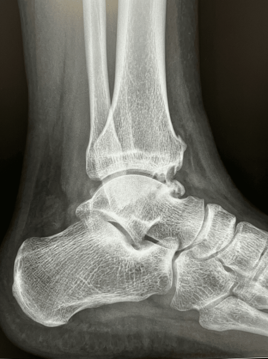
- Os trigonum: Accessory ossicle posterior to talus (10-25% prevalence)
- Stieda process: Elongated lateral talar tubercle (unfused os trigonum)
- FHL tendon: Travels between talar tubercles, at risk during posterior procedures
Posterior portal anatomy (endoscopy):
- Posterolateral portal: Lateral border Achilles, anterior to sural nerve
- Posteromedial portal: Medial border Achilles, protects tibial nerve and vessels
- Working space: Behind ankle joint and superior to calcaneal tuberosity
Biomechanics of Impingement
Normal ankle dorsiflexion range where anterior structures approximate without impingement. Plantarflexion range where posterior structures may impinge between tibia and calcaneus. Extreme plantarflexion in ballet dancers maximally compresses posterior ankle structures.
Motion Dynamics
- Normal dorsiflexion: Anterior joint space opens, posterior space narrows
- Normal plantarflexion: Posterior space opens, anterior space closes
- Impingement mechanism: Hypertrophied tissue or bone caught during end-range motion
- Synovitis cascade: Repetitive impingement causes inflammation, further hypertrophy, worsening symptoms
Pathophysiology
General Mechanism of Ankle Impingement
Ankle impingement occurs when soft tissue or osseous structures become trapped between bones during terminal motion, causing pain and functional limitation. The pathophysiology differs between anterior and posterior impingement:
- Mechanical impingement: Physical entrapment of tissues at end-range motion
- Inflammatory cascade: Repetitive impingement causes synovitis, tissue hypertrophy, and worsening symptoms
- Osseous vs soft tissue: Osseous lesions from chronic trauma/degeneration; soft tissue from acute injury or inflammation
- Activity-specific: Pattern relates to sport demands (kicking = anterior, en pointe = posterior)
- Repetitive forced dorsiflexion causes anterior capsular traction
- Tibial and talar osteophytes form at sites of capsular avulsion
- Soft tissue hypertrophy (synovitis, meniscoid lesion) after ankle sprains
- Extreme plantarflexion compresses posterior structures between tibia and calcaneus
- Os trigonum (unfused secondary ossification center) or Stieda process becomes symptomatic
- FHL tendon can become entrapped in the posterior compartment
Anterior Impingement Pathophysiology
Anterior impingement results from osseous or soft tissue pathology limiting dorsiflexion.
Footballer's Ankle (Osseous)
- Mechanism: Repetitive forced dorsiflexion causes capsular avulsion and osteophyte formation
- Tibial spurs: Anterior distal tibia develops traction spurs from capsular pulling
- Talar spurs: Anterior talar neck develops spurs that articulate with tibial osteophytes
- Kissing lesions: Matching tibial and talar spurs that interlock during dorsiflexion
- Sports: Soccer (kicking), running, basketball (jumping), gymnastics
Soft Tissue Impingement
- Anterolateral soft tissue: Hypertrophied synovium or meniscoid lesion after ankle sprain
- Bassett's ligament: Fascial band from AITFL to talus, may become pathologic
- Synovitis: Chronic inflammation from repetitive impingement creates hypertrophic tissue
- Scarring: Post-traumatic fibrosis in anterior gutter after injury
- Distal fascicle AITFL: Anterior inferior tibiofibular ligament fascicle impingement
Classification Systems
- osseous
- Small tibial or talar spurs less than 3 mm
- softTissue
- Minimal synovitis, no meniscoid lesion
- symptoms
- End-range dorsiflexion discomfort only
- treatment
- Conservative management usually successful
- osseous
- Moderate spurs 3-5 mm, not yet kissing
- softTissue
- Moderate synovitis or small meniscoid lesion
- symptoms
- Pain with activities requiring dorsiflexion
- treatment
- May respond to conservative care or injection
- osseous
- Large spurs greater than 5 mm, kissing lesions
- softTissue
- Severe synovitis, large meniscoid lesion
- symptoms
- Rest pain, significant motion loss
- treatment
- Surgical debridement typically required
- osseous
- Extensive osteophytes with articular damage
- softTissue
- Dense fibrosis, loose bodies
- symptoms
- Daily symptoms, marked functional limitation
- treatment
- Arthroscopic debridement, may progress to arthritis
Risk Factors
- Soccer players: Kicking motion with forced dorsiflexion
- Runners: Repetitive dorsiflexion during push-off and landing
- Previous ankle sprains: Soft tissue hypertrophy and scarring
- Chronic ankle instability: Abnormal motion causing impingement
- High-impact sports: Basketball, volleyball, gymnastics
- Anatomical factors: Limited native dorsiflexion, tight Achilles
Posterior Impingement Pathophysiology
Posterior impingement occurs from compression of structures between tibia, talus, and calcaneus during plantarflexion.
Os Trigonum Syndrome
- Anatomy: Accessory ossicle posterior to lateral talar tubercle
- Prevalence: Present in 10-25% of population (bilateral in 50% of those affected)
- Pathologic mechanism: Becomes symptomatic from repetitive compression or synchondrosis injury
- Nutcracker effect: Os compressed between tibia and calcaneus in plantarflexion
- FHL involvement: Tendinitis may coexist from adjacent inflammation
Stieda Process
- Definition: Elongated lateral talar tubercle (unfused os trigonum)
- Fracture: Can fracture at junction with talus (Shepherd fracture)
- Impingement: Functions similarly to os trigonum causing posterior compression
- Imaging: Appears as elongated posterior process on lateral radiograph
Soft Tissue Impingement
- Posterior capsule: Hypertrophic synovitis from repetitive compression
- FHL tendinopathy: Tenosynovitis from adjacent inflammation or direct compression
- Posterior talofibular ligament: Thickened ligament may contribute to symptoms
- Posterior intermalleolar ligament: Hypertrophy from chronic loading
Dancer's Syndrome
Special consideration for ballet dancers:
- En pointe position: Extreme plantarflexion (90+ degrees) maximally narrows posterior space
- Relevé repetition: Thousands of repetitions weekly in professional dancers
- FHL involvement: Concurrent FHL tendinitis in 40-60% of cases
- Career impact: May be career-ending if conservative management fails
Risk Factors
Prevalence of os trigonum in general population, small fraction become symptomatic. Ballet dancers with posterior ankle pain have coexistent FHL tendinopathy requiring treatment. En pointe position requires extreme plantarflexion compressing posterior structures maximally.
High-risk activities:
- Ballet dancing (especially en pointe work)
- Soccer (plantarflexion during kicking follow-through)
- Downhill running (repetitive plantarflexion)
- Gymnastics (landing positions)
- Figure skating (toe pointing during jumps)
Clinical Presentation
Clinical presentation varies based on impingement type, with anterior impingement causing dorsiflexion-related symptoms and posterior impingement affecting plantarflexion activities.
Anterior Impingement Clinical Features
History
- Pain location: Anterior ankle joint line, worse with dorsiflexion
- Timing: End-range dorsiflexion activities (kicking, squatting, stairs)
- Insidious onset: Gradual progression over months to years
- Previous injury: History of ankle sprain in 60-70% of soft tissue cases
- Sports impact: Reduced performance in kicking, jumping, cutting activities
- Morning stiffness: Common, improves with activity initially
Physical Examination
- Tenderness: Anterolateral or anteromedial joint line palpation
- Range of motion: Reduced dorsiflexion (normal 10-20 degrees)
- Impingement test: Forced passive dorsiflexion reproduces anterior pain
- Palpable spurs: Large osteophytes may be palpable anteriorly
- Swelling: Anterior ankle fullness from synovitis
- Gait: May have shortened stride or altered heel strike
Special tests:
- Anterior impingement test: Passive forced dorsiflexion with tibial translation elicits pain
- Molloy test: Palpation of anterolateral gutter with dorsiflexion and inversion
- Single leg squat: Pain at bottom of deep squat position
- Hop test: Pain with landing from single leg hop
Differential Diagnosis
- keyFeature
- Catching/fullness in anterolateral gutter, post-sprain, normal radiographs
- distinguisher
- Positive Molloy test, MRI/US shows meniscoid lesion; pain reproduced in dorsiflexion
- keyFeature
- Dorsiflexion-limited pain in kicking/squatting athletes
- distinguisher
- Tibial/talar osteophytes on weight-bearing lateral radiograph
- keyFeature
- Deep posterior pain on plantarflexion, en pointe or kicking follow-through
- distinguisher
- Positive plantarflexion (nutcracker) test; os trigonum with marrow oedema on MRI
- keyFeature
- Recurrent giving-way, often coexists with anterolateral impingement
- distinguisher
- Positive anterior drawer / talar tilt; stress radiographs abnormal
- keyFeature
- Diffuse pain, stiffness, crepitus, older or post-traumatic patient
- distinguisher
- Global joint-space narrowing on weight-bearing views, not focal impingement
- keyFeature
- Posteromedial pain, hallux triggering, dancers
- distinguisher
- Pain on resisted/passive hallux motion; FHL fluid on MRI; often coexists with os trigonum
- keyFeature
- Deep aching, catching, post-sprain, mechanical symptoms
- distinguisher
- Subchondral defect on MRI/CT; not motion-position specific
- keyFeature
- Acute posterior pain after forced plantarflexion injury
- distinguisher
- Fracture line at lateral tubercle on CT; acute onset versus chronic os trigonum
Posterior Impingement Clinical Features
History
- Pain location: Posterior ankle, may radiate to heel or plantar foot
- Character: Deep aching pain, worse with plantarflexion activities
- Onset: Gradual in athletes, may be acute after forced plantarflexion injury
- Activities: En pointe (dancers), kicking (soccer), downhill running
- Functional loss: Cannot maintain extreme plantarflexion positions
- FHL symptoms: Triggering or weakness if concurrent tendinopathy
Physical Examination
- Tenderness: Posterior ankle, lateral or medial to Achilles tendon
- Range of motion: Pain at end-range plantarflexion
- Posterior impingement test: Forced passive plantarflexion reproduces pain
- Palpable os trigonum: May feel prominence posterior to talus
- FHL assessment: Check for triggering, weakness, stretch test positive
Special tests:
- Posterior impingement test: Passive forced plantarflexion elicits posterior pain
- Nutcracker test: Palpate os trigonum while passively plantarflexing ankle
- FHL stretch test: Dorsiflexion with hallux extension (positive if concurrent FHL issue)
- Resisted plantarflexion: Usually pain-free (differentiates from Achilles pathology)
Imaging Findings
- findings
- Os trigonum or Stieda process visible posterior to talus
- utility
- Initial screening, confirms osseous pathology
- limitations
- Cannot assess soft tissue or bone edema
- findings
- Bone marrow edema in os trigonum, joint effusion, FHL tenosynovitis
- utility
- Differentiates symptomatic from incidental os trigonum, shows soft tissue
- limitations
- Static images, cannot assess dynamic impingement
- findings
- 3D reconstruction of os trigonum size and position, synchondrosis detail
- utility
- Pre-operative planning for bone excision
- limitations
- Poor soft tissue detail, radiation exposure
- findings
- Dynamic FHL assessment, posterior soft tissue thickening
- utility
- Dynamic evaluation, can guide injection
- limitations
- Operator dependent, limited bone detail
- findings
- Increased uptake in symptomatic os trigonum
- utility
- Confirms symptomatic versus incidental finding
- limitations
- Non-specific, radiation, rarely used now with MRI available
Diagnostic Injection
- Technique: Posterior ankle injection with local anesthetic under ultrasound guidance
- Targets: Peri-os trigonum region or posterior joint recess
- Interpretation: Significant pain relief confirms posterior impingement as pain generator
- Therapeutic: May add corticosteroid for temporary symptom relief
- Caution: Avoid FHL tendon sheath (rupture risk with steroid)
Investigations
Standard Imaging Protocol
Plain Radiographs:
- Weight-bearing lateral: Essential first-line imaging for both types
- Anterior impingement: Tibial and talar osteophytes visible on lateral view
- Posterior impingement: Os trigonum or Stieda process visible posteriorly
- AP and mortise views: Rule out arthritis, loose bodies, other pathology
MRI Indications:
- Confirmation of symptomatic pathology (bone marrow edema indicates active disease)
- Soft tissue assessment (synovitis, meniscoid lesion, FHL tendinopathy)
- Differentiate incidental anatomical variants from symptomatic pathology
- Pre-operative planning to identify concurrent pathology
CT Scan:
- Large osteophytes requiring 3D planning for resection
- Complex osseous anatomy for surgical planning
- Assessment of articular surface integrity
Diagnostic Injection:
- Ultrasound-guided injection confirms impingement as pain generator
- Local anesthetic provides diagnostic information
- Corticosteroid may provide temporary therapeutic benefit
Management
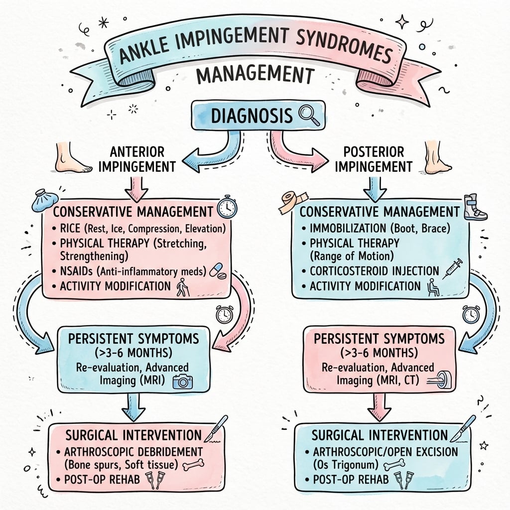
Initial management is conservative for both anterior and posterior impingement.
Activity Modification
- Relative rest: Reduce or eliminate aggravating activities 4-8 weeks
- Cross-training: Maintain fitness with low-impact alternatives (swimming, cycling)
- Sport modification: Avoid extreme dorsiflexion (anterior) or plantarflexion (posterior)
- Gradual return: Progressive loading protocol over 8-12 weeks
- Technique adjustment: Biomechanical correction to reduce impingement forces
Immobilization
- CAM boot: 2-4 weeks for severe acute symptoms
- Position: Neutral ankle position to reduce end-range compression
- Duration: Minimum necessary to avoid stiffness (2-4 weeks maximum)
- Gradual weaning: Transition to supportive athletic taping
Pharmacological Interventions
- NSAIDs: Oral (naproxen 500 mg BD) or topical for 2-4 week courses
- Analgesics: Paracetamol for pain control without inflammation
- Ice therapy: 15-20 minutes multiple times daily for acute symptoms
- Topical treatments: Anti-inflammatory gels or patches
Most patients require 3-6 months of conservative management before considering surgical intervention.
Surgical Management
Surgical Complications
Intraoperative Complications
- anteriorArthroscopy
- Superficial peroneal (2%), deep peroneal (less than 1%)
- posteriorEndoscopy
- Sural (2%), tibial (less than 1%)
- openSurgery
- Higher rates (5-8%), same nerves at risk
- management
- Most resolve spontaneously, neuroma excision if painful
- anteriorArthroscopy
- Anterior tibial artery (rare, less than 0.5%)
- posteriorEndoscopy
- Posterior tibial vessels (rare, less than 0.5%)
- openSurgery
- Higher risk with open dissection
- management
- Immediate vascular surgery consultation if identified
- anteriorArthroscopy
- Not applicable
- posteriorEndoscopy
- Laceration or scarring (1-2%)
- openSurgery
- Similar risk (1-2%)
- management
- Repair if identified, may need FHL release or transfer
- anteriorArthroscopy
- Residual osteophytes (3-5%)
- posteriorEndoscopy
- Incomplete os trigonum excision (2-4%)
- openSurgery
- Lower risk with direct visualization
- management
- Revision surgery if symptomatic, confirm on imaging
Post-Operative Complications
- Infection: 1-2% arthroscopy, 3-5% open surgery
- Stiffness: More common after anterior procedures and with prolonged immobilization
- Recurrence: 5-10% may have persistent or recurrent symptoms
- CRPS: Rare (less than 1%) but devastating complication
- Arthrofibrosis: Excessive scar formation limiting motion
- Wound problems: Delayed healing, dehiscence more common in open procedures
Failure Management
Diagnostic approach:
- Repeat imaging (CT or MRI) to assess adequacy of resection
- Consider alternative diagnoses (arthritis, instability, tarsal coalition)
- Assess rehabilitation compliance and progression
- Diagnostic injection to confirm pain generator
Revision surgery indications:
- Confirmed incomplete osteophyte resection on CT
- Residual os trigonum fragment on imaging
- Progressive symptoms despite appropriate rehabilitation
- New pathology identified (arthritis, loose body)
Revision technique:
- May require open approach after failed arthroscopy for better visualization
- More aggressive debridement of residual pathology
- Address any concurrent pathology missed initially
- Consider salvage options if severe arthritis present (fusion, arthroplasty)
Complications
Arthroscopic Procedure Complications
Nerve Injury (1-5%):
- Superficial peroneal nerve: most common with anterolateral portal
- Sural nerve: risk with posterolateral portal
- Tibial nerve: rare, posterolateral approach
- Prevention: portal placement under direct vision, mark superficial peroneal nerve preoperatively
Vascular Injury (less than 1%):
- Anterior tibial artery: anterior approach
- Posterior tibial artery: posterior approach
- Prevention: avoid deep aggressive resection, know safe zones
Tendon Injury:
- FHL: highest risk during posterior procedures (4-7% transient weakness)
- Tibialis anterior: anterior approach
- Prevention: protect FHL during os trigonum excision, visualize tendons
Instrument Breakage:
- Burr or shaver blade breakage in joint
- Management: immediate arthroscopic retrieval
Infection (less than 1%):
- Superficial wound infection: oral antibiotics
- Deep joint infection: rare, requires washout
Condition-Specific Complications
Recurrence of Impingement (5-15%):
- Incomplete resection of pathology
- Reformation of scar tissue
- Management: revision arthroscopy vs open procedure
Ankle Instability:
- Excessive lateral ligament release during anterior debridement
- Prevention: preserve ATFL fibers during capsular work
Progression to Arthritis:
- Pre-existing cartilage damage may progress despite successful debridement
- Higher risk with Scranton Grade 3-4 lesions
Postoperative Care
Immediate Postoperative (0-2 Weeks)
Weight Bearing:
- Anterior arthroscopy: Weight bear as tolerated in protective boot
- Posterior endoscopy: Protected weight bearing for 2 weeks
- Os trigonum excision: Partial weight bearing for 2 weeks
Wound Care:
- Keep dressings dry for 48-72 hours
- Portal wounds: steri-strips, minimal suturing
- Compression bandage to minimize swelling
Early Mobilization:
- Ankle pumps and gentle ROM from day 1
- Ice and elevation to control swelling
- DVT prophylaxis per surgeon preference (aspirin typically adequate for low-risk patients)
Rehabilitation Phase (2-6 Weeks)
Physiotherapy Initiation:
- Formal physiotherapy from week 2
- ROM exercises: full dorsiflexion/plantarflexion goals
- Scar mobilization at portal sites
- Proprioceptive training: wobble board progression
Strengthening:
- Week 2-4: isometric exercises
- Week 4-6: progressive resistance (theraband)
- Calf strengthening: eccentric loading progression
Return to Activity (6-12 Weeks)
Sport-Specific Rehabilitation:
- Week 6-8: jogging on flat surfaces
- Week 8-10: sport-specific drills
- Week 10-12: return to training (non-contact)
- Week 12+: return to full competition
Dancers (Ballet):
- Anterior: relevé progression from week 6
- Posterior: en pointe work from week 8-10 (os trigonum excision)
- Full performance: 3-4 months anterior, 3-5 months posterior
Follow-Up Schedule
- 2 weeks: wound check, suture removal if needed
- 6 weeks: clinical review, progress assessment
- 3 months: final review for straightforward cases
- 6-12 months: if persistent symptoms or elite athletes
Outcomes and Return to Sport
Success Rates by Procedure
Anterior arthroscopic debridement achieves good-to-excellent outcomes in appropriately selected patients.
Posterior endoscopic os trigonum excision with high success rates, best for isolated posterior pathology.
Return to pre-injury sport level achievable in most athletes with appropriate rehabilitation.
Return to Sport Timeline
- anteriorImpingement
- 4-6 months to full performance
- posteriorImpingement
- 3-5 months to en pointe work
- prognosis
- Excellent if isolated pathology, may need technique modification
- anteriorImpingement
- 3-5 months to competitive play
- posteriorImpingement
- 2-4 months to competitive play
- prognosis
- Greater than 85% return to pre-injury level
- anteriorImpingement
- 3-4 months to racing
- posteriorImpingement
- 2-3 months to racing
- prognosis
- Greater than 90% return to full training
- anteriorImpingement
- 4-6 months to competitive play
- posteriorImpingement
- 3-4 months to competitive play
- prognosis
- Good return but may have reduced vertical jump initially
Prognostic Factors
Favorable outcomes:
- Isolated anterior or posterior impingement (not combined pathology)
- No significant arthritis at time of surgery
- Young active patient with good rehabilitation compliance
- Complete resection of pathology at surgery
- Early intervention (symptom duration less than 2 years)
Poor prognostic factors:
- Concurrent moderate-severe ankle arthritis
- Combined anterior and posterior pathology
- Chronic symptoms (greater than 5 years duration)
- Previous ankle fracture or surgery
- Worker's compensation or litigation
- Smoking, diabetes, inflammatory arthropathy
Syndesmotic (High-Ankle) Impingement
The PALS classification lists syndesmotic (high-ankle) impingement as a fourth type, and the anterior section notes a pathological distal AITFL fascicle, but the entity itself is never developed.
What it is
- After a syndesmotic (high-ankle) sprain, a hypertrophic or scarred distal fascicle of the anterior-inferior tibiofibular ligament (AITFL) - Bassett's ligament - or synovial/scar tissue in the tibiofibular recess can become entrapped against the anterolateral talar dome
- This produces chronic anterolateral ankle pain in dorsiflexion and external rotation
- It is a soft-tissue impingement without frank diastasis - distinct from syndesmotic instability, where the syndesmosis is widened and unstable (covered in the syndesmotic instability topic)
Presentation and confirmation
- Deep anterolateral pain aggravated by dorsiflexion/push-off and by external-rotation stress, typically after a "high" sprain that recovered more slowly than a simple lateral sprain
- Tenderness over the anterolateral joint line and the distal AITFL
- Plain radiographs and stress views are normal (no diastasis); MRI or diagnostic arthroscopy shows the thickened distal fascicle and recess synovitis, and a diagnostic injection into the recess that relieves the pain confirms the impingement
Management
- A structured non-operative trial as for other soft-tissue impingement (activity modification, physiotherapy, image-guided injection)
- If that fails, arthroscopic debridement/resection of the impinging distal AITFL fascicle and recess synovium gives good results - provided the syndesmosis is stable
- If there is any true instability or diastasis, that must be stabilised rather than simply debrided
Q: What is syndesmotic (high-ankle) impingement and how does it differ from syndesmotic instability? A: It is a soft-tissue anterolateral impingement after a high-ankle sprain - a hypertrophic distal AITFL fascicle (Bassett's ligament) or tibiofibular-recess synovitis entrapped against the talus, causing anterolateral pain in dorsiflexion and external rotation with normal radiographs and stress views. It is confirmed by MRI/arthroscopy and a diagnostic injection and treated by arthroscopic debridement of the impinging tissue. It differs from syndesmotic instability, where the syndesmosis is widened and unstable (diastasis) and requires stabilisation, not just debridement.
Impingement and Coexisting Ankle Instability
The controversies section and Scenario 3 both stress that occult lateral ligament instability frequently underlies anterolateral impingement and is a leading cause of failed debridement, and the Gianakos systematic review found women have higher rates of associated instability - but the parent topic never sets out how to assess for and act on it.
Why they coexist
- Both follow inversion / anterolateral ankle injury: recurrent microinstability lets the talus translate abnormally, which drives the synovial hypertrophy, meniscoid lesion and capsular scarring of anterolateral impingement
- Anterolateral impingement is therefore often a marker of underlying instability rather than an isolated problem
How to assess it
- In every anterolateral impingement, actively look for instability: a history of recurrent sprains and giving-way, examination for anterior drawer and talar tilt (a clinically negative drawer does not fully exclude microinstability), and stress radiographs or MRI of the ATFL/CFL
- Maintain a high index of suspicion in women and in athletes with recurrent sprains
Why it matters at surgery
- Failure to recognise and address coexisting instability is a leading cause of persistent symptoms after debridement
- If instability is confirmed, plan to add a lateral ligament stabilisation (e.g. Broström repair) at the same sitting, and assess the ATFL intra-operatively
- Avoid over-aggressive lateral capsular release that would itself destabilise the ankle (the detailed reconstruction is covered in the lateral ankle instability topic)
Q: Why must you assess for lateral ankle instability in anterolateral impingement? A: The two share an inversion-injury mechanism, and occult microinstability drives the synovitis/meniscoid lesion of anterolateral impingement - so impingement is often a marker of instability. Look for recurrent giving-way, anterior drawer/talar tilt (a negative drawer does not exclude microinstability) and stress imaging. Untreated instability is a leading cause of failed debridement, so confirmed instability should be stabilised (e.g. Broström) at the same operation and the ATFL assessed intra-operatively - while avoiding over-aggressive capsular release that would destabilise the ankle.
Guidelines, Registries & Global Practice
Global Epidemiology
- Anterior impingement accounts for the majority of impingement-related chronic ankle pain in athletes; soccer, basketball, volleyball and gymnastics carry the highest exposure
- Posterior impingement is over-represented in ballet dancers, soccer players (kicking follow-through) and downhill runners
- Os trigonum is present in roughly 10-25% of the general population (bilateral in around half), but only a small fraction become symptomatic
- FHL tendinopathy coexists in 40-60% of posterior impingement cases, particularly in dancers, and must be screened for in every case
Society Guidance Compared
No single high-level society guideline governs ankle impingement; practice is consensus and evidence-driven. Recommendations converge internationally:
- position
- Posterior 2-portal hindfoot endoscopy (van Dijk technique) is the reference standard for os trigonum and posterior impingement
- emphasis
- Endoscopic over open where expertise exists
- position
- Arthroscopic debridement first-line for refractory anterior impingement; address concomitant instability and chondral lesions
- emphasis
- Identify and treat associated pathology
- position
- Minimum 3-6 months structured non-operative care before arthroscopy; ultrasound-guided diagnostic injection to confirm pain generator
- emphasis
- Conservative trial and diagnostic confirmation
- position
- Earlier surgery acceptable for elite athletes after a focused conservative trial; shared decision-making on timing
- emphasis
- Career and demand-adjusted timing
Registry and Evidence Notes
- Ankle impingement procedures are soft-tissue/arthroscopic and are not captured by national arthroplasty registries (NJR, AJRR, AOANJRR, SHAR) the way implant procedures are
- The strongest pooled evidence comes from systematic reviews (Zwiers 2015; Gianakos/Kennedy 2021) rather than registries, reporting around 81% good-to-excellent results with major complications near 1%
- Long-term data (Walsh 2014) show durable symptom relief despite frequent radiographic osteophyte recurrence
High- versus Limited-Resource Practice Variation
- Well-resourced settings: routine MRI to distinguish symptomatic from incidental pathology, ultrasound-guided diagnostic injection, and endoscopic/arthroscopic surgery with dedicated foot-and-ankle expertise
- Limited-resource settings: diagnosis relies on weight-bearing plain radiographs and clinical impingement tests; open excision remains a valid alternative where arthroscopy towers, fluid management or trained personnel are unavailable, accepting longer recovery and higher wound morbidity
- Universal principles: a structured conservative trial first (except high-demand athletes), confirm the pathology is symptomatic before operating, and protect the FHL and posterior neurovascular bundle whatever the approach
Controversies & Areas of Uncertainty
- Open versus endoscopic posterior excision: posterior hindfoot endoscopy is the de facto reference standard with faster recovery and lower wound morbidity, but no adequately powered randomised trial proves superiority over open excision; the open posteromedial approach remains defensible where endoscopic equipment or expertise is lacking.
- Osteophyte recurrence and its meaning: radiographic osteophytes recur in up to 84% after anterior debridement (Walsh 2014), yet functional gains persist - the clinical relevance of recurrence is debated, and motion gains are typically modest.
- Role of co-existing instability in anterolateral impingement: how often "anterolateral impingement" is actually driven by occult lateral ligament instability is unresolved; failure to address instability is a leading cause of failed debridement, prompting calls for routine intra-operative ATFL assessment.
- Symptomatic versus incidental os trigonum: there is no universal threshold for marrow oedema or injection response that reliably confirms a symptomatic ossicle, so patient selection remains partly clinical.
- Timing of surgery in elite athletes: how short a conservative trial can safely be before operating on a professional athlete (career impact versus avoidable surgery) is a matter of shared decision-making, not high-level evidence.
- Corticosteroid and biologic injections: corticosteroid gives only short-term relief and risks FHL/tendon harm; PRP and other biologics for impingement have minimal supporting data and no established role.
Key Mnemonics
PALSAnkle Impingement Types
Hook:Your PALS get ankle impingement syndromes
SPURAnterior Impingement Clinical Features
Hook:Think of the anterior ankle SPUR causing impingement
DANCERPosterior Impingement Features
Hook:Think of DANCER syndrome for posterior ankle impingement
MCQ Practice Points
Q: What MRI finding confirms a symptomatic os trigonum versus incidental finding? A: Bone marrow edema within the os trigonum indicates active inflammation and symptomatic impingement. Os trigonum is present in 10-25% of population - most are asymptomatic. MRI showing edema within the ossicle, along with posterior ankle effusion, confirms the diagnosis.
Q: What nerve is at greatest risk during anterior ankle arthroscopy and how is it protected? A: Superficial peroneal nerve is at greatest risk (crosses anterolateral portal path in 5-28% of patients). Protect by: (1) marking nerve course before incision, (2) making incision with blade perpendicular to skin only, (3) spreading subcutaneously with hemostat.
Q: In what percentage of posterior impingement cases does FHL tendinitis coexist? A: 40-60% of posterior ankle impingement cases have concurrent FHL tendinopathy, particularly in ballet dancers. Both must be addressed surgically for optimal outcome - failure to release FHL will result in persistent symptoms despite os trigonum excision.
Q: What type of ankle impingement has the best response to conservative management? A: Anterior soft tissue impingement has 67% success with conservative treatment (vs 41% for osseous anterior and 45% for posterior). Conservative measures include activity modification, NSAIDs, physiotherapy, and corticosteroid injections for 3-6 months trial.
Q: What is the Scranton and McDermott classification for anterior tibiotalar osteophytes? A: Grade 1: Less than 3mm tibial spur. Grade 2: 3-5mm tibial spur without talar involvement. Grade 3: Greater than 5mm tibial spur with secondary talar spur. Grade 4: Pantalar arthritic changes. Grades 3-4 require surgical intervention; Grade 4 may need fusion rather than debridement.
Clinical Decision Scenarios
Practise clinical reasoning and management decisions out loud
“A 24-year-old semi-professional soccer player presents with 18 months of progressive anterior ankle pain. He describes pain when kicking the ball and during deep squatting drills. He has had 4 months of physiotherapy and two corticosteroid injections with temporary relief only. Examination reveals anterolateral joint line tenderness, reduced dorsiflexion to 5 degrees (normal 15 degrees contralateral), and pain with forced dorsiflexion. Weight-bearing lateral radiograph shows 6 mm tibial and talar osteophytes with kissing lesions. MRI confirms moderate anterior impingement with grade 2 cartilage changes but no significant arthritis.”
“A 19-year-old professional ballet dancer presents with 12 months of posterior ankle pain preventing en pointe work. She describes deep aching pain during plantarflexion that has progressively worsened. She occasionally feels triggering with great toe flexion. Examination shows posterior ankle tenderness lateral to Achilles, painful forced plantarflexion, and positive FHL stretch test with triggering. MRI demonstrates os trigonum with bone marrow edema, posterior joint effusion, and FHL tenosynovitis in zone 1. She has a major audition in 4 months and asks if surgery can get her ready in time.”
“A 28-year-old recreational netballer has had anterolateral ankle pain for 10 months following an inversion sprain. She reports catching and a feeling of fullness in the anterolateral gutter when she pushes off, but denies true giving-way. Examination shows anterolateral joint-line tenderness, a positive Molloy impingement test, a negative anterior drawer and a negative talar tilt. Weight-bearing radiographs are normal with no osteophytes. She has completed 4 months of physiotherapy including proprioceptive work.”
Must-Know Anatomy
- Anterior portals: anteromedial (medial to TA), anterolateral (lateral to EDL/PT)
- Posterior portals: posterolateral and posteromedial, 1-2 cm proximal to superior calcaneus
- Superficial peroneal nerve: crosses anterior ankle, mark before anterolateral portal
- Os trigonum: posterior to talus, present 10-25% population, bilateral 50% of cases
- FHL tendon: between talar tubercles, at risk during posterior procedures
Anterior vs Posterior Features
- ANTERIOR: dorsiflexion pain, soccer/running, tibiotalar spurs (footballer's ankle)
- POSTERIOR: plantarflexion pain, ballet/soccer, os trigonum (dancer's syndrome)
- Anterior test: forced passive dorsiflexion reproduces pain
- Posterior test: forced passive plantarflexion (nutcracker test) elicits pain
- Imaging: lateral XR shows osseous pathology, MRI shows soft tissue and edema
Conservative Management
- Activity modification 3-6 months first-line treatment
- NSAIDs and physiotherapy addressing contributing factors
- Corticosteroid injection: 40-60% temporary relief, max 2-3 injections
- Success rates: 50-60% anterior (better soft tissue), 40-50% posterior
- Predictors of failure: large osteophytes (greater than 5 mm), mechanical symptoms, professional athletes
Surgical Indications
- Failed 3-6 months appropriate conservative treatment
- Persistent symptoms limiting activities or sport participation
- Large osteophytes (greater than 5 mm) or symptomatic os trigonum on imaging
- Professional athletes after 6-12 weeks conservative trial
- Mechanical symptoms (locking, catching) suggesting structural lesion
Arthroscopic Technique Pearls
- ANTERIOR: supine, non-invasive distraction, AM/AL portals, shaver for synovium, burr for osteophytes
- POSTERIOR: prone position, posterolateral/posteromedial portals, excise os trigonum, protect FHL
- Superficial peroneal nerve: anterolateral portal risk, mark pre-op with dorsiflexion
- Tibial nerve: posteromedial portal risk, stay posterior and use blunt dissection
- Complete resection: confirm no residual impingement with ROM testing under visualization
Viva Traps
- Don't rush to surgery - 3-6 months conservative trial required unless professional athlete
- Recognize concurrent FHL pathology in posterior impingement (40-60% coexist in dancers)
- Endoscopic approach preferred when available: faster recovery, lower complications
- Realistic timeline: 3-4 months anterior, 2-3 months posterior to return to sport
- Professional ballet dancers: 4-6 months to en pointe, technique modification may be needed
Critical Numbers
- Osteophyte size: less than 3 mm (mild), 3-5 mm (moderate), greater than 5 mm (severe requiring surgery)
- Conservative success: 50-60% anterior, 40-50% posterior with appropriate treatment
- Surgical success: 85-90% anterior, 90-95% posterior with endoscopic approach
- Return to sport: 3-5 months anterior, 2-4 months posterior arthroscopy
- Complication rate: 4% anterior, 3-4% posterior endoscopy (nerve injury most common)
Evidence Base
Original 2-Portal Hindfoot Endoscopy (Landmark Technique)
- First description of the 2-portal posterior (hindfoot) endoscopic approach in the prone position
- Posterolateral and posteromedial portals give access to posterior ankle, subtalar joint and peri-articular structures
- Index case: professional ballet dancer with bilateral os trigonum and FHL tendinitis treated by ossicle excision and FHL release
- Patient resumed professional dancing within 2 months of endoscopic treatment
Arthroscopic Treatment of Anterior Ankle Impingement (Systematic Review)
- Systematic review of 20 studies on arthroscopic treatment of anterior ankle impingement
- Good-to-excellent patient satisfaction in 74% to 100% across reporting studies
- 94.3% to 97.5% of patients would undergo the same procedure again
- Overall complication rate 4.6%, major complications only 1.1%
Outcomes of Arthroscopy for Anterior Ankle Impingement (Sex Differences)
- Systematic review of 28 articles evaluating 1,506 patients
- Good-to-excellent results with an overall success rate of 81.0%
- Average complication rate 4.0%; commonest were mild nerve symptoms and superficial infection
- Female patients had higher rates of traumatic sprain, chondral injury and chronic instability than males
Arthroscopic Debridement for Anterior Impingement: 5-Year Prospective Outcomes
- Prospective series of 46 patients without osteoarthritis, mean age 29, minimum 5-year follow-up
- Foot Function Index improved from 20.5 pre-op to 2.7 at final follow-up (p less than 0.001)
- Dorsiflexion gain was small (24.7 to 27.0 degrees) and clinically modest
- 84% showed radiographic recurrence of osteophytes, yet functional gains were maintained
Endoscopic Excision of Os Trigonum in Recreational Athletes
- Retrospective series of 81 recreational athletes (mean age 27.8) with posterior impingement from os trigonum
- AOFAS hindfoot score improved from 39.4 pre-op to 97.7 at 1 year
- VAS pain fell from 7.5 to 0.6 at 1 year; only 5 patients dropped to a lower activity level
- 5 complications (4 transient); no permanent neurovascular injury
Posterior Ankle Arthroscopy for Hindfoot Impingement
- 15 patients (16 ankles) with posterior impingement, mean follow-up 32 months
- Procedures: os trigonum excision (11), posterior process decompression (5), FHL tenolysis (5)
- Mean AOFAS hindfoot score 91; mean return to sport at 5.8 months; 14 returned to pre-injury level
- Only transient scar numbness (5) and transient stiffness (1); no permanent neurovascular injury