Chronic medial heel pain | Differentiate from plantar fasciitis | Three compression sites | EMG diagnosis
- Differentiation from plantar fasciitis is critical - Baxter's has less morning pain and more neuritic quality
- Nerve runs deep to abductor hallucis, superficial to quadratus plantae, then turns laterally to innervate ADM
- Three common compression sites: deep fascia of abductor hallucis, plantar fascia origin, fibrous arch of ADM origin
- EMG is diagnostic gold standard showing prolonged distal motor latency greater than 6.2ms
- Surgical release must identify and decompress all three potential compression sites
- “Minimal morning pain and neuritic quality distinguish from plantar fasciitis
- “All three compression sites (abductor hallucis fascia, plantar fascia, ADM arch) require release
- “EMG gold standard with DML >6.2 ms; ~15% false negative rate
- “Anatomic variation in ~15%: proximal origin within tarsal tunnel
Anatomy & Pathophysiology

Nerve Anatomy
Origin and Branching:
- Terminal branch of tibial nerve in tarsal tunnel
- Divides into medial and lateral branches
- First branch (Baxter's nerve) arises 5-8mm distal to medial malleolus
- Runs anterolaterally between muscle layers
- Innervates abductor digiti minimi (ADM) muscle
- May give sensory branches to medial calcaneal periosteum
Muscle Compartments:
- Superficial: deep fascia of abductor hallucis
- Deep: superior surface of quadratus plantae
- Lateral turn: passes under plantar fascia origin
- Terminal: enters ADM through fibrous arch
- Proximity to calcaneal spur formation site
- Adjacent to medial calcaneal neurovascular bundle
Clinical Presentation
History
- Chronic medial heel pain (duration typically greater than 6 months)
- Pain worse after prolonged standing or walking
- Minimal to no morning pain (contrast to plantar fasciitis)
- Neuritic quality: burning, tingling, electric-like sensations
- Pain radiates along medial heel, may extend to medial arch
- Symptoms worsen throughout day with activity
- Night pain uncommon unless severe
- Duration and onset of symptoms (gradual onset typical)
- Quality of pain (neuritic vs mechanical)
- Diurnal variation (morning vs evening worse)
- Response to previous plantar fasciitis treatments
- Occupational demands (prolonged standing, walking)
- History of trauma or previous heel surgery
- Medical history (diabetes, peripheral neuropathy, inflammatory arthritis)
- Acute onset suggests alternative diagnosis (stress fracture, rupture)
- Progressive weakness of foot musculature suggests severe neuropathy
- Night pain suggests tumor or infection
- Bilateral symptoms suggest systemic cause or tarsal tunnel syndrome
- Constitutional symptoms suggest systemic disease
Physical Examination
- Evaluate hindfoot alignment (varus or valgus)
- Assess for cavus foot deformity
- Look for muscle atrophy of abductor digiti minimi (late finding)
- Check for signs of prior surgery or trauma
- Observe for pes planus or high arch
- Antalgic gait with shortened stance phase on affected side
- Reduced push-off power
- May demonstrate lateral weight shift to avoid medial heel pressure
- Assess cadence and step length symmetry
Muscle atrophy is a late finding and indicates chronic severe denervation. Compare the bulk of the lateral foot musculature to the contralateral side. Subtle atrophy may only be apparent with careful side-to-side comparison.
Investigations - EMG and Nerve Conduction Studies
Electrodiagnostic testing is the gold standard for confirming Baxter's nerve entrapment and should be performed before surgical intervention.
- Motor Study: Stimulate tibial nerve at ankle, record from abductor digiti minimi
- Normal Distal Motor Latency (DML): 4.0-6.2 milliseconds
- Abnormal DML: Greater than 6.2 milliseconds diagnostic of entrapment
- Amplitude: Reduced compared to contralateral side suggests axonal loss
- Abductor digiti minimi: Abnormal spontaneous activity (fibrillations, positive sharp waves)
- Other lateral plantar nerve muscles: Should be normal in isolated Baxter's
- Medial plantar nerve muscles: Normal (helps exclude tarsal tunnel syndrome)
- Proximal muscles: Normal (excludes lumbosacral radiculopathy)
- Prolonged DML with normal amplitude: demyelination (neurapraxia)
- Prolonged DML with reduced amplitude: axonal injury (axonotmesis)
- Comparison to contralateral side helpful if unilateral symptoms
- False negative rate approximately 15% especially in early or mild cases
Imaging
Imaging Modality Comparison
IMAGEIMAGE - Diagnostic Workup Sequence
Hook:IMAGE your diagnostic pathway from basic to advanced studies — Exam Tip: The key to diagnosis is systematic clinical examination with specific attention to quality of pain (mechanical vs neuritic), location of tenderness (fascia vs nerve course), and response to standard plantar fasciitis treatments.
Diagnostic Injection
Local Anesthetic Injection
Diagnostic injection can be both confirmatory and therapeutic, though it should be performed with caution and proper technique.
- Identify maximal point of tenderness (abductor hallucis-quadratus plantae interval)
- Use 25-27 gauge needle
- Inject 2-3mL of local anesthetic (lidocaine 1% or bupivacaine 0.25%)
- Can add 20-40mg methylprednisolone acetate for therapeutic effect
- Complete relief: Confirms diagnosis of Baxter's nerve entrapment
- Partial relief: May indicate coexisting pathology (plantar fasciitis)
- No relief: Questions diagnosis, consider alternative causes
- Temporary relief only: Suggests nerve compression rather than irreversible damage
- Avoid multiple injections (risk of fat pad atrophy, plantar fascia rupture)
- Use ultrasound guidance when available for accurate placement
- Counsel patient regarding potential complications
- Maximum 2-3 injections with minimum 6-week intervals
Management Algorithm
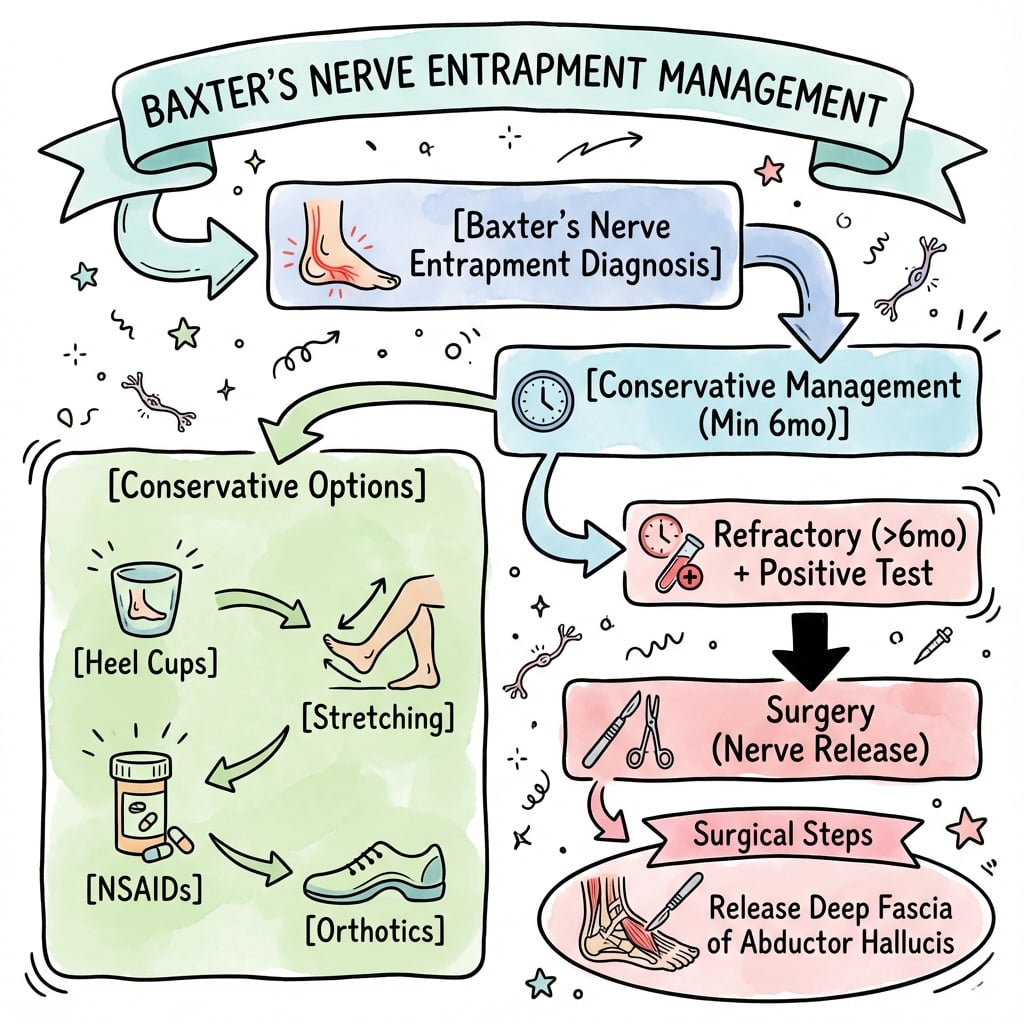
Conservative Treatment
- Reduce prolonged standing and walking
- Low-impact exercise alternatives (cycling, swimming)
- Weight loss if BMI greater than 25
- Avoid barefoot walking on hard surfaces
- Job modification if occupational demands excessive
- Cushioned heel cups with medial arch support
- Custom orthotics to improve foot biomechanics
- Medial heel wedge if valgus hindfoot alignment
- Soft insoles to reduce heel strike impact
- Intrinsic foot muscle strengthening
- Plantar fascia stretching (towel stretch)
- Gastrocnemius and soleus stretching
- Nerve gliding exercises for tibial nerve
- Ultrasound therapy to reduce inflammation
- Iontophoresis with dexamethasone
- Ice massage for acute flares
- Night splints to maintain ankle dorsiflexion
- NSAIDs: 2-4 week course for anti-inflammatory effect
- Neuropathic pain medications: Gabapentin 300-900mg daily or pregabalin 75-150mg twice daily
- Topical treatments: Capsaicin cream or compound topical analgesics
- Avoid: Oral corticosteroids (limited evidence, systemic side effects)
- Seated position with knee extended
- Dorsiflex ankle to neutral, invert foot
- Plantarflex ankle while maintaining inversion
- Return to neutral position
- Repeat 10 times, 3 sets daily
- Promotes nerve mobility and reduces adhesions
Original Description of Calcaneal Nerve Entrapment in Athletes
Surgical Treatment
Technique: Open Decompression via Medial Incision
- Supine position with bump under ipsilateral hip
- Thigh tourniquet application
- Ensure access to medial heel and ankle
- Fluoroscopy available but typically not required
-
Incision: 4-6cm curvilinear incision centered 2cm distal and plantar to medial malleolus, along posterior border of abductor hallucis
-
Exposure: Incise deep fascia, identify and protect posterior tibial nerve branches, retract abductor hallucis muscle inferiorly
-
Nerve Identification: Locate first branch of lateral plantar nerve between abductor hallucis (superficial) and quadratus plantae (deep), typically 5-8mm distal to medial malleolus
-
Site 1 Decompression: Release deep fascia of abductor hallucis from its calcaneal origin, free nerve from fascial tunnel
-
Site 2 Decompression: Follow nerve laterally to plantar fascia origin, partially release plantar fascia (medial 30%) if compressing nerve, protect nerve during fascia release
-
Site 3 Decompression: Trace nerve to ADM muscle, release fibrous arch at muscle origin, ensure complete nerve mobility
-
Inspection: Verify nerve freely mobile along entire course, no residual compression points, check for intraneural scarring or nerve thickening
-
Closure: Repair deep fascia loosely, dermal and skin closure, soft dressing, posterior splint in neutral
- Nerve may be small and easily missed (2-3mm diameter)
- Complete decompression requires release of all three sites
- Avoid excessive traction on nerve during dissection
- Partial plantar fascia release only when necessary (30% medial release safe)
- Mark nerve with vessel loop to avoid inadvertent injury
The key to successful surgery is identifying the nerve and systematically releasing all three compression sites. Incomplete decompression is the most common cause of surgical failure.
Landmark Surgical Series (Baxter & Pfeffer)
Combined Fascia Release plus Nerve Decompression
Rehabilitation Protocol
Post-Operative Rehabilitation Timeline
Nerve Gliding Post-Operatively:
- Begin at 2-3 weeks after surgery
- Promotes nerve mobility and prevents adhesions
- Technique same as conservative treatment protocol
- Perform 2-3 times daily throughout recovery
- Critical for optimal nerve healing and function
Complications
Surgical Complications
- Failure to identify nerve (most common technical error)
- Inadvertent transection or excessive traction
- Injury to other tibial nerve branches
- Prevention: meticulous dissection, loupe magnification, vessel loop marking
- Missed compression sites (especially Site 3)
- Inadequate fascial release
- Failure to trace nerve to ADM muscle
- Prevention: systematic approach to all three sites
- Delayed healing due to tension or hematoma
- Superficial infection
- Dehiscence requiring revision closure
- Prevention: careful incision placement, hemostasis, minimal tension closure
- Numbness from calcaneal sensory branch injury
- Painful neuroma formation
- Medial heel dysesthesias
- Treatment: desensitization, neuropathic medications, neuroma excision rarely
- Rare if complete decompression achieved (less than 5%)
- Usually due to incomplete initial decompression
- May result from scar tissue reformation
- Treatment: revision surgery with neurolysis, consider nerve transposition
- Risk with excessive plantar fascia release (greater than 50%)
- Lateral column overload syndrome
- Gradual development of flatfoot deformity
- Prevention: limit plantar fascia release to 30% medial portion
- Treatment: arch support orthotics, rarely requires surgical reconstruction
Differential Diagnosis
Chronic Heel Pain Differential Diagnosis
The MRI Sign: Abductor Digiti Minimi Denervation - and Its Limits
Two of this topic's own EvidenceCards (Chen 2025; Tedeschi 2025) and the MCQ section all invoke the MRI sign of Baxter's neuropathy, but the imaging section never develops it - and the MCQ's claim that it is the "most specific finding" actually sits in tension with the Chen systematic review that headlines this page.
- The sign is denervation of the abductor digiti minimi (ADM). Because Baxter's nerve is (largely) the motor nerve to the ADM, entrapment produces denervation change confined to the ADM with the medial-plantar-innervated muscles spared. Acute/subacute denervation shows as muscle OEDEMA (T2/STIR hyperintensity in the ADM); chronic denervation shows as FATTY INFILTRATION and ATROPHY (T1 fatty replacement and loss of bulk). Isolated ADM change points to the first branch of the lateral plantar nerve.
- But it is common and non-specific. The Chen 2025 systematic review (4 studies, 1052 participants) found ADM fatty infiltration in 4-11% of the general population, at similar rates in people with and without foot pain (about 8% vs 6%), with no data specific to plantar heel pain or confirmed Baxter's - so the association is unproven. This reconciles the "most specific finding" claim: ADM fatty infiltration is the classic sign but is not diagnostic in isolation and must be correlated with the clinical picture and electrodiagnostics (Tedeschi 2025 makes the same point).
- Practical use. MRI supports the diagnosis and, importantly, excludes the mimics (calcaneal stress fracture, mass, plantar fasciitis) - but you should not offer decompression on an incidental ADM fatty-infiltration finding alone.
Q: What is the MRI sign of Baxter's neuropathy and how specific is it? A: Denervation of the abductor digiti minimi - oedema (T2/STIR) acutely, fatty infiltration/atrophy (T1) chronically, with the medial-plantar muscles spared. It is the classic sign, but the 2025 systematic review found it in 4-11% of the general population and equally in people with and without foot pain, so it is common and non-specific - correlate with the clinical picture and EMG, and do not operate on an incidental finding.
Ultrasound-Guided Perineural Hydrodissection
The Guidelines section lists "image-guided injection or hydrodissection before surgery", but the topic never explains what hydrodissection is - and it is the modern minimally-invasive step that now sits between failed simple conservative care and open surgery.
- What it is. An ultrasound-guided injection in which a volume of fluid (normal saline or local anaesthetic, with or without corticosteroid; some use dextrose for perineural injection therapy) is delivered into the fascial plane immediately around the nerve to mechanically separate ("hydro-dissect") it from the adjacent abductor-hallucis fascia and adhesions, relieving compression while the needle and fluid spread are watched in real time.
- Where it sits. A minimally-invasive option between failed conservative treatment and open decompression: it is both diagnostic (like a targeted local-anaesthetic block confirming the nerve as the pain source) and therapeutic, and is safer than a blind heel steroid injection because seeing the plane avoids depositing steroid into the fat pad or plantar fascia (the source of the fat-pad-atrophy and plantar-fascia-rupture complications).
- Caveats. The technique is increasingly used and is supported by the image-guided-injection literature for entrapment neuropathies generally, but high-quality evidence specifically for Baxter's is still limited; the usual steroid-volume and frequency cautions apply, and it does not replace surgery for a truly recalcitrant, electrodiagnostically-confirmed entrapment.
Q: What is ultrasound-guided hydrodissection and where does it fit? A: A real-time ultrasound-guided injection of fluid (saline/LA ± steroid) into the plane around the nerve to mechanically free it from the compressing abductor-hallucis fascia and adhesions. It is both diagnostic and therapeutic, sits between failed conservative care and open decompression, and is safer than a blind heel steroid injection (the plane is seen, avoiding fat-pad/plantar-fascia complications). Evidence specific to Baxter's is limited, and it does not replace surgery for a recalcitrant EMG-confirmed entrapment.
Expected Outcomes
- Success rate: 50-60% with comprehensive conservative management
- Time to improvement: 3-6 months typical
- Better outcomes if treated early (symptoms less than 12 months)
- Recurrence uncommon if modifiable risk factors addressed
- Good to excellent results: 75-85% at 2-5 year follow-up
- Symptom improvement: 90% achieve significant pain reduction
- Complete resolution: 60-70% have no residual symptoms
- Time to maximal improvement: 3-6 months post-operatively
- Return to pre-symptom activity level: 70-80%
- EMG confirmation of diagnosis (prolonged DML)
- Symptom duration less than 2 years
- Positive response to diagnostic injection
- No workers' compensation claim
- Isolated Baxter's without complex plantar fasciitis
- Nerve identified and decompressed at all three sites
- Symptom duration greater than 2 years
- Workers' compensation or litigation pending
- Multiple prior heel surgeries
- Failure to identify nerve during surgery
- Coexisting tarsal tunnel syndrome
- Peripheral neuropathy from diabetes or other causes
- Incomplete decompression at initial surgery
- Return to provocative activities without modification
- Failure to address biomechanical factors (obesity, foot alignment)
- Development of scar tissue or adhesions
- Progression of underlying neuropathy
Long-Term Outcomes:
- Majority maintain good results at 5+ years if successful initially
- Small percentage develop recurrent symptoms (5-10%)
- Some patients develop plantar fasciitis or other heel pathology later
- Importance of continued biomechanical optimization with orthotics and weight management
Contemporary Review: An Under-Recognised Cause of Heel Pain
Guidelines, Registries & Global Practice
Global Epidemiology
- Plantar heel pain affects roughly 1 in 10 people over a lifetime and is one of the commonest foot complaints presenting to primary care worldwide
- Baxter's neuropathy is widely cited as accounting for up to 20% of chronic heel pain, though this figure derives from expert review rather than population studies
- It frequently coexists with plantar fasciitis rather than occurring in isolation
- A systematic review (1052 participants) found fatty infiltration of abductor digiti minimi on MRI in 4-11% of the general population, with similar rates in people with and without foot pain - so the imaging sign is common and not specific
- True population prevalence of symptomatic Baxter's neuropathy remains undefined
- Higher BMI and occupations requiring prolonged standing or walking
- Runners and athletes (the original athletic cohorts of Henricson and Westlin)
- Cavus or pronated foot morphology and coexisting plantar fasciitis
- Diabetes and other causes of peripheral neuropathy lower the threshold for symptoms and worsen outcomes
First Report of the Muscle-Branch Entrapment
MRI Fatty Infiltration of ADM: Systematic Review
MCQ Practice Points
Q: What is Baxter's nerve and what is its anatomical course?
A: Baxter's nerve is the first branch of the lateral plantar nerve (inferior calcaneal nerve). Course: Arises from lateral plantar nerve in tarsal tunnel, passes between abductor hallucis and quadratus plantae, then changes direction 90° to run laterally toward the abductor digiti minimi. Entrapment sites: (1) Between abductor hallucis fascia and quadratus plantae (most common); (2) Medial calcaneal tuberosity (plantar fascia origin).
Q: What are the clinical features of Baxter's nerve entrapment and how does it differ from plantar fasciitis?
A: Baxter's nerve: Burning/neuralgic medial heel pain; May radiate laterally; Worse with activity; Tenderness at abductor hallucis origin (more medial than plantar fascia); Potential abductor digiti minimi weakness/atrophy; Negative Tinel's at tarsal tunnel. Plantar fasciitis: Localized tenderness at plantar fascia origin (medial calcaneal tubercle); Worse with first steps in morning; No neurogenic character. The conditions often coexist (chronic fasciitis can cause secondary nerve compression).
Q: What diagnostic studies are useful for Baxter's nerve entrapment?
A: MRI: May show atrophy and fatty infiltration of abductor digiti minimi (denervation changes) - most specific finding; May also show plantar fasciitis if coexistent. Electrodiagnostic studies: Prolonged distal motor latency to abductor digiti minimi; Fibrillations/positive sharp waves in ADM; Technically challenging due to anatomical access. Ultrasound: Can assess for abductor hallucis hypertrophy or plantar fascia thickening. Diagnosis often clinical.
Q: What is the conservative management of Baxter's nerve entrapment?
A: Initial management (similar to plantar fasciitis): Activity modification, night splints, orthotic heel cups with medial arch support, stretching (plantar fascia, Achilles), NSAIDs. Specific measures: Avoid compressive footwear; Medial heel wedge to offload compression site; Corticosteroid injection (cautious - may cause plantar fascia rupture or fat pad atrophy). Response to conservative treatment less reliable than for plantar fasciitis alone.
Q: What is the surgical treatment for refractory Baxter's nerve entrapment?
A: Nerve release via medial heel incision. Technique: Incision posterior to medial malleolus to plantar aspect; Identify lateral plantar nerve and first branch; Release abductor hallucis fascia (deep and superficial); Follow nerve distally, release any impinging structures including plantar fascia if thickened. Combined procedure: If coexistent plantar fasciitis, perform partial plantar fasciotomy (medial 1/3). Success rate 80-90% for isolated Baxter's release.
At a Glance
Baxter's nerve entrapment is compression of the first branch of the lateral plantar nerve (nerve to abductor digiti minimi), a common cause of chronic medial heel pain often misdiagnosed as plantar fasciitis. Key differentiator: Baxter's has less morning pain, more neuritic quality, and tenderness at the abductor hallucis-quadratus plantae interval rather than the medial calcaneal tuberosity. The nerve courses between abductor hallucis (superficial) and quadratus plantae (deep), with three compression sites: deep fascia of abductor hallucis, plantar fascia origin, and fibrous arch of ADM. EMG is diagnostic (motor latency over 6.2ms to ADM). Conservative treatment succeeds in 50-60%; surgical release requires identifying all three compression sites.
BAXTERBAXTER - Nerve Anatomic Course
Hook:Remember BAXTER's path from medial to lateral under the arch of the foot — Exam Tip: Always assess for tarsal tunnel syndrome in addition to isolated Baxter's entrapment, especially if symptoms extend proximal to the heel or involve other branches of tibial nerve.
Compression Sites
Three Sites of Nerve Compression
Pathophysiology
Compression Neuropathy Mechanism
The first branch of the lateral plantar nerve is susceptible to compression due to its anatomic course through multiple fibromuscular tunnels. Chronic compression leads to focal demyelination and axonal injury.
Stages of Nerve Injury (Seddon Classification):
- Neurapraxia - Early stage with focal demyelination, reversible with decompression
- Axonotmesis - Axonal injury with intact epineurium, potential for recovery after decompression
- Neurotmesis - Complete nerve disruption, rare in chronic compression syndromes
The compression is exacerbated by:
- Repetitive stress during heel strike phase of gait
- Prolonged standing increasing weight-bearing pressure
- Plantar fascia inflammation pulling on nerve at compression sites
- Calcaneal spur formation narrowing available nerve space
- Muscle hypertrophy in active individuals reducing tunnel diameter
COMPRESSCOMPRESS - Risk Factors for Baxter's Nerve Entrapment
Hook:Remember what can COMPRESS the nerve in its tight anatomical course — Exam Tip: Key distinguishing features from plantar fasciitis: less morning pain, worsens with activity duration, neuritic quality (burning/tingling), poor response to plantar fasciitis treatments
Viva Scenarios
Practise clinical reasoning and management decisions out loud
“A 48-year-old obese mail carrier presents with 2-year history of medial heel pain that has failed orthotics, physical therapy, NSAIDs, and one corticosteroid injection. Pain is described as burning, worse after long postal route, minimal in morning. Tenderness at abductor hallucis-quadratus plantae interval. EMG shows prolonged distal motor latency of 7.8ms to abductor digiti minimi. How would you manage this patient?”
“You performed an open Baxter's nerve decompression 6 months ago. The patient initially improved for 2 months but now reports recurrent symptoms identical to pre-operative pain. What are the potential causes and how would you manage this situation?”
Definition
- Compression neuropathy of first branch of lateral plantar nerve (nerve to ADM)
- Presents as chronic medial heel pain with neuritic quality
- Often misdiagnosed as plantar fasciitis
Anatomy
- Nerve arises 5-8mm distal to medial malleolus
- Runs between abductor hallucis (superficial) and quadratus plantae (deep)
- Turns laterally to innervate abductor digiti minimi (ADM)
- Three compression sites: deep fascia of abductor hallucis, plantar fascia origin, fibrous arch at ADM
Clinical Diagnosis
- Medial heel pain worse with prolonged standing
- Minimal morning pain (unlike plantar fasciitis)
- Neuritic quality: burning, tingling
- Tenderness 2-3cm distal to medial malleolus at muscle interval
- Positive nerve percussion test (Tinel sign)
Investigations
- EMG/NCS gold standard: prolonged DML to ADM greater than 6.2ms diagnostic
- Plain radiographs to rule out fracture and bone pathology
- MRI shows muscle edema, not required if EMG positive
- Diagnostic injection confirms nerve source of pain
Conservative Treatment
- Success rate 50-60%
- Orthotics with medial arch support
- Activity modification, NSAIDs, neuropathic pain meds (gabapentin)
- Nerve gliding exercises and physical therapy
- Trial 3-6 months minimum before surgery
Surgical Treatment
- Open medial approach: identify nerve between abductor hallucis and quadratus plantae
- Decompress all three compression sites
- Partial plantar fascia release if compressing nerve
- Success rate 75-85%; complication rate 9%
Outcomes
- Good-excellent results in 78% at 2 years
- Better: symptom duration less than 18 months, positive EMG, positive injection response
- Worse: workers' comp, obesity, diabetes, symptom duration greater than 2 years
Differential
- Plantar fasciitis: morning pain worse, mechanical not neuritic
- Tarsal tunnel syndrome: multiple nerve branches, positive Tinel at tunnel
- Calcaneal stress fracture: squeeze test positive, MRI diagnostic
- Fat pad atrophy: thin heel pad, direct calcaneal pressure pain