Posterior Tibial Nerve Compression | Flexor Retinaculum | Medial Ankle
- Posterior Tibial Nerve Branches: Medial plantar (abductor hallucis), lateral plantar (intrinsics), medial calcaneal (heel sensation)
- Tinel Sign: Most reliable clinical test - tap posterior to medial malleolus
- Space-Occupying Lesions: 20-30% have identifiable cause (ganglion, lipoma, varicosities)
- Double Crush: Exclude proximal nerve compression (L4-S2 radiculopathy)
- Surgical Release: Must decompress entire tunnel including abductor hallucis fascia
- “Burning plantar pain worse at night = classic presentation
- “Always examine for intrinsic muscle weakness (toe spread)
- “MRI before surgery to identify space-occupying lesions
- “Incomplete release = recurrence - extend distally through abductor tunnel
Posterior tibial nerve posterior to medial malleolus. Passes beneath flexor retinaculum with posterior tibial artery and tendons (Tom, Dick, And Very Nervous Harry).
Medial calcaneal first, then bifurcation. Medial plantar (larger, sensory dominant), lateral plantar (motor dominant). All may be affected.
Tinel most reliable. Dorsiflexion-eversion stress test positive in 80%. Check intrinsic muscle power and sensation.
Complete release essential. Must decompress flexor retinaculum AND abductor hallucis origin. Identify and protect all branches.
- Investigations
- NCS baseline
- Treatment
- Orthotics, activity modification
- Key Pearl
- Trial 3 months conservative treatment
- Investigations
- MRI to exclude lesion
- Treatment
- Corticosteroid injection trial
- Key Pearl
- Injection confirms diagnosis if relief
- Investigations
- Urgent MRI and NCS
- Treatment
- Surgical decompression
- Key Pearl
- Complete release including distally
TOM, DICK And Very Nervous HARRYTarsal Tunnel Contents
Hook:Anterior to posterior order - Tom, Dick And Very Nervous Harry. The NERVE is vulnerable between the vessels and FHL!
MLCPTN Terminal Branches
Hook:MLC = Medial, Lateral, Calcaneal - the three branches you must identify and decompress!
Overview and Epidemiology
TTS is the lower limb analogue of carpal tunnel syndrome but is much less common. Understanding the anatomy and terminal branches is essential for exam and surgical planning.
Tarsal Tunnel Syndrome is compression of the posterior tibial nerve or its terminal branches beneath the flexor retinaculum on the medial aspect of the ankle.
- Female predominance: 2:1 ratio
- Peak age: 40-60 years
- Bilateral: 25% of cases
- Associated conditions: Pes planus, diabetes, RA
Less common than carpal tunnel syndrome but important to recognise.
- Trauma: Ankle fractures, sprains (most common)
- Pes planus: Increased nerve tension
- Space-occupying lesions: Ganglion, lipoma
- Systemic: Diabetes, hypothyroidism, RA
20-30% have identifiable mass lesion causing compression.
Anatomy and Pathophysiology
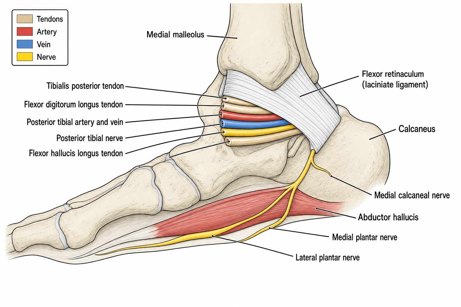
The tarsal tunnel is a fibro-osseous tunnel on the medial ankle. The ROOF is the flexor retinaculum (laciniate ligament) extending from medial malleolus to calcaneus. The FLOOR is the medial talus, sustentaculum tali, and medial calcaneus.
- Roof: Flexor retinaculum (laciniate ligament)
- Floor: Medial surface of talus, sustentaculum tali, medial calcaneus
- Anterior: Medial malleolus
- Posterior: Medial calcaneal tuberosity
- Tibialis posterior tendon
- Flexor digitorum longus tendon
- Posterior tibial artery and veins
- Posterior tibial nerve
- Flexor hallucis longus tendon
- Medial calcaneal nerve: First branch, sensory to medial heel
- Medial plantar nerve: Larger terminal branch, sensory to medial 3.5 toes, motor to abductor hallucis, FHB, FDB, first lumbrical
- Lateral plantar nerve: Smaller terminal branch, motor to intrinsics, sensory to lateral 1.5 toes
- Increased pressure within the tarsal tunnel (greater than 30 mmHg)
- Venous congestion and nerve ischaemia
- Demyelination (reversible in early stages)
- Axonal damage (irreversible - motor weakness, atrophy)
- Fibrosis and adhesions (chronic cases)
Aetiology and Classification
Aetiological Classification
- Examples
- Ankle fractures, sprains, dislocation
- Frequency
- Most common
- Examples
- Ganglion, lipoma, neurilemoma, varicosities
- Frequency
- 20-30%
- Examples
- Pes planus, hindfoot valgus, tarsal coalition
- Frequency
- Common
- Examples
- Diabetes, hypothyroidism, RA, amyloidosis
- Frequency
- Variable
- Examples
- No identifiable cause
- Frequency
- 30-40%
Always investigate for underlying cause - MRI before surgery.
SPACECauses of Tarsal Tunnel Syndrome
Hook:SPACE = Something is taking up SPACE in the tunnel! Look for mass lesions.
Biomechanical TTS: The Traction Neuropathy of the Valgus Hindfoot
The topic repeatedly names pes planus and hindfoot valgus as causes ("increased nerve tension") and tells you to assess hindfoot alignment, but the mechanism — and what it means for treatment — is never developed. It matters because it is a fundamentally different problem from a space-occupying lesion.
The mechanism is traction, not compression. A planovalgus hindfoot (collapsed medial arch with heel valgus) places the posterior tibial nerve and its branches under stretch. As the heel everts and the arch falls, the nerve is tensioned and angulated as it curves around the medial malleolus and sustentaculum tali. Tellingly, dorsiflexion-eversion — the position of the most sensitive provocative test — is essentially the resting posture of the valgus foot, so the nerve is chronically loaded. This is a traction (stretch) neuropathy, distinct from the pressure of a ganglion or lipoma.
What it changes:
- Assessment: always examine and document standing hindfoot alignment. A flexible planovalgus foot that corrects on tiptoe (double-heel-rise) flags a biomechanical, potentially correctable contributor; a rigid valgus (e.g. tarsal coalition) does not.
- Conservative care: for biomechanical TTS, orthotic correction of the valgus (medial arch support, medial heel posting) is genuinely therapeutic — it offloads the traction and can relieve symptoms without surgery, which is why orthotics feature so prominently in the conservative plan.
- Surgical nuance (the unresolved point): a tunnel release that leaves the deforming traction force uncorrected may fail. For a fixed symptomatic valgus, some surgeons advocate realigning the hindfoot (e.g. a medialising calcaneal osteotomy or flatfoot reconstruction) alongside, or instead of, a simple release — recognising that you must address the cause of the stretch, not just the tunnel. This is not standardised, but it is why the traction mechanism is worth understanding. (General flatfoot reconstruction is developed in the pes-planus / flatfoot topics.)
Q: Why does a flatfoot cause tarsal tunnel syndrome, and how does that change management? A: A planovalgus hindfoot puts the tibial nerve under traction as the heel everts and the arch collapses (dorsiflexion-eversion, the provocative position, is its resting posture) — a stretch neuropathy, not a compression. So assess standing alignment, correct the valgus with orthotics first, and for a fixed deformity consider hindfoot realignment rather than relying on release alone.
Clinical Assessment
- Burning pain: Plantar foot and toes
- Nocturnal symptoms: Worse at night, wake from sleep
- Aggravating factors: Prolonged standing, walking
- Radiation: Along medial arch to toes
- Associated: Weakness of toe flexion
Burning plantar pain worse at night is virtually diagnostic.
- Tinel sign: Tap posterior to medial malleolus
- Dorsiflexion-eversion test: Positive in 80%
- Two-point discrimination: Greater than 6mm abnormal
- Intrinsic muscle testing: Toe spread, FHB power
- Hindfoot alignment: Check for pes planus
Always compare to contralateral side.
Provocative Tests:
- Technique
- Tap posterior to medial malleolus
- Sensitivity
- 58%
- Specificity
- 92%
- Technique
- Hold 30 seconds, reproduces symptoms
- Sensitivity
- 81%
- Specificity
- 85%
- Technique
- 30 seconds over tunnel
- Sensitivity
- 50%
- Specificity
- 90%
- Technique
- DF + eversion + compression
- Sensitivity
- 85%
- Specificity
- 88%
Consider: Plantar fasciitis (different location), Morton neuroma (forefoot), L5-S1 radiculopathy (check back), peripheral neuropathy (bilateral, diabetics), Baxter neuropathy (first branch LPN).
Double Crush Syndrome: The Proximal Lesion You Must Not Miss
The must-know list and a viva trap both warn you to "exclude proximal nerve compression (L4-S2 radiculopathy)" and "consider double crush", but the concept is never explained — and it is one of the commonest reasons a tarsal tunnel release fails.
The double-crush hypothesis (Upton and McComas). A nerve compressed at one point along its length is rendered more susceptible to symptomatic dysfunction from a second, often subclinical, compression elsewhere — the two lesions summate rather than acting independently. The proposed mechanism is that the proximal compression impairs axoplasmic flow, lowering the threshold at which a distal site becomes symptomatic.
Why it applies to TTS. The tibial nerve carries L4-S3 fibres, so a coexisting lumbosacral radiculopathy (disc or foraminal stenosis) — or a more proximal sciatic/tibial lesion — can "prime" the very axons that are then finished off at the tarsal tunnel. The same logic links TTS to a coexisting diabetic polyneuropathy, which is itself a diffuse "crush".
What it changes in practice:
- It explains some "idiopathic" and "failed-release" TTS — a distal decompression cannot fix a missed proximal contributor.
- It mandates a proximal screen before committing to surgery: examine the lumbar spine, straight-leg raise, reflexes and proximal myotomes, and ask the electrodiagnostician to sample proximal muscles and paraspinals, not just the foot.
- It supports treating both sites (and tempering surgical expectations) when two lesions coexist — and raises the threshold for releasing a tunnel when the dominant problem is proximal.
Q: A patient with clinical TTS has back pain and bilateral symptoms; the distal nerve conduction study is borderline. What concept must you apply? A: Double crush. A proximal lesion (L4-S2 radiculopathy, or a diffuse diabetic neuropathy) lowers the threshold for symptomatic distal compression via impaired axoplasmic flow. Screen and image the spine, sample proximal muscles on EMG, treat both sites, and do not expect a tarsal tunnel release alone to fix a missed proximal cause.
Investigations
Nerve Conduction Studies
Gold standard for confirmation but false negative rate 30-50%.
- Normal
- Less than 4.4 ms
- Abnormal
- Greater than 4.4 ms
- Normal
- Less than 4.6 ms
- Abnormal
- Greater than 4.6 ms
- Normal
- Greater than 5 mcV
- Abnormal
- Reduced or absent
EMG findings in motor involvement:
- Fibrillation potentials in intrinsics
- Positive sharp waves
- Reduced recruitment
NCS may be normal in 30-50% of clinical TTS.
Management
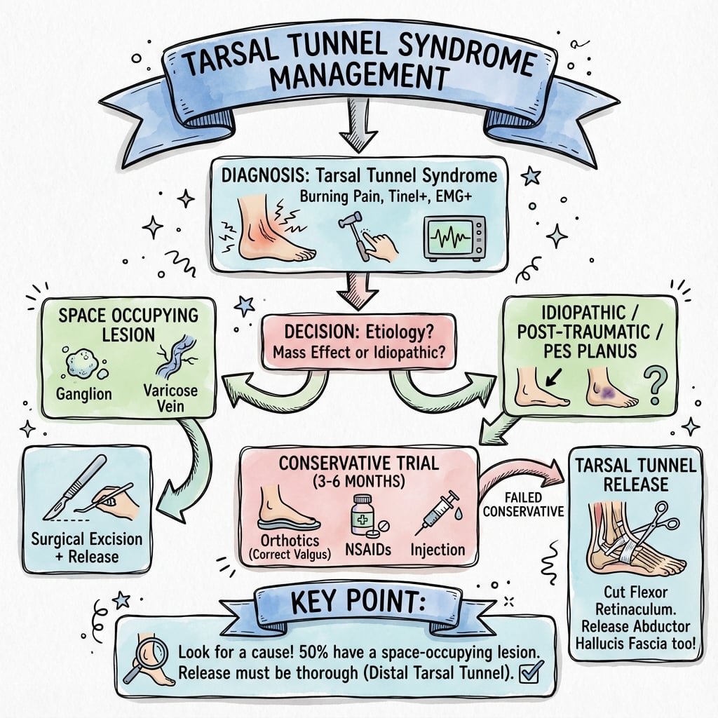
Non-Operative Management
First-line treatment for 3-6 months.
- Avoid prolonged standing
- Limit high-impact activities
- Comfortable, supportive footwear
- Medial arch support for pes planus
- Heel cushioning
- Custom orthotics if required
- NSAIDs for pain relief
- Gabapentin for neuropathic pain
- Topical capsaicin
- Nerve gliding exercises
- Stretching programme
- Strengthening
Success rate 40-50% with conservative treatment.
Surgical Technique
Complete Tarsal Tunnel Release
- Failed conservative treatment (3-6 months)
- Space-occupying lesion
- Progressive motor deficit
- Intractable symptoms
- Supine with leg externally rotated
- Thigh tourniquet
- Foot at end of table
- Curvilinear incision posterior to medial malleolus
- Extend distally along abductor hallucis
- Length 6-8 cm for adequate exposure
- Identify and protect posterior tibial vessels
- Incise flexor retinaculum completely
- Identify main PTN trunk
- Trace nerve proximally and distally
- Identify and release all three branches
- Release medial calcaneal nerve
- Continue release through abductor hallucis tunnel
- Excise any mass lesions
- Leave retinaculum open
- Close subcutaneous tissue and skin
- Bulky dressing
- Must release distally through abductor tunnel
- Identify and protect all branches
- Excise mass lesions completely
- Neurolysis if fibrosis present
These principles ensure complete decompression and optimal outcomes.
Complications
- Recurrence: 10-20% (incomplete release)
- Wound complications: Delayed healing, infection
- Nerve injury: Damage to branches
- Scar tethering: Nerve adhesions
- Persistent symptoms: Incomplete decompression
Most complications from inadequate release.
- Complete release: Include abductor tunnel
- Meticulous technique: Identify all branches
- MRI preoperatively: Plan for mass lesions
- Gentle handling: Minimise nerve trauma
- Early mobilisation: Reduce adhesions
Attention to detail prevents recurrence.
- Incidence
- 10-20%
- Prevention
- Full exposure distally
- Management
- Revision surgery
- Incidence
- 5%
- Prevention
- Careful closure, offload
- Management
- Wound care, possible grafting
- Incidence
- 15-30%
- Prevention
- Proper patient selection
- Management
- NCS, consider revision
- Incidence
- 2-5%
- Prevention
- Early mobilisation
- Management
- Pain management, therapy
Postoperative Care and Rehabilitation
- Bulky dressing and posterior splint
- Elevation above heart level
- Non-weight bearing 2 weeks
- Rest, ice, elevation
- Ankle pumps
- Wound check at 2 weeks
- Transition to weight-bearing as tolerated
- Gentle range of motion
- Scar massage once healed
- Progressive strengthening
- Return to normal footwear
- Gradual return to activity
- Burning pain relief: 2-4 weeks
- Sensory improvement: 3-6 months
- Motor recovery: 6-12 months (if present)
- Full recovery: 6-12 months
Outcomes and Prognosis
Prognostic Factors:
- Poor Prognosis
- Idiopathic TTS
- Poor Prognosis
- Chronic symptoms over 12 months
- Poor Prognosis
- Failed injection
- Poor Prognosis
- Motor deficit present
- Poor Prognosis
- Denervation on EMG
Guidelines, Registries & Global Practice
There are no dedicated national-society clinical practice guidelines specific to tarsal tunnel syndrome from the major bodies, reflecting its rarity and the limited high-level evidence. Practice is instead shaped by foot-and-ankle society consensus, electrodiagnostic practice parameters and surgical experience, and is broadly consistent worldwide.
- Region
- International / US
- Position
- NCS may confirm tibial neuropathy at the ankle (Level C); diagnosis remains clinical
- Region
- US
- Position
- Stepwise care - conservative first, image to exclude mass lesion, release for refractory or compressive cases
- Region
- UK
- Position
- Similar stepwise pathway; MRI and electrodiagnostics used selectively before surgery
- Region
- Europe
- Position
- Emphasise excluding differentials (polyneuropathy, radiculopathy) and identifying a structural cause
- Region
- International
- Position
- No robust evidence base; calls for a structured, step-wise approach and RCTs
Global epidemiology and practice variation:
- TTS is uncommon and consistently under-recognised; precise population incidence is unknown across all regions.
- No arthroplasty/implant-style registries track TTS, as treatment is soft-tissue decompression rather than implantation.
- High-resource settings: routine pre-operative MRI and electrodiagnostics, ultrasound-guided diagnostic injection, and access to microsurgical neurolysis.
- Limited-resource settings: diagnosis is predominantly clinical with provocative tests; MRI and nerve conduction studies may be unavailable, so identifiable causes (post-traumatic deformity, large masses) are prioritised for surgery.
- Across all settings the surgical principle is identical: complete release of the flexor retinaculum, decompression of all branches, distal release through the abductor hallucis fascia, and excision of any space-occupying lesion.
Controversies and Areas of Uncertainty
TTS is a high-yield viva topic precisely because much of its management rests on low-level evidence. Be ready to defend a position while acknowledging the uncertainty.
No accepted diagnostic gold standard exists. Clinical criteria, provocative tests and electrodiagnostics each have limitations, and there is no validated case definition - so the same foot may be labelled TTS by one clinician and plantar heel pain or polyneuropathy by another.
True sensitivity/specificity of NCS could not be determined in the AANEM review, and a normal study does not exclude TTS. Some surgeons require electrodiagnostic confirmation before release; others operate on a strong clinical picture plus imaging.
Endoscopic release offers smaller incisions but cannot address mass lesions and risks incomplete decompression. There are no high-quality randomised comparisons; open complete release remains the reference standard, especially when a space-occupying lesion is present.
Outcomes are substantially worse without an identifiable cause. Whether truly idiopathic cases benefit from surgery at all is debated, and many advocate exhausting conservative options and confirming with diagnostic injection first.
MCQ Practice Points
Q: What is the order of structures in the tarsal tunnel from anterior to posterior? A: Tom, Dick And Very Nervous Harry - Tibialis posterior, Flexor Digitorum longus, Artery (posterior tibial), Vein, Nerve (posterior tibial), Flexor Hallucis longus. The posterior tibial nerve lies between the vessels anteriorly and FHL posteriorly.
Q: A patient has burning pain in the plantar foot. Which test is most sensitive for tarsal tunnel syndrome? A: Dorsiflexion-eversion stress test has highest sensitivity (81%) and specificity (85%). Hold position for 30 seconds to reproduce symptoms. Tinel sign is more specific (92%) but less sensitive (58%).
Q: What is the most common cause of recurrence after tarsal tunnel release? A: Incomplete release, particularly failure to decompress distally through the abductor hallucis tunnel. Complete release must include the flexor retinaculum proximally and extend through the abductor hallucis fascia distally to fully decompress the medial and lateral plantar nerves.
Q: Which patient with tarsal tunnel syndrome is most likely to have a good surgical outcome? A: Patient with space-occupying lesion (ganglion, lipoma) has approximately 90% success rate. Idiopathic cases have only 50% success. Other good prognostic factors include short duration of symptoms, sensory only symptoms, and positive response to diagnostic injection.
Q: Which branch of the posterior tibial nerve branches first within the tarsal tunnel? A: Medial calcaneal nerve branches first (proximal in the tunnel), providing sensory innervation to the medial heel. The nerve then bifurcates into medial plantar (larger, sensory dominant) and lateral plantar (motor dominant) branches more distally.
Clinical Decision Scenarios
Practise clinical reasoning and management decisions out loud
“How would you assess this patient?”
“What is your management plan?”
“How would you approach this?”
Anatomy
- Tom, Dick And Very Nervous Harry = contents anterior to posterior
- Three terminal branches: medial calcaneal (first), medial plantar, lateral plantar
- Flexor retinaculum = roof; medial talus, sustentaculum tali and medial calcaneus = floor
Clinical Features
- Burning plantar pain worse at night
- Tinel positive posterior to medial malleolus
- Dorsiflexion-eversion test most sensitive
- Intrinsic weakness = advanced disease
Investigations
- NCS: prolonged distal motor latency (less than 4.4ms MPN)
- MRI: essential to identify mass lesions (20-30% have one)
- NCS false negative rate 30-50%
Treatment Algorithm
- Conservative 3-6 months: orthotics, activity modification, medications
- Injection: diagnostic and therapeutic, 50-60% temporary relief
- Surgery if failed conservative or progressive motor deficit
Surgical Pearls
- Curvilinear incision posterior to medial malleolus
- Complete release of flexor retinaculum
- MUST extend through abductor hallucis tunnel distally
- Identify and protect all three branches
Outcomes
- 60-85% overall success
- 90% success with mass lesion
- 50% success idiopathic
- Recurrence 10-20% (incomplete release)
Evidence Base
Surgical Release Outcomes (Objective vs Subjective)
- 60 patients (68 feet) undergoing tarsal tunnel release; all had positive Tinel sign and abnormal motor nerve conduction
- 85% complete symptom relief by objective assessment, but only 51% by subjective patient-reported assessment
- Significant improvement in work quality, productivity and interpersonal relationships
- Highlights a dichotomy between objective and patient-perceived outcomes - counsel realistically
Anatomic Pain Scale & Predictors of Non-operative Failure
- Prospective evaluation of 46 patients (56 feet) treated non-operatively or surgically
- Surgery improved medial calcaneal and medial plantar - but not lateral plantar - nerve region pain
- Predictors of failed conservative treatment: longer motor nerve conduction latency and more foot comorbidities
- Anatomic pain mapping aids pre- and post-treatment assessment
Electrodiagnostic Testing in TTS (AANEM Evidence-Based Review)
- Systematic review of 317 articles; only 4 met methodological criteria, all Class III evidence
- Sensory NCS more often abnormal than motor NCS, but true sensitivity and specificity could not be determined
- NCS may help confirm tibial neuropathy at the ankle (Level C recommendation)
- Diagnosis remains primarily clinical; well-designed studies still needed
Dorsiflexion-Eversion Provocative Test
- Test described in 50 normal volunteers (100 feet) and 37 patients (44 feet) treated operatively
- Maximal ankle dorsiflexion + eversion + MTPJ dorsiflexion held 5-10 s stretches and compresses the nerve beneath the laciniate ligament
- Reproduced or intensified symptoms in the large majority of affected feet; induced no symptoms in any normal foot
- Symptoms abolished post-release (mean 2.9 months) except in calcaneal fracture-related cases
Variable Tibial Nerve Branching in the Tarsal Tunnel
- Gross anatomical study of 20 cadaveric lower limbs using the malleolar-calcaneal axis as reference
- Bifurcation proximal to the axis (within/above the tunnel) in 55%, at the axis in 30%, distal in 15%
- High and variable bifurcation means branches must be sought across the whole tunnel at surgery
- Knowledge of branching reduces risk of incomplete release and iatrogenic nerve injury
Narrative Review of Diagnosis and Management
- Narrative review identifying 88 relevant articles across multiple databases
- TTS is rare and frequently under-diagnosed; the optimal stage for conservative vs surgical care remains unclear
- Limited high-level evidence exists to guide management; no robust RCTs available
- Calls for a structured, step-wise, evidence-based treatment pathway